1429
MS
Three Pathways between the Sacroiliac Joint
and Neural Structures
Joseph D. Fortin, William J. Washington, and Frank J. E. Falco
BACKGROUND AND PURPOSE: Despite ongoing clinical suspicion regarding the relation-ship between sacroiliac joint (SIJ) dysfunction and lower extremity symptoms, there is a paucity of scientific literature addressing this topic. The purpose of this study was to describe patterns of contrast extravasation during SIJ arthrography and postarthrography CT in patients with lower back pain and to determine whether there are pathways of communication between the SIJ and nearby neural structures.
METHODS: Fluoroscopically guided SIJ arthrography was performed on 76 SIJs. After the injection of contrast medium, anteroposterior, lateral, and oblique radiographs as well as 5-mm contiguous axial and direct coronal CT images were obtained. Contrast extravasation patterns were recorded for each joint. These observations included a search for contrast ex-travasation from the SIJ that contacted nearby lumbosacral nerve roots or structures of the plexus.
RESULTS: Sixty-one percent of all joints studied revealed one of five contrast extravasation patterns. Three of these observed patterns show a pathway of communication between the SIJ and nearby neural structures. These included posterior extravasation into the dorsal sacral foramina, superior recess extravasation at the sacral alar level to the fifth lumbar epiradicular sheath, and ventral extravasation to the lumbosacral plexus.
CONCLUSION: Three pathways between the SIJ and neural structures exist.
At the turn of the century, strain of the sacroiliac joint (SIJ) was regarded as the primary etiologic factor leading to symptoms of sciatica (1). Later, in 1936, Pitkin and Pheasant (2) described lower ex-tremity pain as originating in the sacroiliac and lumbosacral joints and their accessory ligaments and coined the term sacroarthrogenic telalgia. For decades, SIJ fusion was a common surgical pro-cedure used to alleviate symptoms of sciatica (3). After Mixter and Barr’s (4) initial description of the herniated nucleus pulposus, focus on the SIJ as a mediator of lower extremity symptoms fell from favor.
Since that time, however, there has been a grow-ing understandgrow-ing that morphologic changes are not necessarily predictive of the symptom complex of any individual with lower back pain (527). For
Received, June 13, 1997; accepted after revision, March 9, 1999.
From Spine Technology and Rehabilitation, Indiana Uni-versity School of Medicine (J.D.F.), Fort Wayne, IN, the Neu-ro Care Center (W.J.W.), Canton, OH, and Comprehensive Spine & Sports Medicine (F.J.E.F.), Wilmington, DE.
Address reprint requests to Joseph D. Fortin, DO, Division of Spine Technology and Rehabilitation, 7230 Engle Road, Suite 210 Fort Wayne, IN 46804.
qAmerican Society of Neuroradiology
instance, structural changes of the intervertebral disk, including some disk herniations causing neu-ral compression, are seen in asymptomatic individ-uals (5, 7). Despite ongoing clinical suspicion re-garding the relationship of the SIJ to lower extremity symptoms, there is a paucity of scientific literature addressing this topic.
re-FIG1. ‘‘Normal’’ right SIJ arthrograms of a 32-year-old woman with right-sided lower back and hip pain after a motor vehicle accident. A, Anteroposterior view. Note the characteristic coin-shaped inferior recess (arrowhead) and the bead of contrast within the joint margin (closed arrow). Symbols: S, sacrum; IL, ilium; 3, S3 dorsal sacral foramina.
B, Right anterior oblique projection delineates the full extent of the contrast medium within the joint space (wavy arrow). The closed arrow is directed to the needle tip.
C, Left anterior oblique view. This ‘‘en face’’ projection reveals the auricular configuration of the SIJ surface (dark arrows). Symbols: 4, pedicle of L4; 5, pedicle of L5; 1, pedicle of S1.
D, The ventral capsule of the joint is well demarcated (arrows) in this lateral, plain-film arthrogram.
lationship between the SIJ and nearby neural structures.
Methods
Forty-three consecutive patients with lower back pain (mean age, 33 years; age range, 24248 years), were enrolled in the study after receiving Internal Review Board approval. The par-ticipants met the following criteria: back pain present for lon-ger than 2 months; lower back pain:lower extremity pain ratio of greater than or equal to 75%:25%; no evidence of motor or sensory changes revealed by examination to suggest radicu-lopathy; no allergy to contrast media or iodine; Beta human chorionic gonadotropin-negative; and willingness to participate in the study. Investigators chose to study the SIJs on the symp-tomatic sides of the participants. Thirty-one of the 43 partici-pants had bilateral complaints, which allowed for examination of 76 SIJs.
SIJ arthrography was performed as described in a previous study by Fortin and Tolchin (16). Complete arthrograms were obtained in all cases, and no complications resulting from the procedure were observed. No complications were anticipated because previous studies suggested that arthrography is a safe procedure in carefully selected participants (8, 9, 16). None-theless, patients were monitored for allergy to contrast medi-um, postprocedural infection, and bleeding. These problems were minimized by excluding those with histories of allergy to iodinated materials or contrast medium, using a sterile tech-nique and 25-gauge spinal needles. Moreover, there are no ma-jor vessels along the needle pathway when using the technique previously described by Fortin et al (8, 9). After the injection of contrast medium, anteroposterior, lateral, and oblique radi-ographs were obtained for each SIJ. Postarthrography CT was performed on the same joints. Five-millimeter contiguous transaxial and direct coronal images were acquired. A 2208
gantry tilt was used for the transaxial scans, and a1208gantry angle was used for the coronal scans. Window-level settings as well as bone-detail algorithms were adjusted for optimal visualization of soft tissue and osseous structures. Two ob-servers (J.F., F.F.), both of whom are experienced interpreters of SIJ arthrograms, graded the arthrograms and postarthrog-raphy CT findings. The joints were divided into anterior, pos-terior, superior, and inferior components for regional recording of contrast patterns from both techniques. Choices of contrast patterns included anterior, posterior, superior, and inferior ex-travasation. Other findings, such as capsular attenuation (ie, capsular bulge without associated extravasation) or diverticula, were noted by the observers but were not the focus of this study. In addition to recording the region of contrast extrava-sation from the joints, the observers were asked to search for extravasation of contrast medium from the SIJ that contacted nearby lumbosacral nerve roots or structures of the sacral plexus.
There was complete agreement between both reviewers re-garding all but one case in which extension of contrast medium from the superior aspect of the joint had occurred. Both re-viewers observed the leakage of contrast medium, yet one failed to document the extension of contrast medium into the adjacent L5 root canal, which was undoubtedly present on postarthrography CT scans (Fig 5). Complete concordance of findings between plain films and postarthrography CT was achieved in terms of the region of extravasation. CT was nec-essary to visualize the neural elements and to determine wheth-er contrast medium contacted them.
Results
Pattern of extravasation No. observed Percentage
Ventral
Dorsal to first sacral foramina Dorsal subligamentous Superior to sacral ala Inferior
None
12 6 18 2 9 29
16% 8% 24% 3% 12% 38%
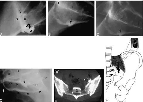
FIG2. Images of a 36-year-old man with left-sided lower back, posterior hip, and thigh pain, who sustained an offshore work-related
lifting injury.
A, Anteroposterior arthrogram of the inferior aspect of the left SIJ shows a normal inferior recess (r), bead of contrast within the joints margins (arrow), and collection of contrast medium escaping through a ventral tear (VT).
B, Lateral view confirms that the collection of contrast medium escaping through a ventral tear (arrow) is remote to the needle tip, which is in the inferior aspect of the capsule.Symbols: 1, body of S1; 2, body of S2; 3, body of S3.
C, Same projection as that shown in B except with a wider field of view. Arrow indicates ventral tear.
D, Offset opposite lateral arthrogram discloses an intact ventral capsule (arrowheads) on the contralateral side compared with a disrupted capsule with a ventral tear (arrows). White arrow points to the needle in inferior aspect of right SIJ.
E, Post-arthrography axial CT scan obtained at the S2 level (bone window/level settings), with contrast medium in both SIJs. Presacral collection of contrast medium is evidence of a left ventral capsular tear (open arrow). Contrast medium contracts the lumbosacral plexus elements (arrowheads).
F, Line drawing of the ventral hemipelvis, which allows contrast medium to escape and contact the neural elements of the sacral plexus (interrupted lines). The inset shows the torn fibers of the ventral capsule allowing contrast medium to leak slowly.
of all SIJs studied revealed extravasation of con-trast medium. Observed concon-trast medium extrava-sation patterns from the ventral, dorsal, superior, and inferior aspects of the 76 SIJ capsules are dis-played in Table 1.
(Fig 2). Other ventral capsular findings, not includ-ed as frank contrast minclud-edium extravasation, deserve special mention. Six arthrograms revealed ventral capsular attenuation without extravasation, whereas nine others exhibited a wispy pattern of extrava-sation through a small ventral schism (Fig 3).
Dorsal leakage of contrast medium was seen in 24 (32%) of all SIJs. In six of these, contrast me-dium was observed to enter the first dorsal sacral foramen, which overlies the first sacral nerve root (Fig 4). In the others, contrast medium layered be-tween the dorsal sacroiliac ligament and the sacrum.
[image:3.612.61.541.266.609.2]cap-FIG 3. Capsular attention (ie, focal
cap-sular bulge) and schism in a 28-year-old man with an insidious onset of lower back pain.
A, Concentric area of ventral capsular attenuation is displayed at soft-tissue set-tings (arrowheads) of this postarthrogra-phy axial CT scan obtained at the S2 level. B, Markedly attenuated area of the ven-tral capsule and sacroiliac ligament allows seepage of contrast medium in a feathery, wispy dispersal pattern (ie, schism with in-distinct margins) into the presacral region (arrow).
FIG4. A 42-year-old laborer with back pain and occasional posterior right lower extremity pain to the calf. A potential pathway between the SIJ and anterior ramus of S1 is present.
A, Arthrogram of the anteroposterior view reveals contrast medium extending (dotted line) into the S1 dorsal foramina (1). Symbols: 2, S2 dorsal foramina; 5, pedicle of L5.
B and C, Axial and direct coronal postarthrography CT scan through the S1 foramina (soft-tissue window/level settings). Contrast is observed in the right S1 dorsal foramina on both scans (arrowheads).
D, Compare this axial CT scan at the S1 level in another patient after bilateral SIJ arthrography to the above case. The arrowheads point to the S1 anterior rami bilaterally. Notice on the right how contrast medium encircles the S1 segmental root.
year-old patient with lower back pain and left lower extremity paresthesias.
A, Arthrogram of the anteroposterior view allowed visualization of contrast me-dium extravasating from the superior re-cess (wavy line) of the SIJ toward the L5 root canal. Symbols: 5, L5 vertebral body; S, sacral ala; IL, ilium.
B, Postarthrography axial CT scan ob-tained at the L52S1 level reveals contrast medium extending to the L5 epiradicular sheath (arrows). The arrowhead points to the contralateral L5 anterior ramus. Sym-bols:S, sacral ala; IL, ilium. Refer to Figure 4E for a comparative line drawing.
sular recess along the sacral ala to the fifth lumbar epiradicular sheath (Fig 5).
Discussion
During the past several decades, there have been numerous reports of patients suspected of having idiopathic SIJ pain who also have associ-ated lower extremity symptoms, including radi-cular pain (2, 10, 11). The mechanism by which lower extremity pain occurs in patients with SIJ dysfunction remains unknown. Unfortunately, spi-nal morphologic studies often fail to be adequately predictive of the true pain generator in any given patient. Studies that seek to explain morphologic and physiologic changes and how they relate to the evolution of spinal pain syndromes are much needed.
In this study, five principal patterns of extracap-sular contrast extravasation from the SIJ were ob-served using plain-film arthrography and postar-thrography CT. Three of these patterns reveal a potential pathway of communication between the SIJ and nearby neural structures. These include posterior extension into the dorsal sacral foramina, superior recess extravasation at the sacral alar level to the fifth lumbar epiradicular sheath, and ventral leakage to the lumbosacral plexus.
Frank extravasation of contrast medium is easily visible on both plain-film arthrograms and thrography CT scans. The added value of postar-thrography CT lies in its greater sensitivity for de-tection of subtle capsular changes and extravasation of small amounts of contrast medium. This is es-pecially true for the ventral capsule in which changes of normal capsular structure are seen. The range of ventral capsular findings may represent points on a continuum of the same process. The patterns of ventral capsular attenuation and schism later might become frank ventral tears.
The large percentage of observed dorsal extrav-asation is not surprising considering that the dorsal capsule is discontinuous compared with the ventral capsule, which is a continuous sheet of connective tissue (10). The dorsal capsule allows contrast me-dium to track along the posterior aspect of the
sa-crum and enter the dorsal sacral foramen relatively unimpeded.
Our findings raised some intriguing questions that have provided the substrate for investigations that are currently underway. Is it plausible that in the setting of capsular disruption, intra-articular contents, including inflammatory chemical media-tors in symptomatic patients, could leak from the SIJ in a manner similar to the extracapsular contrast extravasation patterns observed in this study? If so, irritation of adjacent neural structures could mani-fest as lower extremity symptoms.
The role of contrast-enhanced anatomic studies as progenitors in revealing spine pain mechanisms bears historical significance. For example, Lind-blom (18) provided the foundation for the under-standing of discogenic pain when he injected red dye into the nucleus pulposus of cadaveric disks and observed it leaking through annular rents. Similarly, we think that patterns of communica-tion from the SIJ to nearby neural structures may have significance. There is evidence of a substan-tial role for biochemical or inflammatory media-tors in discogenic and facet joint pain (12215). Accordingly, the pathophysiologic mechanism by which SIJ dysfunction leads to lower extremity pain may include biochemical factors that affect regional nerves via direct communication from the SIJ.
Conclusion
Contrast extravasation after SIJ arthrography can be visualized in patients with lower back pain. Five extravasation patterns were observed, including three pathways of communication between the SIJ and nearby neural structures.
References
1. Goldthwaite GE, Osgood RB. Consideration of the pelvic artic-ulations from an anatomical, pathological, andchemical stand-point. Boston Med Surg J 1905;152:593–601
2. Pitkin HC, Pheasant HC. Sacroarthrogenic telalgia. J Bone Joint Surg Am 1936;18:111–133
4. Mixter WJ, Barr JS. Rupture of the intervertebral disc with involvement of the spinal canal. N Engl J Med 1934;211:210– 215
5. Bodin SD, David DO, Dina TS. Abnormal magnetic resonance scans of the lumbar spine in asymptomatic subjects. A pro-spective investigation. J Bone Joint Surg Am 1990;72:403–408 6. Hitselberger WE, Witten RM. Abnormal myelograms in
asymp-tomatic patients. J Neurosurg 1968;28:204–206
7. Jensen MC, Brant-Zawadski MN, Obuchowski N, Modic MT, Malkasian D, Ross JS. Magnetic resonance imaging of the lum-bar spine in people without back pain. N Engl J Med 1994;2: 69–73
8. Fortin JD, Aprill CN, Ponthieux B, Pier J. Sacroiliac joint. Pain referral maps upon applying a new injection/arthrography technique. Part II. Clinical evaluation. Spine 1994;19:1483– 1489
9. Fortin JD, Dwyer AP, West S, Pier J. Sacroiliac joint. Pain re-ferral maps upon applying a new injection/arthrography tech-nique. Part I. Asymptomatic volunteers. Spine 1994;19:1475– 1482
10. Bernard TN Fr, Cassidy DJ. SIJ syndrome. In: Frymoyer J, ed. The Adult Spine. Principles and Practice. New York: Raven Press 1991;2107–2130
11. Bernard TN Jr, Kirkaldy-Willis WA. Recognizing specific char-acteristics of nonspecific low back pain. Clin Orthop 1987;217: 266–280
12. Beaman DN, Graziano CP, Glover RA, Wojtys EM, Chang V. Substance P innervation of lumbar spine facet joints. Spine 1993;18:1044–1049
13. Saal JS, Franson RC, Dobrow R, Saal JA, White AH, Goldthwaite N. High levels of inflammatory phospholipase A2 activity in lumbar disc herniations. Spine 1990;15:674–678
14. Willburger RE, Wittenberg RH. Prostaglandin release from lum-bar disc and fat tissue. Spine 1994;19:2068–2070
15. Wittenberg RH, Willburger RE, Kleemeyer KS, Peskar BA. In vitro release of prostaglandins and leukotrienes from synovial tissue, cartilage, and bone in degenerative joint diseases.
Ar-thritis Rheum 1993;36:(10)1444–1450
16. Fortin JD, Tolchin R. Sacroiliac arthrograms and post-arthrog-raphy CT. Arch Phys Med Rehabil 1993;74:1259 [Abstract] 17. Willard FH. The muscular, ligamentous and neural structure
of the low back and its relation to back pain. In: Vleeming A, Mooney V, Dorman T, Snijders C, Stoeckart R, eds. Movement Stability and Low Back. New York: Churchill Livingstone; 1997; 14–17