The organisation and function of the brain serotonergic system seems to be highly conserved across the vertebrate subphylum (Parent et al., 1984). Serotonin (5-hydroxytryptamine, 5-HT) is involved in the regulation of the hypothalamic–pituitary–adrenocortical (HPA) axis in mammals (Chaoulof, 1993; Dinan, 1996) as well as in the control of the hypothalamic–pituitary–interrenal (HPI) axis (the teleostean homologue of the HPA axis) in fish (Winberg and Lepage, 1998; Øverli et al., 1999; Höglund et al., 2000). In several studies, the ratio of 5-hydroxyindoleacetic acid (5-HIAA, the major 5-HT metabolite) to 5-HT brain concentrations has been found to correlate with plasma levels of cortisol, suggesting that the action of brain 5-HT on the HPI axis is stimulatory (Øverli et al., 2000; Höglund et al., 2000; Winberg and Lepage, 1998). Moreover, treatment with 8-OH-DPAT, a selective 5-HT1Areceptor agonist, elevates plasma
levels of cortisol in cannulated rainbow trout (Winberg et al., 1997). The role of the brain 5-HT system in the control of the HPI axis is, however, not clear. Brain serotonergic pathways in mammals are important when coping with stress, not only by initiating the adrenocortical stress response but also terminating it (Markus et al., 2000b). The brain 5-HT system is not a unitary system and, in mammals, 5-HT pathways terminating in the hypothalamic paraventricular nucleus stimulate HPA axis activity, whereas those terminating in the
hippocampus inhibit it (Jacobson and Sapolsky, 1991; Deakin, 1991; Deakin and Graeff, 1991; Maes and Meltzer, 1995; Summers et al., 1998; Markus et al., 2000b).
Serotonin is synthesized from the essential amino acid
L-tryptophan (TRP), and the first and rate-limiting step in
the biosynthesis of 5-HT is the hydroxylation of TRP to 5-hydroxytryptophan (5-HTP). Since the enzyme tryptophanhydroxylase (TPH), catalysing the hydroxylation of TRP, does not seem to be saturated by TRP in vivo, the rate of this reaction appears to be restricted by TRP availability both in mammals (for a review, see Boadle-Biber, 1993) and in teleost fish (Aldegunde et al., 1998, 2000; Winberg et al., 2001). Elevated dietary intake of TRP has been reported to result in increased brain levels of TRP and elevated rates of 5-HT synthesis and metabolism (Johnston et al., 1990; Aldegunde et al., 1998, 2000; Winberg et al., 2001). In mammals, increased functional release of 5-HT following elevated dietary intake of TRP has been confirmed using microdialyses and in vivo voltametry (for a review, see Boadle-Biber, 1993).
Serotonin seems to inhibit aggressive behaviour in all vertebrates, and the fact that elevated dietary intake of TRP suppresses aggressive behaviour in rainbow trout
Oncorhynchus mykiss (Winberg et al., 2001) suggests that
elevated dietary intake of TRP in fish also results in increased synaptic 5-HT release.
JEB4243
Juvenile rainbow trout were isolated in individual compartments and allowed to acclimate for 1 week, during which they were fed commercial trout pellets. The feed was then replaced by pelleted feed supplemented with L-tryptophan (TRP) at two, four or eight times the concentration in the commercial feed. Fish were fed these supplemented feeds daily to satiety for 1 week, after which half of the fish were stressed, by lowering the water level for 2 h, while the remaining fish were left undisturbed. In undisturbed fish, supplementary dietary TRP resulted in slightly elevated plasma cortisol levels. In response to the stress, fish that had been fed control feed showed elevated plasma cortisol levels, but fish fed the TRP-supplemented
feed displayed a significant reduction in this stress-induced elevation of plasma cortisol levels. Plasma and brain TRP levels were elevated in fish fed TRP-supplemented feed. TRP is the precursor of the monoamine neurotransmitter serotonin. Brain serotonergic activity was elevated by stress and also tended to be increased by elevated dietary TRP intake. The central serotonergic system is involved in the control of the hypothalamic–pituitary–interrenal axis, the action of serotonin being to stimulate or inhibit this neuroendocrine axis through different projections.
Key words: Serotonin, brain, fish, feed, amino acids, stress, Salmonidae, rainbow trout, Oncorhynchus mykiss, aquaculture. Summary
Introduction
Elevated dietary intake of
L-tryptophan counteracts the stress-induced elevation
of plasma cortisol in rainbow trout (Oncorhynchus mykiss)
Olivier Lepage, Olof Tottmar and Svante Winberg*
Evolutionary Biology Centre, Department of Comparative Physiology, Uppsala University, Norbyvägen 18A, SE-752 36, Sweden
*Author for correspondance (e-mail: Svante.Winberg@ebc.uu.se)
The carrier transporting TRP across the blood–brain barrier is non-specific, also transporting other large neutral amino acids (LNAA; i.e. tyrosine, phenylalanine, leucine, isoleucine, valine). Brain levels of TRP will thus not only depend on plasma levels of TRP, but also on plasma levels of other LNAA competing for the same carrier (Boadle-Biber, 1993; Aldegunde et al., 1998, 2000). However, Aldegunde et al. (2000) obtained results suggesting that the competition between TRP and other LNAA for uptake into the brain is less important in rainbow trout than in mammals. One reason for this could be that the total plasma pool of TRP is directly available for uptake to the brain, since TRP is largely found in the free state in rainbow trout plasma (Rozas et al., 1990). In mammals, on the other hand, TRP in plasma is primarily bound to albumin, and only the small fraction of free TRP is directly available for uptake into the brain.
The fact that TRP is primarily albumin-bound in blood plasma of mammals spares it from insulin-mediated uptake by muscle, and as a consequence, a meal rich in carbohydrate will increase brain 5-HT synthesis by inducing insulin secretion, which lowers the blood concentration of LNAA other than TRP (Fernstrom, 1983). Markus et al. (2000b) found that a carbohydrate-rich, protein-poor (CR/PP) food diminished the depressive mood and cortisol response to controllable as well as uncontrollable laboratory-induced stress in highly stress-prone human subjects. Assuming that the central 5-HT system is involved, Markus et al. (2000b) hypothesised that the effect of CR/PP food on the induced cortisol response in stress-prone subjects is mediated by a stimulation of the 5-HT pathway connecting the raphe nucleus to the hippocampus, which inhibits HPA activation (Deakin, 1991; Deakin and Greaff, 1991).
The aim of the present study was to explore whether a stimulation of the brain 5-HT system by dietary supplementation of TRP affects plasma cortisol concentrations in stressed and non-stressed fish. To this end, isolated juvenile rainbow trout were fed feeds containing different levels of TRP for 7 days, after which they were sampled directly or subjected to a standardised stressor.
Acute stress elevates brain TRP concentrations both in mammals (Curzon et al., 1972; Dunn, 1988) and teleost fish (Winberg and Nilsson 1993a), an effect that at least in mammals appears to be mediated by a stress-induced elevation of sympathetic activity and circulating plasma catecholamines (Dunn and Welch, 1991). An activation of the sympathetic system stimulates lipolysis, resulting in elevated plasma levels of non-esterified fatty acids, competing with TRP for binding to albumin and thus elevating the plasma pool of free TRP available for uptake into the brain (reviewed by Chaouloff, 1993). Sympathetic activation may also increase brain TRP uptake by affecting the permeability of the blood–brain barrier (Chaouloff, 1993). The mechanisms mediating the stress-induced elevation of brain TRP levels in rainbow trout, where TRP is not transported in the plasma bound to albumin, are not known.
In order to further clarify the effects of stress on TRP uptake
to the brain, plasma and brain levels of LNAA other than TRP were also analysed in the present study.
Materials and methods
Fish
Experimental fish were juvenile (2-year-old) rainbow trout
Oncorhynchus mykiss Walbaum, weighing 98.8±47.9 g (mean
± S.D., N=50). Prior to the experiment, the fish were kept indoors in a 1 m3holding tank at a density of approximately
0.02 kg l–1for more than 1 month. The light/dark regime (see
below) was continuously and automatically adjusted to the conditions at latitude 51°N. When in the holding tank, fish were hand-fed with commercial trout pellets (Ewos ST40) at 1–2% of the body mass per day.
Experimental protocol
The experiment was performed in eight 250 l glass aquaria, continuously supplied with aerated Uppsala tapwater (0.8 l min–1, 8–10°C). Light (12 h:12 h light:dark) was provided
by two 20 W warm white fluorescent tubes placed 100 mm above the water surface. Each aquarium was divided into four individual 65.5 l compartments by removable PVC walls. At the start of the experiment, fish were selected from the holding tank, weighed and transferred to individual, visually isolated, compartments within the experimental aquaria. The fish were kept visually isolated and allowed to acclimate to the experimental environment for 1 week, during which they were hand-fed commercial trout pellets (Ewos ST40) once a day to satiation. Food intake of individual fish was quantified by counting the number of pellets consumed. For quantification of food intake, an individual fish was hand-fed with one pellet at the time until it rejected three pellets. Pellets not consumed were removed after feeding. Following this week of acclimation, commercial feed was exchanged for an experimental feed, supplemented with TRP at a level corresponding to two (2× feed), four (4× feed) or eight (8× feed) times the TRP content of the commercial feed, but otherwise identical to this feed (Table 1). Control fish received feed that was not supplemented with TRP (1×feed). Fish were fed once a day to satiation and individual food intake was quantified (as described above). After receiving TRP-supplemented feed for 1 week, half of the fish in each group were exposed to a standardised stressor by lowering the water level until the dorsal fin of the fish was above the water surface. Following 2 h of exposure to this stressor, fish were killed (see below), and blood and brain tissues collected. Blood samples and brain tissue were also collected from undisturbed fish, fed control feed (1×feed) or feed supplemented with TRP (2×, 4× or 8×feed) and held visually isolated during the experimental period.
Blood and brain tissue sampling
Following stress exposure, the fish were anaesthetised (500 mg l–1 ethyl-m-aminobenzoate methanesulphonate) and
vasculature, using a syringe pre-treated with heparin. Blood samples were rapidly transferred to Eppendorf tubes and centrifuged at 1500 g for 10 min at 4°C. The blood plasma was then separated, divided into samples, frozen on dry ice and stored at –80°C. Following blood sampling, the fish were killed by decapitation, and the brain was rapidly removed (within 2 min) and divided into telencephalon (excluding olfactory bulbs), hypothalamus (excluding the pituitary gland), optic tectum, cerebellum and brain stem (including the medulla and part of the spinal cord). Each brain part was wrapped into aluminium foil, frozen in liquid nitrogen and stored at –80°C.
Assays
The frozen brain samples were homogenised in 0.4 mol l–1
ice-cold perchloric acid (PCA) containing 0.2% EDTA and 40 ng ml–1 epinine (deoxyepinephrine, the internal standard),
using a Potter–Elvehjem homogeniser (optic tectum, cerebellum and brain stem) or an MSE 100 W ultrasonic disintegrator (telencephalon and hypothalamus).
Brain levels of 5-HT and 5-HIAA were quantified using high-performance liquid chromatography (HPLC) with electrochemical detection, as described by Øverli et al. (1999). Plasma and brain levels of TRP were analysed using the same HPLC system with a mobile phase consisting of 75 mmol l–1
sodium phosphate in deionized water containing 15% methanol and brought to pH 3.1, and an oxidation potential of 600 mV (Winberg et al., 2001). Plasma samples used for TRP analysis were deproteinized and extracted in 0.4 mol l–1PCA
containing 0.2% EDTA.
The amount of other LNAA was measured using HPLC and fluorometric detection of the derivative formed between the amine and naphtalene-2,3-dicarboxaldehyde (NDA), as described by de Montigny et al. (1987). The derivatization consisted of adding 50µl of brain or plasma extract to 200µl of borate buffer (0.1 mol l–1, pH 10.1) followed by the addition
of 50µl of sodium cyanide (25 mmol l–1) and 200µl NDA
(10 mmol l–1). After a reaction time of 15 min, the reaction
mixture was analyzed by gradient-elution HPLC. The HPLC apparatus consisted of a multiple solvent delivery system (CM 4000, LDC/Milton Roy, Riviera Beach, USA), a refrigerated autoinjector (CMA/2000, Carnegie Medecin, Stockholm, Sweden), a Nucleosil RP-18 column (3µm particle size, 0.4 cm i.d.×10 cm length; Duren, Germany), and a JASCO FP-920
fluorescense Detector (JASCO Corporation, Tokyo, Japan) set at excitation/emission wavelengths of 420/490 nm. The mobile phase was delivered at 1 ml min–1and consisted of 50 mmol l–1
potatium phosphate in deionised water containing 10 % tetrahydrofuran, pH 6.8 (solvent A), or 55% acetonitrile and 10% methanol, pH 6.8 (solvent B). A linear gradient from 100% solvent A/0% solvent B to 40% solvent A/60% solvent B over a 60 min period was used. Samples were analysed by comparison with standard solutions of known concentrations, and corrected for recovery of the internal standard (chlorophenylalanine, 10 mmol l–1) using the HPLC software
BORWIN (FMPS developpements, France).
Cortisol analysis was performed directly on rainbow trout plasma without extraction, using a validated radioimmunoassay (RIA) modified from Olsen et al. (1992), as described by Winberg and Lepage (1998).
Statistics
All data are presented as means ±S.E.M. Effects of feed and treatment (stress versus non-stress) were examined using two-way analysis of variance (ANOVA) followed by the least significant difference (LSD) post-hoc test. Correlations were tested using Spearman rank-correlation coefficients. All statistical analyses were performed using STATISTICA statistical software.
Results
Feed intake
During acclimation to the experimental aquaria, food intake gradually increased to reach a constant level after 7 days (0.988±0.070 of body mass). When switching to experimental feed [control feed (1×feed) as well as feed supplemented with TRP (2×, 4×and 8×feed)], food intake remained at the same level as observed for standard feed at the end of the acclimation period. There was no difference in food intake between fish receiving control feed and fish fed feed supplemented with 2×, 4×and 8×TRP (Fig. 1).
Blood plasma TRP, LNAA and cortisol levels
[image:3.612.55.568.87.168.2]Stressing the fish by lowering the water levels in the aquaria had a significant effect on plasma [cortisol] (F1,41=5.82, P=0.0204); fish subjected to stress showed elevated plasma Table 1. Concentration of large neutral amino acids in experimental feeds
Amino acids (g 100 g–1)
Feed Tyrosine Valine Isoleucine Leucine Phenylalanine Tryptophan
1× 1.44 2.13 1.84 3.30 1.87 0.44
2× 1.41 2.11 1.82 3.27 1.86 0.95
4× 1.39 2.05 1.77 3.19 1.82 1.84
8× 1.40 2.05 1.75 3.17 1.78 3.57
The feeds were produced from the same batch of standard feed (Ewos ST40) but the 2×, 4× and 8× feeds were supplemented with
[cortisol] compared to undisturbed fish (Fig. 2). The stressed fish fed control feed displayed significantly higher plasma [cortisol] (P=0.0002) than non-stressed controls. There was no significant effect of feeding TRP-feed on plasma cortisol levels but there was a significant interaction between treatment (stress
versus non-stress) and feed (F3,41=3.96, P=0.0144). Basal plasma [cortisol] was elevated after feeding the fish TRP-supplemented feed, and non-stressed fish fed 2×, 4× and 8× TRP-feed showed significantly higher plasma [cortisol] than non-stressed fish fed control feed (P=0.0416, P=0.0288,
P=0.0071, respectively) (Fig. 2). By contrast, stressed fish fed
4× TRP-feed displayed significantly lower plasma [cortisol] than stressed fish fed control feed (P=0.0357). Interestingly, in fish fed 4× and 8× TRP-feed, stress did not result in any significant elevation of plasma [cortisol] (Fig. 2).
Dietary TRP supplementation had an effect on plasma [TRP] (F3,38=11.21, P<0.0001), plasma [TRP] increasing with
increasing feed TRP levels (Table 2). Since elevated dietary TRP caused an increased plasma [TRP] without affecting [LNAA] in the plasma, feeding the fish TRP-supplemented feed had a significant effect on plasma [TRP]/[LNNA] ratios (F3,39=10.24, P<0.0001) (Table 2). Stress had no significant
effect on [TRP], [LNAA] or [TRP]/[LNAA] in the plasma (Table 2). Stress or dietary TRP did not have significant effects on the plasma concentration of individual LNAAs, other than 0
0.5 1.0 1.5 2.0
1
0 2 3 4 5 6 7 8 9 10 11 12 13 14 Day
F
ood intak
e
(percentage of body mass)
[image:4.612.338.518.73.256.2]1× feed (control) 2× feed 4× feed 8× feed
Fig. 1. Food intake, as percentage of body mass, of isolated juvenile rainbow trout after being transferred to observation aquaria. On day 7, the commercial trout feed was exchanged for feeds supplemented with L-tryptophan to a level corresponding to two (2×, open squares), four (4×, open circles), or eight (8×, open triangles) times of that of the non-supplemented control feed (1×, filled triangles). Values are means ±S.E.M. from 5–7 individuals.
0 5 10 15 20 25 30
1× 2× 4× 8×
a
a,c a,c
b,c a,c
a,c
b a,c
[Cortisol] (ng ml
–1)
Feed TRP level
[image:4.612.57.278.75.208.2]Non-stressed fish Stressed fish
Fig. 2. Plasma levels of cortisol in isolated juvenile rainbow trout fed feeds supplemented with L-tryptophan to a level corresponding to two (2×), four (4×) and eight (8×) times the L-tryptophan content of the non-supplemented control feed (1×). Stressed fish (filled squares) were exposed to a standardized stressor (lowering the water level), whereas non-stressed fish were kept undisturbed (open triangles). Different letters indicate a significant difference at the P<0.05 level (LSD post-hoc test). Values are means ±S.E.M. from 5–7 individuals.
Table 2. Effect of dietary L-tryptophan (TRP) and stress on the plasma concentration of TRP and large neutral amino acids (LNAA), and ratios of TRP to LNAA concentrations in blood plasma of rainbow trout
Plasma
[TRP] [LNAA]
Feed Treatment (mg ml–1) (mg ml–1) [TRP]/[LNAA]
1× Non-stressed 0.06±0.005a 24.44±3.59a 0.0024±0.0005a
Stressed 0.06±0.03a 24.73±5.59a 0.0025±0.0002a
2× Non-stressed 0.12±0.20a 21.59±4.84a 0.0054±0.0007a
Stressed 0.09±0.018a 34.09±11.34a 0.0027±0.0012a
4× Non-stressed 0.44±0.17a 21.71±5.39a 0.0202±0.0032a
Stressed 0.49±0.18a,b 39.59±22.59a 0.0124±0.0110a,b
8× Non-stressed 0.72±0.18b 33.98±14.89a 0.0214±0.0350b
Stressed 1.02±0.28b 19.54±2.22a 0.0521±0.0180b
Fish were fed feeds supplemented with TRP to a level corresponding to two (2×), four (4×) and eight (8×) times the TRP content of non-supplemented control feed (1×) for 7 days.
[image:4.612.42.562.534.684.2]TRP (i.e. leucine, isoleucine, phenylalanine, tyrosine or valine, data not shown).
Brain [ TRP], [LNAA], [5-HIAA] and [5-HT], and brain [5-HIAA]/[5-HT] ratios
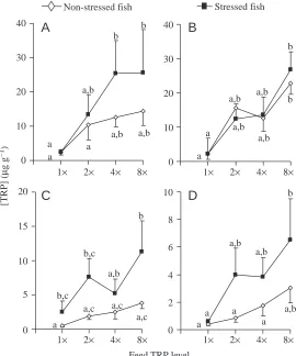
Stress had a significant effect on [TRP] in the optic tectum (F1,39=7.68, P=0.0085) and brain stem
(F1,38=4.24, P=0.0465), stressed fish showing elevated
[TRP] (Fig. 3C,D). Moreover, feeding the fish TRP-supplemented feed had a significant effect on [TRP] in the telencephalon (F3,36=2.88, P=0.0492), hypothalamus
(F3,40=4.60, P=0.0074) and optic tectum (F3,39=2.30, P=0.0085) (Fig. 3A–C). TRP supplementation resulted in
elevated [TRP] in these brain areas and a similar but non-significant trend (F3,38=0.2.68, P=0.0605) was also
observed in brain stem (Fig. 3D). Stressed fish fed 4× (P=0.0245) and 8× TRP-feed (P=0.0243) displayed significantly higher telencephalic [TRP] than stressed fish fed control feed (Fig. 3). Similarly, in the hypothalamus, brain stem and optic tectum [TRP] was significantly higher in stressed fish fed 8× TRP-feed (P=0.0079,
P=0.0103, P=0.0121, respectively) than in stressed fish
fed control feed (Fig. 3B–D). Moreover, in the hypothalamus, undisturbed fish fed 8×TRP-feed showed higher [TRP] than undisturbed fish fed control feed (P=0.0198) (Fig. 3B). In optic tectum, stressed fish fed 8× TRP-feed had significantly higher [TRP] than non-stressed fish fed 8×TRP-feed (P=0.0301) (Fig. 3C).
Brain [TRP]/[LNAA] ratios were not significantly affected by stress or dietary TRP in any part of the brain (Table 3). Similarly, there were no significant effects of stress or dietary TRP on brain [LNAA] (Table 3), or on the concentration of individual LNAAs, other than TRP (i.e. leucine, isoleucine, phenylalanine, tyrosine or valine; data not shown).
Highly significant correlations were found between plasma [TRP] and [TRP] in the telencephalon (rs=0.45, P=0.0027), hypothalamus (rs=0.52, P=0.0003), brain stem (rs=0.44, P=0.0033) and optic tectum (rs=0.39, P=0.0104). Similar significant correlations were also observed
between plasma [TRP]/[LNAA] ratios and [TRP] in telencephalon (rs=0.73, P=0.0036), hypothalamus (rs=0.68, P=0.0005), brain stem (rs=0.69, P=0.0138), and optic tectum (rs=0.78, P=0.0236).
[5-HT] was not affected by stress or dietary TRP supplementation in any part of the brain (Table 3).
Stress had a significant effect on [5-HIAA] in the telencephalon (F1,37=16.82, P=0.0002), hypothalamus
(F1,40=16.94, P=0.0002), and brain stem (F1,32=2.28, P=0.0366), and a similar but not significant trend was observed
in the optic tectum (F1,41=3.17, P=0.0826) (Table 3). In each
of these brain parts, stressed fish displayed elevated [5-HIAA] as compared to undisturbed fish. However, hypothalamic [5-HIAA] levels were significantly increased only in stressed fish fed control feed (P=0.0425) (Table 3).
Stress had a significant effect on [5-HIAA]/[5-HT] ratios in
the telencephalon (F1,37=15.67, P=0.0004), hypothalamus
(F1,40=8.85, P=0.0049) and brain stem (F1,32=11.20, P=0.0021) (Fig. 4A,B,D), but there was no significant effect
in the optic tectum (Fig. 4C). In each of these brain parts, stressed fish displayed elevated [5-HIAA]/[5-HT] ratios as compared to undisturbed fish. In the brain stem, stressed fish fed control feed showed an elevation of [5-HIAA]/[5-HT] ratios as compared to undisturbed controls (P=0.0130). Telencephalic [5-HIAA]/[5-HT] ratios were significantly elevated in stressed fish fed 2×and 4×TRP-feed compared to undisturbed controls fed 2× and 4×TRP-feed (P=0.0339 and
P=0.0096, respectively). There was no significant effect of
elevated dietary TRP levels on the brain [5-HIAA]/[5-HT] ratio, but there was a tendency towards an elevated [5-HIAA]/[5-HT] ratio in all brain areas of fish fed TRP-supplemented feed (Fig. 4).
A correlation was found in stressed fish between [TRP] Non-stressed fish Stressed fish
0 10 20 30 40
b
a
b
a,b
a,b a,b a a
0 10 20 30 40
b
a,b b a,b a,b
a,b a
a
[TRP]
(
µ
g
g
–
1)
0 2 4 6 8 10
a,b b
a,b
a
a a,b a
a 15
0 a
b
a,b b,c
b,c
a,c a,c a,c
Feed TRP level
1× 2× 4× 8× 1× 2× 4× 8×
1× 2× 4× 8× 1× 2× 4× 8×
5 10 20
A
C
B
[image:5.612.296.566.73.397.2]D
and [5-HIAA]/[5-HT] only in the telencephalon (rs=0.53, P=0.0097).
Discussion
[image:6.612.44.562.110.418.2]The present study reveals a significant interaction between dietary TRP supplementation and stress, reflecting the attenuation by TRP of the stress-induced elevation of plasma [cortisol]. At the highest levels of TRP supplementation, dietary TRP more-or-less blocked the increase in plasma [cortisol] induced by stress. However, increased dietary TRP tended to elevate basal levels of plasma cortisol in non-stressed fish. As will be discussed below, these effects of dietary TRP could both be mediated by the brain 5-HT system, TRP being the amino acid precursor of 5-HT. Still, feeding the fish TRP-supplemented feed for 7 days only resulted in a small, not quite significant, elevation of brain [5-HIAA] and [5-HIAA]/[5-HT] ratios. However, in the study by Winberg et al. (2001), the lowest level of dietary supplementation of TRP (8.38 mg TRP g–1dry feed), which was approximately fourfold
higher than the highest dose in the present study, also had only
small effects on brain [5-HIAA] and [5-HIAA]/[5-HT] ratios in fish fed this feed for 7 days, but pronounced effects on aggressive behaviour.
In accordance with previous studies, stress resulted in elevated brain [5-HIAA]/[5-HT] ratios (Winberg and Nilsson, 1993b; Winberg and Lepage, 1998; Øverli et al., 1999; Höglund et al., 2000). The central 5-HT system has been suggested to stimulate the HPI axis, and Winberg et al. (1997) showed that treatment with 8-OH-DPAT, a specific 5-HT1A
-receptor agonist, results in a dose-dependent elevation of plasma [cortisol] in dorsal aorta-cannulated rainbow trout. In mammals, 5-HT terminals make synaptic contact with corticotropin-releasing hormone immunoreactive neurons within the hypothalamic paraventricular nucleus (Liposits et al., 1987), and treatment with 5-HT precursors, such as TRP or 5-HTP, as well as 5-HT receptor agonists, has been reported to stimulate HPA axis activity, elevating plasma levels of glucocorticoids (Chaouloff, 1993). Thus, the elevation of plasma [cortisol] in non-stressed fish fed TRP-supplemented feed observed in the present study could well have been mediated by a stimulation of the brain 5-HT system.
Table 3. Effect of dietaryL-tryptophan (TRP) and stress on the concentration of large neutral amino-acids (LNAA) and ratios of
TRP to LNAA concentrations in the plasma and brain, and concentration of serotonin (5-HT) and its metabolite 5-hydroxyindolacetic acid (5-HIAA) in the brain of rainbow trout
Telencephalon Hypothalamus
[LNAA] [5-HIAA] [5-HT] [LNAA] [5-HIAA] [5-HT]
Feed Treatment (mg g–1) [TRP]/[LNAA] (ng g–1) (ng g–1) (mg g–1) [TRP]/[LNAA] (ng g–1) (ng g–1)
1× Non-stressed 1.75±0.41a 0.0100±0.0095a 356±39a 1479±429a 1.37±0.39a 0.0110±0.0095a 61±13a 748±86a
Stressed 2.34±0.39a 0.0014±0.0004a 541±95b 1318±138a 1.91±0.59a 0.0014±0.0004a 174±46b 1008±123a
2× Non-stressed 2.47±1.09a 0.0060±0.0017a 348±37a 942±82a 1.67±0.54a 0.0160±0.0093a 81±18a,b 969±117a
Stressed 1.97±0.42a 0.0120±0.0075a 544±37b 997±99a 2.16±0.71a 0.0120±0.0075a 187±43b 1308±123a
4× Non-stressed 1.92±0.24a 0.0066±0.0012a 478±65a,b 1282±144a 1.71±0.47a 0.0200±0.012a 104±12a,b 1069±89a
Stressed 1.94±0.32a 0.0180±0.0081a 632±46b 1168±228a 1.47±0.24a 0.0150±0.0078a 298±76c 1424±269a
8× Non-stressed 2.66±0.64a 0.0062±0.0025a 459±32a,b 1063±59a 2.84±0.35a 0.0081±0.0026a 181±46b,c 1012±160a
Stressed 1.63±0.19a 0.0130±0.0061a 628±66b 1052±73a 1.72±0.27a 0.0150±0.0060a 233±34b,c 1303±210a
Brain Stem Optic tectum
[LNAA] [5-HIAA] [5-HT] [LNAA] [5-HIAA] [5-HT]
Feed Treatment (mg g–1) [TRP]/[LNAA] (ng g–1) (ng g–1) (mg g–1) [TRP]/[LNAA] (ng g–1) (ng g–1)
1× Non-stressed 0.39±0.09a 0.0100±0.0095a 75±22a,c 220±38a 0.33±0.08a 0.0110±0.0095a 66±19a 264±40a
Stressed 0.46±0.05a 0.0013±0.0004a 123±23a.b 262±48a 0.63±0.12a 0.0046±0.0034a 121±20a 349±64a
2× Non-stressed 0.42±0.11a 0.0036±0.0015a 54±8c 163±19a 0.70±0.23a 0.0052±0.0018a 51±6a 257±35a
Stressed 0.40±0.08a 0.0140±0.0080a 88±14a–c 192±13a 0.90±0.27a 0.0150±0.0088a 86±11a 269±19a
4× Non-stressed 0.33±0.07a 0.0170±0.0130a 87±12a–c 212±19a 0.51±0.10a 0.0052±00013a 85±20a 288±39a
Stressed 0.31±0.05a 0.0180±0.0081a 113±20a–c 225±32a 0.54±0.08a 0.0150±0.0078a 93±17a 289±32a
8× Non-stressed 0.34±0.04a 0.0093±0.0043a 114±29a–c 291±73a 0.55±0.05a 0.0078±0.0024a 106±26a 348±86a
Stressed 0.33±0.06a 0.0150±0.0060a 149±27b 300±50a 0.45±0.07a 0.0220±0.0084a 107±30a 339±68a
Fish were fed feeds supplemented with TRP to a level corresponding to two (2×), four (4×), and eight (8×) times the TRP content of non-supplemented control feed (1×) for 7 days.
In this light, the observation that supplementary dietary TRP attenuated the stress-induced elevation of plasma [cortisol] may seem contradictory. One explanation for this effect could of course be elevated negative feedback as a result of increased basal plasma [cortisol], as indicated by elevated plasma [cortisol] in non-stressed fish receiving TRP-supplemented feed. However, even though significant, the elevation of plasma [cortisol] in non-stressed fish fed TRP-supplemented feed was small, suggesting that other mechanisms are also involved in mediating the effects of TRP on stress responsiveness.
The facilitating effects of 5-HT agents on plasma cortisol concentrations may not be in conflict with the observation that, under acute stress, 5-HT activity could improve ability to cope with stress and contribute to reducing a cortisol response (Markus et al., 2000b; Höglund et al., 2002). The serotonergic system is not a unitary system and different 5-HT pathways seem to be involved in both initiating and terminating the adrenocortical stress response in mammals (Deakin and Graeff, 1991; Graeff et al., 1996). For instance, 5-HT pathways terminating in the hippocampus are believed to inhibit HPA axis activity (Summers et al., 1998; Markus et al., 2000b). Recently, Höglund et al. (2002) reported that treatment with the 5-HT1A agonist, 8-OH-DPAT,
elevates plasma [cortisol] in undisturbed Arctic charr
Salvelinus alpinus, whereas the same drug, if
administered in connection with stress, suppresses the stress-induced elevation of plasma [cortisol].
Moreover, treatments elevating plasma [TRP] and/or [TRP]/[LNAA] ratios have been reported to counteract stress-induced elevations of plasma cortisol in mammals (Morméde and Dantzer, 1979), including humans (Markus et al., 1998, 1999, 2000a,b). For instance, in pigs preloaded with 50–200 mg kg–1 TRP, activation of the
HPA axis in response to the experience of an unfamiliar environment, in combination with unavoidable shocks, is decreased (Morméde and Dantzer, 1979). Moreover, carbohydrate-rich, protein-poor food, which causes an elevation of plasma [TRP]/[LNAA] ratios, prevents the elevation of plasma cortisol induced by uncontrollable (Markus et al., 1998) and controllable stress (Markus et al., 1999) in stress-prone human subjects. Similarly, a diet enriched in α-lactalbumin, a TRP-rich protein, reduces depressive mood and plasma cortisol concentrations in stress-prone subjects under acute stress (Markus et al., 2000a).
In a recent study we showed that dietary supplementation of TRP for 7 days results in an inhibition of aggressive behaviour in rainbow trout, whereas 3 days of TRP supplementation have no effect on aggressive behaviour (Winberg et al., 2001). Since the effect of elevated dietary TRP intake on 5-HT synthesis and release could be expected to be very rapid, other mechanisms are likely to be involved in mediating effects of elevated dietary TRP on aggressive behaviour, and possibly also in mediating the effects of TRP on stress responsiveness, since in the present study the fish were fed TRP-supplemented
feed for 7 days. Interestingly, the anti-depressive effect of specific 5-HT re-uptake inhibitors (SSRI), such as fluoxetine (Prozac), is only evident after long-term treatment (Mongeau et al., 1997). Initially SSRI treatment markedly decreases the firing activity of HT neurones. However, the firing rate of 5-HT neurones recovers if the SSRI treatment continues for 1–3 weeks, and this recovery seems to be associated with a desensitisation of 5-HT1A somato-dendritic autoreceptors
(Mongeau et al., 1997; Nutt et al., 1999). Moreover, long-term SSRI treatment has been found to desensitise presynaptic 5-HT autoreceptors located at nerve terminals, and to increase the baseline level of 5-HT in the hippocampus (Kreiss and Lucki, 1995). Possibly, long-term effects of elevated dietary intake of TRP on stress responsiveness and aggression in rainbow trout are mediated by similar mechanisms, resulting in elevated 5-HT release in areas of the trout brain homologous to the mammalian hippocampus.
According to Northcutt and Davis (1983), the dorsal and ventral parts of the dorsal area of the teleostean telencephalon are a putative homologue of the mammalian hippocampus.
a,c
a,c a
b b,c
a,b a,b a b,c
b,c,d
a,c a,b
a,c
a,d a,d a
a
a a
a
a a a
a c,a c
c,a
c,b b b a,b
b
[5-HIAA
]/[5-HT]
0.3
0
Feed TRP level
1× 2× 4× 8× 1× 2× 4× 8×
1× 2× 4× 8× 1× 2× 4× 8×
0.1 0.2 0.4
C
0.60 0.2 0.4 0.8
A
Non-stressed fish Stressed fish
0.3
0 0.1 0.2 0.4
B
0.6
0 0.2 0.4
[image:7.612.296.566.72.375.2]D
Notably, in the present study, following 7 days of TRP supplementation, a significant correlation between [TRP] and [5-HIAA]/[5-HT] ratios was observed only in the telencephalon of stressed fish.
Serotonin may also suppress HPI axis activity by inhibiting central norepinephrine (NE) activity. In mammals, NE stimulates hypothalamic corticotropin-releasing hormone, which in turn has a stimulatory effect on NE activity (Huether, 1996), creating a positive feedback loop, which seems to be counterbalanced by an inhibition of the NE system by 5-HT (Aston-Jones et al., 1991; Engberg, 1992). The central NE system has been suggested to stimulate HPI axis activity in teleost fish (Øverli et al., 1999; Höglund et al., 2000). Consequently, by elevating brain 5-HT activity, increased dietary intake of TRP may suppress the stress-related activation of the brain NE system, and by that inhibit the stressed-induced activation of the HPI axis.
As expected, feeding the fish TRP-supplemented feed resulted in elevated plasma [TRP]. Plasma [TRP]/[LNAA] ratios were also elevated in fish fed TRP-supplemented feed since the amount of other LNAA were similar in the feeds (Table 1). Increasing dietary levels of TRP were also reflected in elevated brain [TRP] and a non-significant trend towards increased brain [TRP]/[LNAA]. Moreover, as reported previously, stress resulted in an elevation of brain [TRP] (Winberg and Nilsson 1993a,b) but had no effect on brain levels of other LNAA, nor did stress affect plasma levels of TRP, other LNAA, or plasma [TRP]/[LNAA]. Thus, since in rainbow trout the TRP plasma pool is not protein-bound (Rozas et al., 1990) and is completely available for uptake to the brain, a stress-mediated effect on the availability of TRP for uptake to the brain could not explain the stress-induced elevation of brain [TRP]. It is more likely that the elevation of brain [TRP] observed in stressed rainbow trout is mediated by stress-induced effects on the blood–brain barrier permeability. In mammals, the increase in brain [TRP] during stress seems to involve the sympathetic nervous system, and is attenuated by the β-adrenergic receptor antagonist, propranolol, but not by the α-adrenoreceptor antagonist phentolamine (Dunn and Welch, 1991). Dunn (1999) showed that treatment with the nitric oxide (NO) synthase inhibitor N-nitro-L-arginine (L-NAME) attenuates the stress-induced elevation of brain [TRP] in mice, suggesting that NO is involved in mediating an elevation of brain [TRP] in response to stress in mammals.
In conclusion, the results of the present study show that dietary supplementation of TRP for 7 days attenuates the stress-induced increase in plasma cortisol levels in juvenile rainbow trout, an effect that is most likely to be mediated by the brain serotonergic system. Supplementing feed with TRP could be a promising strategy in aquaculture management, not only making the fish more stress-resistant, but also decreasing aggressive behaviour, and thus the tendency to develop strong dominance hierarchies, resulting in stress, reduced disease resistance and highly variable growth rates of fish in aquaculture (Winberg et al., 2001).
This study was supported by grants from the Swedish Research Council for Environment, Agricultural Sciences and Spatial Planning (FORMAS), the Carl Trygger Foundation, the FACIAS foundation and Ewos International Ltd.
References
Aldegunde, M., Garcia, J., Soengas, J. L. and Rozas, G. (1998). Uptake of
tryptophan into brain of rainbow trout (Oncorhynchus mykiss). J. Exp. Zool.
282, 285-289.
Aldegunde, M., Soengas, J. L. and Rozas, G. (2000). Acute effects of L -tryptophan on -tryptophan hydroxylation rate in brain regions (hypothalamus and medulla) of rainbow trout (Oncorhynchus mykiss). J. Exp. Zool. 286, 131-135.
Aston-Jones, G., Akaoka, H., Charléty, P. and Chouvet, G. (1991).
Serotonin selectively attenuates glutamate-evoked activation of noradrenergic locus coerulus neurones. J. Neurosci. 11, 760-769.
Boadle-Biber, M. C. (1993). Regulation of serotonin synthesis. Prog.
Biophys. Mol. Biol. 60, 1-15.
Chaouloff, F. (1993). Physiopharmacological interactions between stress
hormones and central serotonergic systems. Brain Res. Rev. 18, 1-32.
Curzon, G., Joseph, M. H. and Knott, P. J. (1972). Effects of immobilization
and food deprivation on rat brain tryptophan metabolism. J. Neurochem. 19, 1967-1974.
de Montigny, P., Stobaugh, J. F., Givens, R. S., Carlson, R. G., Srinivasachar, K., Sterson, L. A. and Higuchi, T. (1987).
Naphtalene-2,3-dicarbixaldehyde/cyanide ion: A rationally designed fluorogenic reagent for primary amines. Anal. Chem. 59, 1096-1101.
Deakin, J. W. F. (1991). Depression and 5-HT. Int. Clin. Psychopharmacol. 3, 23-28.
Deakin, J. W. F. and Graeff, F. G. (1991). 5-HT mechanisms of defense. J.
Psychopharmacol. 5, 305-315.
Dinan, T. G. (1996). Serotonin: Current understanding and the way forward.
Int. Clin. Psychopharmacol. 11, 19-21.
Dunn, A. J. (1988). Changes in plasma and brain tryptophan and brain
serotonin and 5-hydroxyindolacetic acid after footshock stress. Life Sci. 41, 1847-1853.
Dunn, A. J. (1999). Brain catecholaminergic and tryptophan responses to
restraint are attenuated by nitric oxide synthase inhibition. Neurochem. Int.
33, 551-557.
Dunn, A. J. and Welch, J. (1991). Stress-induced and endotoxin-induced
increases in brain tryptophan and serotonin metabolism depend on sympathetic nervous system activity. J. Neurochem. 57, 1615-1622.
Engberg, G. (1992). Citalopram and 8-OH-DPAT attenuate nicotine-induced
excitation of central noradrenaline neurons. J. Neur. Transm. 89, 149-154.
Fernstrom, J. D. (1983). Role of precursor availability in control of
monoamine biosynthesis in brain. Physiol. Rev. 60, 484-546.
Graeff, F. G., Guimares, F. S., de Andrade, T. G. C. S. and Deakin, J. F. W. (1996). Role of 5-HT in stress, anxiety and depression. Pharmacol.
Biochem. Behav. 54, 129-141.
Höglund, E., Balm, P. H. M. and Winberg, S. (2000). Skin darkening, a
potential signal in subordinate artic charr (Salvenius alpinus): The regulation role of brain monoamines and pro-opiomelanocortin-derived peptides. J. Exp. Biol. 203, 1711-1721.
Höglund, E., Balm, P. H. M. and Winberg, S. (2002). Stimulatory and
inhibitory effects of 5-HT1Areceptors on ACTH and cortisol secretion in a teleost fish, the Arctic charr (Salvelinus alpinus). Neurosci. Lett. 324, 193-196.
Huether, G. (1996). The central adaptation syndrome: psychosocial stress as
a trigger for adaptive modifications of brain structure and brain function. Prog. Neurobiol. 48, 569-612.
Jacobson, L. and Sapolsky, R. (1991). The role of the hippocampus in
feedback regulation of the hypothalamic-pituitary-adrenocortical axis. Endocrinol. Rev. 12, 118-134.
Johnston, W. L., Atkinson, J. L., Hilton, J. W. and Were, K. E. (1990).
Effect of dietary tryptophan on plasma and brain tryptophan, brain serotonin, and brain 5-hydroxyindoleacetic acid in rainbow trout. J. Nut. Biochem. 1, 49-54.
Kreiss, D. S. and Lucki, I. (1995). Effects of acute and repeated administration
of antidepressant drugs on extracellular levels of 5-hydroxytryptamine measured in vivo. J. Pharmacol. Exp. Ther. 274, 866-876.
serotonergic axons and corticotropin releasing factor (CRF) synthesizing neurons in the hypothalamic paraventricular nucleus of the rat. A light and electron microscopic immunocytochemical study. Histochem. 86, 541-549.
Maes, M. and Meltzer, H. (1995). The serotonin hypothesis of major
depression. In Psychopharmacology: the Fourth Generation of Progress (ed F. E. Bloom and D. J. Kupfer), pp. 933-944. New York: Raven Press.
Markus, C. R., Olivier, B., Panhuysen, G. E. M., Van der Gugten, J., Alles, M. S., Tuiten, A., Westenberg, G. M., Fekkes, D., Koppeschaar, H. F. and de Haan, E. E. H. F. (2000a). The bovine protein α-lactalbumin increases the plasma ratio of tryptophan to the other large neutral amino acids, and in vulnerable subjects raises brain serotonin activity, reduces cortisol concentration, and improves mood under stress. Am. J. Clin. Nutr.
71, 1536-1544.
Markus, C. R., Panhuysen, G., Jonkman, L. M. and Bachman, M. (1999).
Carbohydrate intake improves cognitive performance of stress-prone individuals under laboratory stress. Br. J. Nutr. 82, 457-467
Markus, C. R., Pannhuysen, G., Tuiten, A. and Koppeschaar, H. (2000b).
Effets of food on cortisol and mood in vulnerable subjects under controllable and uncontrollable stress. Physiol. Behav. 70, 333-342.
Markus, C. R., Panhuysen, G., Tuiten, A., Koppeschaar, H., Fekkes, D. and Peters, M. (1998). Does carbohydrate-rich, protein-poor food prevent
a deterioration of mood and cognitive performance of stress-prone subjects when subjected to a stressful task? Appetite 31, 49-65.
Mongeau, R., Blier, P. and de Montigny, C. (1997). The serotonergic and
noradrenergic systems of the hippocampus: their interactions and the effects of antidepressant treatments. Brain Res. Rev. 23, 145-195.
Morméde, P. and Dantzer, R. (1979). Effects of lithium on aggressive
behavior in domestic pigs. J. Vet. Pharmacol. Ther. 2, 299-304.
Northcutt, R. G. and Davis, R. E. (1983). Telencephalic organization in
ray-finned fishes. In Fish Neurobiology, Vol. 2: Brain Stem and Sense Organs (ed. R. E. Davis and R. G. Northcutt), pp. 203-236. Ann Arbor: University of Michigan Press.
Nutt, D., Forshall, S., Bell, C., Rich, A., Sandford, J., Nash, J. and Argyopoulos, S. (1999). Mechanisms of action of selective serotonin
reuptake inhibitors in the treatment of psychiatric disorders. Eur. Neuropsychopharmacol. 3, S81-S86.
Olsen, Y. A., Falk, K. and Reite, O. B. (1992). Cortisol and lactate levels of
Atlantic salmon Salmo salar developing infectious anemia (ISA). Dis. Aquat. Org. 14, 99-104.
Øverli, Ø., Harris, C. A. and Winberg, S. (1999). Short-term effects of fights
for social dominance and the establishment of dominant-subordinate relationships on brain monoamines and cortisol in rainbow trout. Brain Behav. Evol. 54, 263-275.
Parent, A., Poiras, D. and Dubé, L. (1984). Comparative neuroanatomy of
central monoaminergic systems. In Handbook of Chemical Neuroanatomy (ed. A. Björklund and T. Hökfelt), pp. 409-439. Amsterdam: Elsevier.
Rozas, G., Rey, P., André, M. D., Rebolledo, E., Morfin, R. and Aldegunde, M. (1990). Distribution of 5-hydroxytryptamine and related compounds in
various brain regions of the rainbow trout (Oncorhynchus mykiss). Fish Physiol. Biochem. 8, 501-506.
Summers, C. H., Larson, E. T., Summers, T. R., Renner, K. J. and Greenberg, N. (1998). Regional and temporal separation of serotonergic
activity mediating social stress. Neurosci. 87, 489-496.
Winberg, S. and Lepage, O. (1998). Elevation of brain 5-HT activity, POMC
expression and plasma cortisol in socially subordinate rainbow trout. Am. J. Physiol. 43, R645-R654.
Winberg, S. and Nilsson, G. E. (1993a). Time course of changes in brain
serotonergic activity and brain tryptophan levels in dominant and subordinate juvenile Arctic charr. J. Exp. Biol. 179, 181-195.
Winberg, S. and Nilsson, G. E. (1993b). Roles of brain monoamine
neurotransmitters in agonistic behaviour and stress, with particular reference to fish. Comp. Biochem. Physiol. C 106, 597-614.
Winberg, S., Nilsson, A., Hylland, P., Söderstöm, V. and Nilsson, G. E.
(1997). Serotonin as a regulator of hypothalamic-pituitary-interrenal activity in teleost fish. Neurosci. Lett. 230, 113-116.
Winberg, S., Øverli, Ø. and Lepage, O. (2001). Suppression of aggression


![Fig. 4. The [5-HIAA]/[5-HT] ratios in the telencephalon (A),hypothalamus (B), optic tectum (C) and brain stem (D) of isolatedValues are means ±the non-supplemented control feed (1exposed to a standardised stressor (lowering the water level); non-stressedfis](https://thumb-us.123doks.com/thumbv2/123dok_us/1134927.633947/7.612.296.566.72.375/telencephalon-hypothalamus-isolatedvalues-supplemented-standardised-stressor-lowering-stresseds.webp)
