The inner ear of teleosts differs from that of other vertebrates mainly in the complete absence of a cochlea. The membranous labyrinth consists of three connecting chambers, the utricule, saccule and lagena, each containing one otolith. These chambers are continuous with three semi-circular canals, and the entire labyrinth is filled with endolymph. This organ is involved in the maintenance of equilibrium and in the detection of gravity and sound (Dale, 1980; Lowenstein, 1971). In the fields of fisheries biology and management, the analysis of otolith microstructure is of prime importance. Fish otoliths are calcium carbonate concretions with annual and daily rhythmic depositions (Panella, 1971). Thus, in addition to their use in the estimation of age, otolith analysis has been used to obtain information about a fish’s somatic growth history from back calculations of fish size (Campana and Neilson, 1985; Jones, 1992).
In higher vertebrates, the labyrinth fluid is always characterized by high K+and low Na+ concentrations, showing a composition like that of intracellular fluid except for its Ca2+ content (total calcium 0.3–0.6 mmol l−1 with approximately
10 % in the ionized form). In fish, the otolith is in direct contact with the endolymph, and only a few studies have examined the composition of endolymph, mainly in the saccule, which is the largest chamber of the labyrinth in most species. By comparison with other vertebrates, fish endolymph shows a higher Na+ concentration (110–140 mmol l−1: Enger, 1964; Fange et al. 1972; Kalish, 1991; Mugiya and Takahashi, 1985; Watanabe and Miyamoto, 1973) compared with 3–40 mmol l−1 in mammals (Sterkers et al. 1988). A more surprising difference described by Mugiya and Takahashi (1985) is the relative alkalinity of the saccular endolymph (plasma 7.56; endolymph 8.06), whereas mammalian endolymph and plasma have the same pH (Sterkers et al. 1988).
In this paper, we re-examine the ionic composition (Na+, K+, Cl−, Ca2+, PO
43−, pH), total CO2and protein contents of the saccular endolymph in a primitive freshwater teleost (trout) and a more highly evolved seawater teleost (turbot). We also analyze the kinetic characteristics of proton secretion using a titration technique in which an isolated saccule is mounted as a closed sac.
JEB0829
Ionic (Na+, K+, Cl−−, PO
43−−, pH), total CO2, total calcium
and protein concentrations in the plasma and endolymph of the inner ear were compared in trout Oncorhynchus
mykiss and turbot Scophthalmus maximus. In both species,
saccular endolymph was characterized by high levels of K+
and total CO2and in trout by an alkaline pH. The kinetic
characteristics of proton secretion across the saccular epithelium of trout were investigated using a titration technique in which isolated saccules were mounted as closed sacs. The rate of proton secretion depends strongly on the pH of the Ringer’s solution and secretion stops at a pH below 7.2. Proton secretion is driven by an energy-dependent mechanism involving basolateral
ouabain-sensitive Na+/K+ exchangers. Proton secretion was
partially inhibited by acetazolamide and completely inhibited in Na+-free Ringer or in the presence of
1 mmol l−−1 amiloride. A cellular model stressing the importance of proton exchange through the saccular epithelium is proposed to explain the regulation of endolymph pH, a crucial factor for the deposition of otolith calcium.
Key words: teleost, inner ear, saccular epithelium, proton secretion, otolith, biocalcification, trout, Oncorhynchus mykiss, turbot, Scophthalmus maximus.
Summary
Introduction
IONIC COMPOSITION OF ENDOLYMPH IN TELEOSTS: ORIGIN AND
IMPORTANCE OF ENDOLYMPH ALKALINITY
P. PAYAN1, H. KOSSMANN2, A. WATRIN3, N. MAYER-GOSTAN3 ANDG. BOEUF2
1Laboratoire de Physiologie Environnementale, Faculté des Sciences, Université de Nice-Sophia Antipolis,
BP 71, 06108 Nice CX2, France, 2Laboratoire de Physiologie des Poissons, IFREMER, Centre de Brest,
BP 70, 29280 Plouzané, France and 3Laboratoire de Physiologie Cellulaire et Moléculaire des Epithéliums et des
Compartiments Calciques, URA CNRS 1938, Faculté des Sciences, Université de Nice-Sophia Antipolis, BP 71, 06108 Nice CX2, France
Accepted 26 March 1997
Materials and methods Fish handling
Approximately 80 trout [Oncorhynchus mykiss (Walbaum)], of about 200 g body mass, 12–13 months old, were supplied by a local fish farmer near Nice, France, and reared in running tap water at approximately 14 °C in circular tanks. Twenty turbot [Scophthalmus maximus (Raf. 1810)], of 100–150 g body mass and 7–8 months old, were obtained from IFREMER Brest, France, and kept in a closed seawater system maintained at 14 °C. Both groups were fed every morning.
Collection of plasma and endolymph
A maximal amount of blood (4–5 ml) was quickly collected with a heparinized syringe by puncturing the caudal vessels. The pH was measured, then the blood sample was centrifuged and the separated plasma kept on ice until analysis. Immediately after blood sampling, the inner ear was exposed to allow collection of the endolymph sample (6–7µl). In both trout and turbot, the delay between blood and endolymph sampling was short (approximately 3 min). The fish was killed by spinal transection, decapitated behind the gill and the head placed on ice. The skull was opened with a frontal incision just behind the eyes, and the saccular membrane was exposed after anterior displacement of the brain. The two species studied differ in the anatomical characteristics of the bony labyrinth and in the viscosity of the endolymph, necessitating different collecting procedures. In the turbot, the bone cavity containing the saccule has a narrow opening and the endolymph was sampled in situ. In order to minimize its contamination by blood, the operative field was rinsed five times with 1 ml of ion-free medium (i.e. 300 mmol l−1glycine treated with
anion-exchange resin; Chelex 100 and Biorad AG50WX8) and adjusted to pH 7.2 with Tris. Before sampling, any fluid remaining in the brain cavity was removed with absorbent paper. Then, under a stereomicroscope, the tip of a calibrated capillary tube (CEI, GC100TF-10), fixed to a micromanipulator (Leitz), was inserted into the saccule and the endolymph slowly sucked up using an infusion and withdrawal syringe pump (Braun). In the trout, the lower parts of the membranous labyrinth were carefully excised after cutting the semicircular canals. The isolated labyrinth was then rinsed five times in ion-free medium and placed in a plastic cup. The surrounding liquid was removed with absorbent paper and the endolymph sampled as for the turbot.
pH determination in blood and endolymph
Blood pH was determined immediately after sampling. To determine the pH of trout endolymph, larger trout (weighing approximately 2 kg and approximately 2 years old) were used since their saccules are large enough to allow the complete insertion of the tip of a needle-like pH electrode (INC microelectrode, MI 407, 0.8 mm in diameter) connected to a pH meter (Tacussel, TT processor 2) into the otosac, thus minimizing air contact and the escape of CO2. pH values were
recorded within 15 s of the incision being made in the saccule
wall. To facilitate handling, the pH electrode was fixed on a micromanipulator (Leitz). Unfortunately, it was not possible to measure the endolymph pH of turbot since no large enough fish were available.
Protein and ion determinations
The volume of each sample was determined by measuring the length of endolymph in the calibrated capillary with a slide caliper. Although the volume obtained from an individual saccule was only a few microlitres, the use of a calibrated capillary tube made possible measurements of ionic (Na+, K+,
Cl−) and protein concentrations in an individual sample. However, for each CO2, Ca2+or PO43−measurement, an entire
sample was needed; thus, it was not possible to obtain data for all the variables from one saccule.
For the protein, Cl−, Na+and K+ measurements, the entire
endolymph sample was injected into 0.5 ml of distilled water and the capillary tube was rinsed repeatedly by aspiration. For each determination of total calcium, total CO2 or PO43−, a
whole endolymph sample was injected directly into the measurement vial. Na+ and K+ were determined by flame
photometry (Eppendorf) after appropriate dilutions. Ca2+, Cl−,
PO43−, total CO2 and protein were measured by
spectrophotometry using Sigma kits for the first four and Commassie Blue for protein with bovine serum albumin as a standard.
Determination of proton secretion by isolated trout saccules A ligature was placed at the base of the semicircular canals of a freshly excised labyrinth, and the saccule sac was immediately immersed in the titration vessel. The labyrinth was held in place by the thread of the ligature. The appearance of protons in the external medium was monitored by automatic titration with a Tacussel titrator (TT Processor 2) coupled to an autoburette Tacussel (EBX3). The pH was continuously recorded using a microelectrode. The solution bathing the labyrinth was 1 ml of modified Ringer (see below), continuously agitated with a magnetic stirrer with renewed air at the surface. At the beginning of each period, the pH of the incubation medium was measured and entered as an end-point in the titration apparatus. The appearance of secreted protons was determined from the amount of titrant delivered into the titration vials by the autoburette, the delivery being recorded graphically, and the initial slope was used for calculations (see Results). Individual experiments included a first period which served systematically as its own control. Both saccules of an individual trout were studied; the second was kept in situ, bathed with an aerated Ringer’s solution and excised just before use.
Statistics
Data are expressed as means ± S.E.M. (N=number of
Chemicals
The modified Ringer was composed of: 135 mmol l−1NaCl,
2.5 mmol l−1KCl, 1 mmol l−1MgCl
2, 1.5 mmol l−1NaH2PO4,
0.4 mmol l−1KH
2PO4, 1.5 mmol l−1CaCl2; buffered with either
1 mmol l−1 NaHCO
3 or 1 mmol l−1 Hepes. The solution also
contained freshly added glucose (1 g l−1) and was aerated
before use. The Na+-free solution was obtained by replacing
NaCl by an equivalent amount of choline chloride.
Pharmacological products (ouabain, amiloride, cyanide, acetazolamide) were purchased from Sigma.
Results
Ion and protein contents in plasma and endolymph The endolymph volumes collected from individual saccules were 5.7±0.5µl (N=9) and 6.3±0.3µl (N=14) in trout and turbot, respectively. Ion contents measured in plasma and endolymph are presented in Table 1. In both species, the endolymph composition differs markedly from that of the plasma, the most striking difference being that of K+content,
with endolymph concentrations being 20 (turbot) to 41 (trout) times higher than those in plasma. The endolymph Na+
concentrations were approximately 50–70 % of those in plasma, while the Cl− concentrations were not significantly different. The total calcium concentration was approximately 40–45 % of that of plasma. Phosphate levels were 2–5 times lower in the endolymph than in the plasma.
Total endolymph protein concentrations in trout and turbot (Table 1) were approximately 2 g l−1, well below the level
measured in plasma (15 times less), but characteristic of the level in cephalic fluids.
In both fish species, the endolymph also differed from the plasma by having a significantly higher total CO2
concentration (2–3 times greater), with higher values in trout than in turbot. Experiments performed in large trout showed that pH in the endolymph was significantly more alkaline than in the plasma (Table 1).
Characteristics of proton secretion by isolated saccules Preliminary experiments were conducted to determine the
optimal buffer conditions for accurate measurement of secreted protons (concentrations from 0 to 10 mmol l−1bicarbonate or
Hepes were tested). It was found that 1 mmol l−1bicarbonate
or Hepes was an appropriate concentration to maintain a stable external pH and was low enough to allow detection of small quantities of protons excreted by an isolated saccule. Thus, the titrant used was a 1 mmol l−1NaOH solution except in Na+-free
experiments, in which 1 mmol l−1KOH was used.
Preliminary experiments showed that proton secretion by an isolated saccule could be followed for 1 h or more. Generally, experiments were divided into three titration periods of 10–15 min. This was long enough to obtain a precise measurement but short enough to obtain a linear proton secretion response. Systematically, at the end of each experiment, the saccule was removed from the titration chamber to check that the secreted protons were of biological origin. Then, 0.05 or 0.1µequiv HCl was added as a standard to quantify H+excretion as nequiv min−1otosac−1.
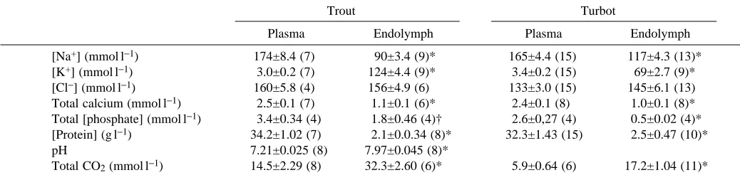
External pH-dependence of proton secretion rate
Fig. 1A illustrates a recording of a typical experiment showing the secretion of protons from one isolated saccule under different pH conditions. For each titration period, after the change of Ringer’s solution, the apparatus was reset to fix the new pH end-point. This experiment shows that increasing the pH of the Ringer’s solution immediately accelerated the rate of proton secretion, which returned to the initial rate when the pH was returned to that of the control period. In contrast (Fig. 1B), acidifying the Ringer’s solution to pH 7.0–7.2 completely stopped proton secretion. After a return to the initial pH, proton secretion restarted after a short delay.
[image:3.609.48.567.584.714.2]Fig. 2 presents the variations in the rates of proton secretion at various Ringer pH values ranging from 7.0 to 7.8. The relationship is complex. Below pH 7.2, no proton secretion was detected, between 7.2 and 7.6, proton secretion increased rapidly; it continued to increase at more alkaline pH values, but slightly less rapidly. Fig. 2 also indicates that the results are similar irrespective of the buffer used (bicarbonate or Hepes). The hypothetical physiological significance of such a relationship for biocalcification will be discussed below.
Table 1. Ion concentrations, pH, total CO2and protein concentration of saccular endolymph and plasma in trout and turbot
Trout Turbot
Plasma Endolymph Plasma Endolymph
[Na+] (mmol l−1) 174±8.4 (7) 90±3.4 (9)* 165±4.4 (15) 117±4.3 (13)*
[K+] (mmol l−1) 3.0±0.2 (7) 124±4.4 (9)* 3.4±0.2 (15) 69±2.7 (9)*
[Cl−] (mmol l−1) 160±5.8 (4) 156±4.9 (6) 133±3.0 (15) 145±6.1 (13) Total calcium (mmol l−1) 2.5±0.1 (7) 1.1±0.1 (6)* 2.4±0.1 (8) 1.0±0.1 (8)* Total [phosphate] (mmol l−1) 3.4±0.34 (4) 1.8±0.46 (4)† 2.6±0,27 (4) 0.5±0.02 (4)* [Protein] (g l−1) 34.2±1.02 (7) 2.1±0.0.34 (8)* 32.3±1.43 (15) 2.5±0.47 (10)*
pH 7.21±0.025 (8) 7.97±0.045 (8)*
Total CO2(mmol l−1) 14.5±2.29 (8) 32.3±2.60 (6)* 5.9±0.64 (6) 17.2±1.04 (11)*
Values are means ±S.E.M. (N).
In order to concentrate the data, most of the following experiments were performed with an external pH of 7.4–7.6 using 1 mmol l−1bicarbonate-buffered Ringer’s solution.
Na+-gradient dependence of proton secretion rate
The Na+ gradient through the basal barrier of the saccular
epithelium was reversed by using a Na+-free Ringer’s solution.
Fig. 3A shows a typical experiment in which Na+ was
substituted by an equivalent concentration of choline. The removal of Na+from the bathing medium caused a rapid and
progressive inhibition of the rate of proton secretion which was complete within 20 min. After returning to a normal Ringer’s solution, proton secretion started again after an approximately
5 min lag. Fig. 3B summarizes experiments performed on seven saccules. To quantify the progressive inhibition, two successive periods of 5 min were selected to calculate proton secretion. Recovery was measured 3–5 min after the return to a normal Ringer’s solution and led to a proton secretion rate not significantly different from that observed during the initial period. Thus, proton secretion is dependent on the presence of Na+on the basolateral side.
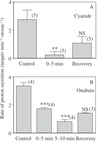
Pharmacological characterization of proton secretion It was only possible to add drugs on the basolateral side of saccular epithelia. Preliminary experiments showed the importance of maintaining a well-aerated air–liquid interface in the titration chamber to allow saccules to excrete protons (results not shown). The addition of 1 mmol l−1 cyanide (a
mitochondrial inhibitor) completely inhibited proton secretion in a non-reversible manner (Fig. 4A). These results suggest that energy-dependent mechanisms are involved in proton secretion. Ouabain (1 mmol l−1), a common inhibitor of Na+/
K+ATPase, added to the normal Ringer’s solution
progressively inhibited the rate of proton secretion in a manner similar to that of a Na+-free Ringer’s solution.
However, unlike the inhibition caused by Na+-free Ringer,
the inhibitory effect of ouabain was not significantly reversible within a 10 min period of recovery after rinsing out the inhibitor (Fig. 4B).
When amiloride, a diuretic inhibiting Na+ transport and
Na+/H+exchange, was used at concentration of 1 mmol l−1, it
immediately decreased the rate of proton secretion by up to 80 %. This inhibitory effect was partially reversible (Table 2). Acetazolamide, a carbonic anhydrase inhibitor, which blocks the hydration of CO2 and consequently perturbs the
acid–base balance of the cell, partially inhibits the secretion of protons in an irreversible manner when used at a concentration of 0.1 mmol l−1(Table 2).
0 30 60
0 100
7.26 7.69 7.24
Otosac removal A
0 30 60
Time (min)
Otosac removal
0 100
7.51 7.03 7.13 7.20 7.50 B
[image:4.609.329.544.70.229.2]Proton secretion (nequiv)
Fig. 1. Effect of the Ringer’s solution pH on proton secretion by isolated saccular sacs. Experimental recordings of typical experiments. (A) Effect of increasing the pH of the Ringer’s solution. (B) Effect of lowering the pH of the Ringer’s solution. The arrows indicate the beginning of each period (after three successive rinsings with the new external medium). The pH of the solutions was held at different values indicated at the beginning of each period. Titrant was 1 mmol l−1NaOH.
6
4
2
0
Rate of proton secretion
(nequiv
min
−
1)
7.0 7.2 7.4 7.6 7.8 8.0
pH
Fig. 2. Relationship between the rate of proton secretion by isolated saccular sacs and the pH of the Ringer’s solution. Experiments were performed with Ringer’s solutions containing 1 mmol l−1bicarbonate
(open symbols) or 1 mmol l−1Hepes (filled symbols). The line was
[image:4.609.56.277.75.470.2]Discussion
Ion distribution between plasma and endolymph Two fluids are in contact with the transporting cells of the saccular epithelium: cephalic fluid and blood. In fish, we think that the exchanges through the saccular epithelium occur preferentially with blood because the connective tissue surrounding the saccular epithelium is highly vascularized, forming a capillary bed (Mayer-Gostan et al. 1997). Furthermore, according to Enger (1964), cephalic fluid and plasma in teleosts do not show substantial differences in their ionic concentrations. Thus, comparisons of ionic distributions can reasonably be made between endolymph and plasma. Our
results show that the differences in the ionic concentrations of plasma and endolymph are similar in the trout and turbot (Table 1) and are in agreement with previously published data. In the trout, the magnitude of the differences observed by Mugiya and Takahashi (1985) between plasma and endolymph concentrations was lower than that found in the present study. This is probably because our technical sampling procedure had
0 30 60
Time (min) 0
100
Otosac removal
7.65 7.73 7.48
Ringer
Na+-free
Ringer
Ringer A
Proton secretion (nequiv)
B
Control 0–5 min 5–10 min Recovery 0
2 4 6
(7)
(7)
(6)
(6)
*
**
†
[image:5.609.58.287.72.439.2]Rate of proton secretion (nequiv min −1 otosac −1)
Fig. 3. Effect of Na+-free Ringer’s solution on the rate of proton
secretion by an isolated saccular sac. Na+ was substituted with
choline. (A) Experimental recording of a typical experiment. The pH of the solutions was held at different values indicated at the beginning of each period. Titrant was 1 mmol l−1 KOH. (B) Rates of proton
secretion in Na+-free Ringer’s solution. Values are means + S.E.M. The
number of experimental values is indicated in parentheses, and the pH of the solutions was held at the following values: 7.56±0.062 (control period); 7.56±0.081 (0–5 min period); 7.57±0.081 (5–10 min period) and 7.57±0.082 (recovery period). Asterisks denote a significant difference from the rate in the control period; *P<0.05; **P<0.01; †P<0.01 compared to preceding period.
Control 0–5 min 5–10 min Recovery 0
2 4
(4)
(4)
(4)
(3)
***
***
NS Ouabain
Control 0–5 min Recovery
0 2 4
(5)
(5)
(5)
**
NS Cyanide
B A
Rate of proton secretion (nequiv min
[image:5.609.356.524.75.320.2]−1 otosac −1)
Fig. 4. Effect of 1 mmol l−1cyanide (A) and 1 mmol l−1ouabain (B)
on the rate of proton secretion by isolated saccular sacs. Values are means + S.E.M. with the number of experiments given in parentheses. The pH of the solutions was held at the following values: (A) cyanide experiments, 7.50±0.036 (control period), 7.53±0.033 (0–5 min period), 7.51±0.043 (recovery period); (B) ouabain experiments, 7.47±0.017 (control period), 7.46±0.017 (0–5 min period), 7.46±0.017 (5–10 min period) and 7.46±0.018 (recovery period). Asterisks denote a significant difference from the rate in the control period; **P<0.01; ***P<0.001; NS, not significantly different from preceding period.
Table 2. Effect of amiloride and acetazolamide on proton secretion by isolated saccules of trout
Rate of proton secretion (nequiv min−1otosac)
Drug Ringer pH Control + Inhibitor Recovery
Amiloride 7.54±0.042 3.6±0.43 0.8±0.44 2.6±0.20
(1 mmol l−1) (8) (8) (8)** (4)†
Acetazolamide 7.40±0.039 3.7±0.27 2.7±0.24 2.71±0.26
(0.1 mmol l−1) (6) (6) (6)* (4)
Values are means ±S.E.M. (N).
[image:5.609.314.568.492.597.2]a low risk of contamination of the endolymph sample by body fluid. Kalish (1991) found a phosphate concentration of approximately 0.5 mmol l−1 in the endolymph of the bearded
rock cod Pseudophycis barbatus. This is the only measurement found in the literature and it is in the range of those found for turbot in the present study.
In vertebrates, the electrical potential of the endolymph side is always positive with respect to the perilymph, the voltage varying from +80 mV in the cochlea to +5 mV in the utriculus (Sterkers et al. 1988). The only published measurements in teleosts give a saccular potential of approximately +10 mV (Enger, 1964). The calculated Nernst potentials for Na+ and
Cl−in our study ranged from +1 mV to +15 mV, values close to the potential measured by Enger (1964), suggesting passive Na+ and Cl− distributions balanced by the transepithelial
potential. The potential for Ca2+ cannot be calculated since
some is bound to proteins. Mugiya and Takahashi (1985) suggested that endolymph Ca2+is probably in equilibrium with
the diffusible form of plasma Ca2+, although this does not
necessarily mean that endolymph Ca2+ is not simply an
ultrafiltrate of Ca2+in the plasma.
The K+ equilibrium potentials are approximately −92 to
−75 mV in trout and turbot, respectively, values very different from the first recorded potential mentioned by Enger (1964). This suggests that energy-dependent mechanisms are involved in maintaining a high K+concentration in the endolymph. The
nature of such a mechanism remains unknown. The very high K+ concentration makes saccular endolymph unique among
extracellular fluids and its functional significance may be related to electrophysiological events taking place in the macula, as has been recorded in mammals (Palmer, 1995; Sterkers et al. 1988).
Other features of the saccular endolymph are the high concentration of total CO2, with a resulting high alkalinity, as
previously reported by Mugiya and Takahashi (1985). Thus, H+, like K+, is not in electrochemical equilibrium (equilibrium
potential approximately +50 mV). This means that energy-dependent mechanisms are involved in maintaining the H+
gradient between plasma and endolymph, which prompted us to investigate the nature of proton transport across the saccular epithelium.
Kinetic characteristics of proton secretion by isolated saccules
Performances of the in vitro saccular sac preparation To measure the secretion of protons, we successively used the two saccules of each fish, the second being kept in situ and bathed in an aerated Ringer’s solution before excision and use. They were taken at random, and the results were averaged because there was no significant difference between values for the left and right saccules. Several points are in favour of the in vitro technique for determining saccular proton secretion: the time the saccule remained active, the immediate termination of proton production following removal of the saccule, the reversibility of many effects (Na+-free
experiments, amiloride, pH) and the effects of the well-known inhibitors used. Thus, the present work provides evidence that the in vitro saccular sac preparation is an adequate tool to study the mechanism of proton secretion.
Hypothetical cellular model for proton secretion by the saccular epithelium
An important advantage of using an isolated saccule mounted as a closed sac is that it reproduces in vivo conditions with regard to the electrochemical gradient across the saccular epithelium. Drugs can only be added to the basal side of the transporting epithelium. The main conclusions from the present study are summarized in Fig. 5, which proposes a hypothetical model of proton-transporting cells.
There are three main characteristics of proton secretion by the isolated saccular epithelium: metabolic energy-dependence, basolateral Na+-dependence and basolateral
pH-[Na+] 90 mmol l−1
[K+] 120 mmol l−1
[Cl−] 150 mmol l−1
pH 8.0
Total CO2 32 mmol l−1 ENDOLYMPH
[Na+] 170 mmol l−1
[K+] 3 mmol l−1
[Cl−] 160 mmol l−1
pH 7.2
Total CO2 14 mmol l−1 PLASMA
Cellular metabolism
Na
H+ +
Amiloride ATP
ADP K
Na+
+ Ouabain
Acetazolamide +
CO2
H2O
HCO3−
+
H +
CA
Basolateral side Apical
side
sensitivity. Proton secretion is driven via an energy-dependent mechanism (revealed by KCN and anaerobic experiments) involving a basolateral ouabain-sensitive Na+/K+-ATPase (see
ouabain experiments). The importance of the presence of external Na+for proton secretion is indicated by the inhibitory
effects of ouabain and amiloride and by the Na+-free
experiments. Once the Na+pump had been exposed to ouabain,
the progressive inhibitory effect on proton secretion probably resulted from a reduction of the Na+gradient and/or electrical
potential across the basolateral barrier of the saccule. At 1 mmol l−1, amiloride, acting as a competitive agent,
immediately blocks Na+conductance and Na+/H+exchange in
most transporting epithelia (Turnheim, 1994). Thus, a Na+/H+
exchanger is probably located at the basolateral barrier of the epithelium; the occurrence of Na+ conductance is more
speculative. The observation that removal of Na+ did not
immediately inhibit H+ release (Fig. 3) is more difficult to
interpret. One possibility is that the connective tissue, with a thickness of 40–70µm (Mayer-Gostan et al. 1997), could maintain a small pool of Na+near the basal membrane of the
saccular epithelium.
The alkalinity of endolymph compared with that of the plasma, which is characteristic of fish, may originate in two ways: from a net transepithelial transport of HCO3− from
plasma to endolymph and/or from CO2. In our experimental
conditions, the first possibility may be discarded as the presence of HCO3−in the Ringer’s solution was not essential
to proton secretion (Fig. 2). The contribution of external CO2
as a source of protons by diffusion into epithelial cells and conversion into H+ + HCO
3−, even if it occurs, must be very
minor as the Ringer’s solution was equilibrated with air (only 0.03 % CO2gas). Although these external plasma sources may
be present in vivo, we propose that, in our in vitro experimental conditions at any rate, excreted protons must have originated from cellular metabolism. The requirement for glucose in the Ringer’s solution and the inhibitory effect of acetazolamide support this hypothesis. This proposition is in agreement with the results of Mugiya et al. (1979), who measured carbonic anhydrase activity in the trout saccule, and Mayer-Gostan et al. (1997), who detected carbonic anhydrase using immunohistology in ionocytes from the saccule.
From these considerations, it appears that the epithelial cells of the trout saccule share numerous functional similarities with the α-cells involved in urine acidification in the proximal tubules of mammalian nephrons (Gluck and Nelson, 1992). The apical membranes of these cells, which are in contact with the urine, correspond to the basal sides of saccular cells. Experiments are now in progress to identitfy the cells responsible for ionic transfer across the saccular wall.
The importance of the plasma pH-dependence of saccular proton secretion
Numerous studies have emphasized the importance of acid–base equilibrium in otolith calcium deposition. Among the most relevant, Mugiya and Takahashi (1985) described diurnal variations of pH and total CO2 in both plasma and
endolymph of the trout, Erulkar and Maren (1961) found that the inner ear of cats contained a very high activity of carbonic anhydrase and Mugiya et al. (1979) noted the importance of this enzyme in otolith formation in trout. In view of the role of carbonic anhydrase, Gauldie and Nelson (1990) suggested the importance of a pH gradient in the endolymph for the calcification of the otolith. All these conclusions emphasize the importance of endolymph acid–base balance and especially of the pH. The present in vitro results clearly demonstrate that the pH of the Ringer’s solution modulates saccular proton secretion and most probably also endolymph pH. Thus, we propose a cellular mechanism to explain the correlation described by Mugiya and Takahashi (1985) between serum and endolymph pH during a 24 h cycle. According to our results, in the physiological range of plasma pH, a change of 0.2 pH units in the Ringer’s solution could induce a change (of up to twofold) in the rate of proton secretion (Fig. 2).
In view of this, an approximate calculation of endolymph total CO2 turnover in the trout is of interest. Taking into
account the proton secretion rate at pH 7.4 in Ringer’s solution (3–4 nequiv min−1otosac−1, Fig. 2), and the pool of total CO
2
in the endolymph of an individual saccule (the product of the concentration, 32 mmol l−1, and the endolymph volume,
approximately 10µl), the turnover of total CO2was found to
be approximately 60 % h−1. This is an extremely high exchange
rate which supports the hypothesis of a relationship between endolymph pH and plasma pH via the rate of proton secretion by the saccule. Thus, in addition to a direct, passive effect via an increase in the total plasma CO2content observed during
light photoperiods by Mugiya and Takahashi (1985), we propose a further effect involving a pH-dependent proton secretory mechanism.
To our knowledge, the presence of a pH gradient in the inner ear of teleosts is unique among the vertebrates and is probably related to biocalcification of otoliths, as pH has been suggested as the major factor affecting the rate of calcium deposition in this organ (Gauldie and Nelson, 1990; Gauldie et al. 1995). The trout saccule, which can be easily isolated and mounted as a sac, offers a good tool for comparative physiological research as it provides a model for studying exchange mechanisms and their regulation that is common to the inner ears of all vertebrates.
This research was supported by IFREMER (programme Régulation de la croissance chez les poissons). H.K. was the recipient of a grant from the Ministère des Affaires Etrangères (programmes ECOS CONICYT de coopération scientifique entre la France et le Chili, C93BO9). We are indebted to Mrs B. Maetz for revising the manuscript.
References
CAMPANA, S. E. ANDNEILSON, J. D. (1985). Microstructure of fish otoliths. Can. J. Fish. aquat. Sci. 42, 1014–1032.
Environmental Physiology of Fishes (ed. M. A. Ali), pp. 387–402. New York, London: Plenum Press.
ENGER, P. R. (1964). Ionic composition of the cranial and labyrinthine fluids and saccular D.C. potentials in fish. Comp. Biochem. Physiol.
11, 131–137.
ERULKAR, S. ANDMAREN, T. H. (1961). Carbonic anhydrase and the inner ear. Nature 189, 549–460.
FANGE, R., LARSSON, A. ANDLIDMAN, U. (1972). Fluids and jellies of the acousticolateralis system in relation to body fluids in Coryphaenoides rupestris and other fishes. Mar. Biol. 17, 180–185. GAULDIE, R. W. AND NELSON, D. G. A. (1990). Otolith growth in
fishes. Comp. Biochem. Physiol. 97A, 119–135.
GAULDIE, R. W., WEST, I. F. ANDCOOTE, G. E. (1995). Evaluating otolith age estimates for Hoplostethus atlanticus by comparing patterns of checks, cycle in microincrement width and cycles in strontium and calcium composition. Bull. mar. Sci. 56, 76–102. GLUCK, S. ANDNELSON, R. (1992). The role of V-ATPase in renal
epithelial H+transport. J. exp. Biol. 172, 205–218.
JONES, C. M. (1992). Otolithic microstructure examination and analysis. Can. spec. Publ. Fish. aquat. Sci. 74, 137–159.
KALISH, J. M. (1991). Determinants of otolith chemistry: seasonal variation in the composition of blood plasma, endolymph and otoliths of bearded rock cod Pseudophycis barbatus. Mar. Ecol. Prog. Ser. 74, 137–159.
LOWENSTEIN, O. (1971). The labyrinth. In Fish Physiology, vol. 5 (ed. W. S. Hoar and D. J. Randall), pp. 207–240. London: Academic Press.
MAYER-GOSTAN, N., KOSSMAN, H., WATRIN, A., PAYAN, P. AND BOEUF, G. (1997). Distribution of ionocytes in the saccular epithelium of the inner ear of two teleosts Oncorhynchus mykiss and Scophtalmus maximus. Cell Tissue Res. (in press).
MUGIYA, Y., KAWAMURAH, H. AND ARATSU, S. (1979). Carbonic anhydrase and otolith formation in the rainbow trout, Salmo gairdneri: enzyme activity of the sacculus and calcium uptake by the otolith in vitro. Bull. Jap. Soc. sci. Fish. 45, 879–882. MUGIYA, Y. ANDTAKAHASHI, T. (1985). Chemical properties of the
saccular endolymph in the rainbow trout, Salmo gairdneri. Bull. Fac. Fish. Hokkaido Univ. 36, 57–63.
PALMER, L. G. (1995). Epithelial Na channels and their kin. News physiol. Sci. 10, 61–67.
PANELLA, G. (1971). Fish otoliths: Daily growth layers and periodical patterns. Science 173, 1124–1127.
STERKERS, O., FERRARY, E. ANDAMIEL, C. (1988). Production of inner ear fluids. Physiol. Rev. 68, 1083–1128.
TURNHEIM, K. (1994). Epithelial sodium transport: basic autoregulatory mechanisms. Physiol. Res. 43, 211–218.