With 8 text-figures printed in Great Britain
VARIABLE RESPONSIVENESS OF A VISUAL
INTERNEURONE IN THE FREE-MOVING LOCUST, AND
ITS RELATION TO BEHAVIOUR AND AROUSAL
BY C. H. FRASER ROWELL
Zoology Department, University of California, Berkeley
(Received 23 March 1971)
INTRODUCTION
The DCMD (descending contralateral movement detector) neurone is a mono-cular visual interneurone which is especially sensitive to movements of small con-trasting objects on the visual field of the eye, and which sends a large axon down the contralateral connective to the thoracic ganglia. Its properties are reviewed in a pre-ceding paper (Rowell, 1971a). In restrained and dissected preparations it shows marked response decrement (habituation) to a repeated stimulus, which is specific to the stimulated area of the retina (Palka, 1967; Rowell & Horn, 1967; Horn & Rowell, 1968). Under these conditions it also shows occasional spontaneous recovery without an intervening rest pause (Horn & Rowell, 1968) and long-term changes in responsiveness which may be associated with sleep and wakefulness (Rowell & Horn, 1968), though no obvious changes of a circadian nature in constant light (Rowell, unpublished). Stimulation of the cervical connective in which the axon runs dis-habituates the response (Rowell & Horn, 1968).
In a variety of other arthropod sensory interneurones (see Discussion) the respon-siveness to direct sensory stimulation seems to be modulated by other inputs from the CNS which do not themselves produce firing in the interneurone. Such modula-tion is sometimes related to other sensory inputs in a more or less direct manner, or may persist in preparations in which all other sensory input has been abolished by cutting the afferent nerves; it may further be either transient or tonic. In view of the detail in which the response characteristics of the DCMD unit are known and the indications that it too might be subject to modulation, it has been examined to see whether such modulation in fact occurs. As such phenomena are more likely to be present, and their behavioural significance more easily understood, in an animal in what approximates to its normal conditions rather than in an immobilized and dis-sected preparation, most of the investigation has been made with chronically implanted electrodes and more or less free-moving animals.
periods of high responsiveness which accompany locomotor activity. These effect are investigated in the results presented here; the paper is principally about sources of dishabituation.
All the effects described hold both for animals which were truly freely moving, and in which the image of the stimulus moved on the retina with the animals' movements, and also in those in which the image was stabilized on the retina, while leaving the animals free to move their limbs though not to change their position. I have a persistent impression, though as yet it is no more, that displacements on the retina brought about by locomotion have less profound effects than would be expected from the results of passive movement of the stimulus on the retina in restrained animals. See, for example, Fig. 7, where the effect of both passive and voluntary movements in a free-moving animal are seen to be minimal, and compare with Fig. 3 (Rowell, 1971 b), where a 30 displacement on the retina of a dissected preparation gives a com-plete recovery.
MATERIALS AND METHODS
Experiments were all performed on Schistocerca vaga Scudder from laboratory culture. Details of techniques and materials are given in a preceding paper (Rowell, 1971a). Electrical stimuli were applied to the antenna in dissected preparations by removing small areas of cuticle from two points along its length and placing silver wire electrodes in contact with these areas. In free-moving implanted animals the antennae were stimulated with a paintbrush, either dry or moistened with dilute potassium hydroxide solution. Some experiments with implanted animals allowed the insect to walk at will during the experiment. This has the disadvantage that some of the variation in the visual neurone can be attributed to the varying gradients of sensitivity known in the visual field and to the displacement of the target on to unhabituated parts of the retina (Palka, 1967; Rowell, 1971a). More critical experi-ments were therefore performed with the animal waxed to a support by the pronotum, holding in its feet a sphere of foam polystyrene of the approximate weight of the animal. In most such experiments the head too was waxed to the support in order completely to eliminate chance movement of the target on the retina. The animal could, however, still eat, drink, fly, jump, walk, kick defensively, etc., and show directional components in all of these. The technique most commonly used was to present to ball-running implanted animals long runs of visual stimuli (movement of a 50 black target back and forward through 280 arc at 56°/s) at repetition rates of 1 every 5 or 10 sec over periods of hours, and to record simultaneously on magnetic tape the DCMD response, the stimulus monitor, and a commentary on the animal's behaviour. Animals with chronically implanted electrodes gave good records for up to 10 days. The results described here are based on some 50 individual animals, many of whom were implants which gave several days of data.
RESULTS
A. Dishabituation by sensory stimuli in the immobilized animal
to antidromic stimulation, which would imply little if any biological significance in the result. These experiments were therefore repeated. Records of the DCMD were made from the tritocerebrum and from the pro/mesothoracic connectives, and stimu-lating electrodes were placed on the cervical connective. Visual stimuli were given to the contralateral eye and the DCMD response was recorded.
Diahabituation was caused by electrical stimulation above a certain level, but the threshold was well below that required to elicit an antidromic spike in the DCMD axon. At higher amplitudes of electrical stimulation the DCMD fired and was able to follow high repetition rates without decrement, showing that it was not activated
K 9
i \ \ \\ iV Y \ \,
A J
* - j *-'j7t
-|— » *—«— 60 78 90
A
Presentation number (i.s.i. 8 sec)
Fig. i. Dishabituation obtained by electrical stimulation of the connective above and below the intensity required to elicit an antidromic potential in the DCMD. The graph shows the DCMD responses to successive movements of the target at an inter-stimulus interval (i.s.i.) of 8 sec. The first electrical stimulus is below DCMD threshold, yet produces a considerable dishabitua-tion. In the second and succeeding electrical stimulations the intensity is progressively in-creased, and all are above DCMD threshold. They produce successively less and less dis-habituation. This effect is characteristic of any repeated dishabituating stimulus (see also Rowell & Horn, 1968, and Fig. 2 below) and suggests that the electrical stimulation does not recruit new dishabituating pathways with increasing amplitude.
via a chemical synapse, and it can be assumed that it was stimulated directly. The effectiveness of the electrical stimulation in producing dishabituation declined with repetition, as described in the original paper. It thus seems certain that it was pro-duced by trans-synaptic activation, and not merely by antidromic stimulation of the axon (Fig. 1).
(ii) Dishabituation of the DCMD neurone can be obtained by a great variety of more or less strong stimuli applied to the dissected animal. These include touch to
730
head, thorax or abdomen; light intensity changes; tactile, chemical or electrica stimulation of the antenna (Fig. 2); reafference from passive movement of the head about the neck (Fig. 9); and currents of air applied frontally to the head. If the legs are freed and prodded, so that vigorous leg movements occur, dishabituation is very marked. Similar stimulation which does not elicit active leg movements is less effective. The dishabituating effects of all these stimuli decline rapidly with repetition, but independently of one another, indicating separate pathways. Stimuli to the head
20
10
20
c 10
8. o
c
:9 < 20
Light Light (K9) Light
190 Light Light
out on
I I
L^fLwyy^^sA—,—^ ^ K*r
w¥-96100 110 127 140 160175 180 Touch Touch Touch
Touch (K9) head abdomen antenna
220 230 240 250 260 270 , 280 290 Elect, stlm. Stimulate antenna
^ of antenna I I I I \ (K12)
Background
30 40 50 60 1130 140
Room light increase
Presentation number (i.s.l. 8 sec)
Fig. 2. Sensory dishabituation of the DCMD in a dissected preparation.
Top Tovi. Increase and decrease in room-light intensity, caused by turning auxiliary lighting
on and off, during the 8 sec inter-stimulus interval (i.s.i.).
Second TOW. Tactile stimulation of various parts of the body. The experimenter's hand was
moved into position during stimuli 225 and 226, and the resulting visual stimulation of the DCMD had no dishabituating effect upon the next response to the test stimulus. During the i.s.i. the tactile stimulus was given to the head (before presentation no. 231), abdomen (no. 250) and antenna (no. 260), and the succeeding responses show dishabituation. Before presentations no. 279 and 280 the hand was moved in as a further control visual stimulus, again without effect.
Third TOW. Constant-current electrical stimulation of the antenna. The dishabituating effect
wanes with repetition, in spite of the constant nature of the stimulus. An increase in general light intensity (compare also row 1) transiently restores the response to the initial level.
remain effective in dishabituation after the neck connectives are cut, showing that the thoracic neuropile is not required for the process, and that the site of dishabituation lies in the head ganglia.
habitu-|ted areas of the retina and that it is not affected by the relative strengths of the habituating stimuli.
(iv) Among the possible mechanisms of dishabituation there is a dichotomy between those which reverse the initial decremental process and thus ' re-set' the system, and those which produce an independent enhancement elsewhere in the pathway which compensates for the initial decrement. It seems possible to distinguish between these alternatives by testing the response of a previously unfatigued area of the retina im-mediately after dishabituation. If the pathway were independently enhanced the response would be greater than the control level for the unfatigued site. If, on the contrary, dishabituation were a re-setting of the fatigued synapses, then no such enhancement would occur. Fig. 3B shows that when the experiment is performed, no enhancement is obtained. The possibility that the unit is already firing at its maximum rate and therefore prevented from showing enhancement is excluded by the fact that the initial response can be greatly increased or decreased by modifying the potency of the stimulus, without otherwise affecting this result. In Fig. 3 B the test stimulus is deliberately made rather weak for this reason. This result suggests strongly that dishabituation is brought about by reversing the habituation process, possibly via epi-synaptic input to the fatigued synapses. Similarly, when the antenna is stimulated simultaneously with, or immediately prior to, the first presentation of the visual stimulus, the response is not above control level. Again no enhancement of response is brought about by a stimulus competent to dishabituate a fatigued preparation.
This conclusion conflicts with previously published data (Rowell & Horn, 1968) showing that electrical stimulation of the nerve cord not only dishabituated the res-ponse, but enhanced it above the initial level. In the light of many subsequent experi-ments, it is now realized that this is a very rare event (three out of several hundred instances). It may be that these cases are in some way artifactual, perhaps representing chance coincidence with an independent incremental process, or an accidental de-pression of the first response; or it may be that under some circumstances an indepen-dent enhancement of the pathway can indeed occur in addition to the re-setting deduced above. The latter certainly seems to be the more frequent.
The hypothesis that dishabituation represents a reversal of the decremental process is further confirmed by its effect upon recovery in the absence of further stimulation. After the unit has habituated to a given stimulus, a recovery curve (though a depressed one) can be obtained by giving test stimuli at intervals after the original habituating sequence has ceased. Such a curve, which is approximately exponential, is seen in the lower line on Fig. 3 C. If the animal is dishabituated (by breaking tarsal contact with the substrate for 10 sec) immediately before the test stimuli are given, it is found (upper line) that the response does not show an exponential recovery curve; instead, the response to the first test stimulus is already at the 'fully recovered' level, which is reached by the control population after some 4 min. (This level is only some 70% of a true first response, because of the depressant effects of the test stimuli.) If dis-habituation represented a transient facilitation elsewhere in the pathway, it would be expected that this transient would be seen to be superimposed on the normal recovery curve, rather than to be by chance of just such an amplitude and duration as to exactly compensate for the recovery curve's deficit.
L18
I 20
8.
10 min
// (1)
/ / i - — - • t
(li)
10 1
Presentation number
(l.s.i. 10 sec) Prodded
L16C
20
10
(i) (111) (iv)
/AT—i H ^ T V # / / T I i — a r
-1 5 -10 66 70 75 -1 5 -10 i 20 Presentation number, stronger stimulus
(i.s.l. 8 sec)
Fig. 3 A. Chronically implanted animal, walking on a ball. Retina fixed. Stimuli are presented to two different areas of the eye, one (heavy line) giving a larger response than the other. The i.s.i. at both these sites is io sec, and the weaker stimulus follows the stronger after 3 sec. Curve (i): a control curve showing the usual habituation in both responses. Curve (ii): the animal is dishabituated after the 15th pair of stimulus presentations, by prodding its abdomen. The resultant response recovery affects both retinal sites equally.
733
B. Sensory dishabituation and other changes in responsiveness
in the intact free-moving animal
(i) In chronically implanted animals which are either free to move or in a ball-running situation, a greater variety of sensory inputs is effective in causing dishabitu-ation. These stimuli remain effective when the eye is fixed so as to eliminate chance movement relative to the target, showing that recovery is not due merely to excitation of a new population of retinula cells. Watching these animals, it is clear that not all sensory inputs affect the DCMD equally; the majority of those which are effective also produce behavioural arousal (as measured by changes in posture or respiratory
Difference between means Significant Not significant
8.
(Mean response after dishabituation
Experimental
0 30 90 150 210 270 330 390 — Seconds after end of habituating sequence-v Dishabituation
1 15 30. 1 2 3 4 5 6 7
Habituating stimulus sequence Test stimulus sequence
Fig. 3C. Chronically implanted animals, running on balls. All are habituated to plateau response level (x = 1-3 action potentials per response) by a series of 30 presentations, i.s.i. 5 sec. After the 30th presentation a single test presentation is made after 30 sec and subsequently every 60 sec. (In a very few cases the first test presentation was made after 15 sec, not 30.) The responses to the test stimuli are presented as a percentage of the response to the first presentation of the habituating sequence. In the ' control' animals the resultant recovery curve is approximately exponential, levelling off at approximately 70%, which is the appropriate plateau level for stimuli given at an i.s.i. of 60 sec (the test sequence).
In the ' experimental' series, the ball was removed from the animal's feet for 10 sec during presentations 28 and 29. It was returned prior to presentation 30, and the response to 30 (and if necessary 31) was checked for dishabituation. This procedure waned in efficacy with repeti-tion, and in trials in which less than 30 % recovery was obtained the results were discarded. In the remaining trials the dishabituated response averaged 52 % of the initial level. There-after, the same test procedure was followed as in the control series.
Control and experimental runs were presented to each animal, with 20 minutes interval between them; the procedure was repeated in the reverse order after some hours, and so on. The means given here are derived from 14 such pairs obtained from 3 individuals (M 1-3).
734
rate) or more obvious motor activity such as walking or defensive kicking with legs. However, the response recovery sometimes precedes and sometimes follows the motor behaviour with which it is associated (Fig. 4).
(ii) Free-moving or ball-running animals spontaneously show periods of rest and periods of activity; activity can also be induced experimentally by a variety of stimuli.
20
10
20 30 40 50 60 70 ' — ' ;" " "m 1 Bright ' — " " ' » • — • Bright
Presentation number (i.s.i. 10 sec)
Fig. 4. Sensory dishabituation in chronically implanted animals. Recordings obtained from intact animals running on a ball, eye fixed with respect to the target.
Row 1. Initial response showing slow decrement and relatively high plateau level characteris-tic of free-moving animals, as opposed to those which are restrained and dissected. During the 52nd to 54th i.s.i. the animal is gently prodded on the thoracic pleuron with a paint-brush handle until it makes a defensive kick with the hind leg. Note that marked dishabituation back to the original level takes place after the kick, but not during the tactile stimulation that elicited it.
Row 2. The same individual as row 1. During the period from the 106th to 114th i.s.i. the thorax is gently tickled with a fine paint brush until the animal kicks. There is a small dis-habituation, again occurring after the kick. In the 119th to 120th i.s.i. the stimulus is repeated. The initial increase in responsiveness comes after the kick, and continues over the succeeding 30 sec to a peak response to the 124th test stimulus. In the 147th to 152nd i.s.i. the tactile stimulus is repeated once again. This time the rise in response follows immediately on the tactile input, and increases as it continues. It has reached and passed its peak and is starting to decline again before the animal kicks, which is the first sign of motor activity.
During active periods (characterized by a spectrum of behaviour including alert posture, higher respiratory rate, sometimes defaecation, movement of antenna and mouthparts (which can be grouped as showing 'arousal'), 'peering' (Wallace, 1959) and locomotion) DCMD responsiveness is quite different from that seen with the animal at rest or in the dissected preparation. In the latter, as described in previous publications, a high initial response and subsequent exponential decrement to a plateau level is typical; responsiveness can be transiently regained by dishabituating stimuli.
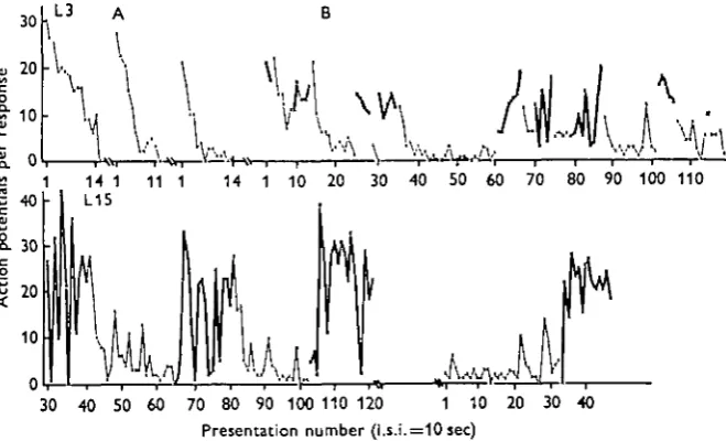
70 80 90 100 110 120 1 10 Presentation number (l.s.i.=10 sec)
[image:9.451.56.386.171.371.2]20 30 40
Fig. 5. Association between maintained high DCMD response and motor activity in spon-taneously active animals. Records taken with implanted electrodes from ball-running animals, retina stabilized. I.s.i., 10 sec. Responses obtained with the animal motionless are shown with a thin line and obtained when it was actively walking with a heavy line. The animals alternate spontaneously between these states, and the transition between them is not related to any ob-vious change in the environment.
Top line: animal L3. A: Three short runs with the animal quiescent, to show characteristic rapid decrement in this condition; rest intervals 10 min. B: Spontaneous alternation between locomotion and rest. Note that responses during the active periods are on average larger than those obtained during rest periods and show no overall change in level; in contrast, responses obtained with the animal stationary start with the first response as high as during the preceding active period, and then undergo a rapid decrement to a low level. The transition upon resuming activity can be either gradual or very abrupt.
Lower line: animal L15. Another individual, showing the same features as in B above in a more extreme form. In this animal a card shield was waxed to the head and prothorax just below the eyes so that the moving legs could not cause visual stimulation during walking. Note that while in most cases the increase in response and the onset of walking take place simultaneously, occasionally (presentation no. 103) walking precedes the response rise by 3O8ec.
736
DCMD responsiveness typically decays exponentially to the low plateau level charac teristic of a resting, habituated animal, and is abruptly raised again on the resumption of motor activity or arousal, spontaneous or evoked (Fig. 5). The increase in visual response during locomotion is not due to increased visual stimulation by the moving legs, as the effect is unchanged when the legs are masked from sight of the eyes (Fig. 5).
(iii) Background activity of the DCMD - in addition to response level - is also significantly higher in aroused or moving animals, relative to quiescent ones. The increase is proportionately comparable (but absolutely small, as background levels are normally very low, rarely reaching a mean frequency of i/sec). This difference in background activity is not merely due to the animal receiving liminal visual stimulation from its own moving appendages. The difference persists in animals which are fitted with a screen waxed to the head so that they cannot see their limbs (Table 1).
Table 1. Correlation of high and low levels of both responsiveness and of background activity of the DCMD with periods of motor activity and quiescence in the behaviour of the animal
These figures are derived from records such as shown in Fig. 5 where the animal is spon-taneously alternating between active and quiet states. The figures for quiet periods omit the first five stimuli following either a first presentation or after a period of activity, during which the response level is declining rapidly.
Each column gives the mean number of action potentials in impulses/sec, the standard error of the mean (not standard deviation) and the number of measurements on which each figure is based. The periods from which the response and background data are derived overlap or are identical.
The differences between the 'active* and 'quiet' columns are significant at P< o-ooi for all pairs of figures.
In the second set of figures for individual L15 the animal was prevented from seeing its own legs by a cardboard screen. The differences between the active and quiet conditions remain unchanged showing that the sight of the moving legs is not responsible for the higher back-ground when the animal is active. The fact that in these figures the masked animal shows higher, not lower, background levels is attributable to the general excitement and arousal caused by the presence of the mask, which the animal struggles to remove.
Response (to 1 sec visual stimulus)
Background (imp./sec during inter-stimulus interval)
Animal
L 3 L i o
LiS
Legs screened from view
Active
X 8 . E .B I I - 7 ± O - S 9
7-o±o-9i
2 O ' 3 4 ± i o i
178 ±i-47 N 73 2 1 79 72 Quiet S S.E.a
343 ± 0 2 8 2-71 ± 0 6 6 3-3010-67
3-8s±°'54
N
1 2 4
17
37 39
Active
S S.B.B
0-I0±0-20 0-75 ±1-20 030 ±067 0-4810-06 I-I2±0-I4 N 65 21 27 140 142 Quiet
X S.B.B
0-05 ±0-09 0-0810-41 O-O2±O-II 0-20±003 o-52±o-o8 N
1 1 6
17 42
116
S9
C. The neural antecedents of dishabituation
fen occur at subliminal levels which do not produce visible movement, yet which have the characteristic output patterns and might well excite proprioceptors. Such subliminal output would explain the variation in precedence and latency seen be-tween movement and DCMD increments; it would also be possible to argue that the entire sensory dishabituation effect was a secondary one, consequent on motor activity initiated by the sensory input.
There are two obvious ways in which such a relationship could be obtained, either by collaterals from the motor system to the visual pathway direct, or by means of sensory reafference, via proprio- or mechano-reception, from movement. These two possibilities are treated experimentally below.
(i) Dishabituation in a de-efferented preparation
To eliminate the possibility of a direct collateral input to the visual system from the motor output system, dishabituation was attempted in preparations with no motor output at all. It has already been noted (§ A( ii) above) that it can be obtained by stimula-tion of the head after the neck connectives are cut, which eliminates a large fracstimula-tion of the total motor apparatus.
Initial attempts to work with a completely excised nerve cord plus the eyes were unsuccessful because of the relatively short life of the preparation. A compromise was found by leaving the CNS in the animal, but working with a totally de-efferented and partially de-afferented brain. All nerves entering or leaving the brain were cut, with the exception of the sensory branch of the antennal nerve and the tegumentary nerve. The retrocerebral complex and frontal ganglion were removed, the circumoesophageal connectives were cut, and the DCMD was recorded from them. Stimulating electrodes were placed on the antennae, and stimulating currents could also be applied via the recording electrode to the circumoesophageal connectives. This preparation would live for some days.
Fig. 6 shows that when the antenna ipsilateral to the seeing eye was stimulated there was a dishabituation of the DCMD. The effect was smaller than usually obtained by similar stimulation in an animal with an intact nervous system, but it was statistically significant and showed the usual decreasing effectiveness with repetition. Stimulation of the contralateral antenna had no effect. It is difficult to be sure of a negative result, but this finding was consistent in three good preparations. Stimulation of the connective containing the DCMD was expected to produce dishabituation, by analogy with the effect of stimulating in the thoracic region in intact animals (see e.g. Rowell & Horn, 1968), but this was not achieved. Instead, stimulation resulted in a long-lasting de-pression of the DCMD, though not a total inhibition, which took between 30 and 120 min to disappear.
which no longer reach the brain after the lesions. This would imply either collateral from the lower motor nervous system, or sensory reafference. In view of the efficacy of sensory input alone in the de-efferented preparation it is simpler to conclude that all dishabituating pathways are basically afferent, and that the input from the body consists of reafferent signal from the animal's movements, rather than of lower motor collaterals; but the latter possibly has not been rigorously excluded. As motor and sensory tracts are not separate in the thoracic nerves, it is difficult to see how this can be done.
L24 and L25
c o
Q.
o .
20
• J ! 10
Stimulate Ipsilateral antenna
Stimulus presentation ( U . i . 5 sec)
Stim.
III
Stimulate contralateral < antenna
Fig. 6. Sensory dishabituation in the absence of any motor output from the CNS. All con-nexions to the brain are cut, with the exception of the sensory branch of the antennal nerve and the tegumentary nerve. The retrocerebral complex, frontal ganglion and all tritocerebral nerves are removed, and the circumoesophageal connectives are cut; the DCMD is recorded from these. Stimulating electrodes are placed on the antennae.
The curve on the left-hand side shows the mean response (and 2 standard deviations) to a stimulus repeated at an interval of 5 sec. In a preparation of this sort the response rapidly reaches a very low plateau level of approximately O'S action potentials per response after the 10th presentation. After the 15th presentation one of the antennal nerves is stimulated for 7 sec.
On the right-hand side is shown the individual responses to the 5 pre-stimulus and 6 post-stimulus responses, at three successive and increasing levels of stimulation of the antenna ipsi-lateral to the seeing eye. It will be seen that the first stimulus at each intensity gives a small but convincing dishabituation, while subsequent presentations of the same stimulus are less effective. At the bottom is shown the response to stimulation of the contralateral antenna (in a different animal, L24), which produces no clear dishabituation.
(ii) Is dishabituation produced by proprioceptive reafference?
Jhese neurones are as yet poorly characterized, but appear to be mechano- and/or proprioceptor intemeurones deriving their input from very large fields, and responding to almost any movement of the body or appendages within that field. At least one neurone with apparently similar properties has been found in the locust thoracic connective, and this is now described.
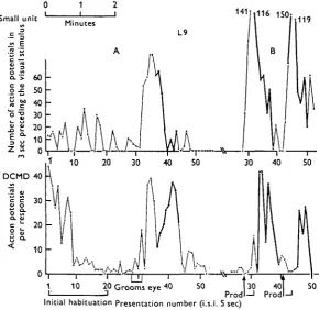
In a number of preparations the record from the nerve cord has included not only the DCMD potentials but also those of another unit, the activity of which is related to the changes in that of the DCMD. It is more or less silent when the animal is at
Smal c al s tent i o CL cio n u "o a . 0 E D 1 unit E > th e DO 0 at L. a. u Of 60 50 40 30 20 10 0
I 16 150; 119 DCMD 40 30 20 10 -50
[image:13.451.68.359.186.468.2]Initial habitation Presentation number (i.s.i. 5 sec)
Fig. 7. Correlation between DCMD responsiveness and activity of small unit in cord, possibly mechanoreceptor interneurone. Chronically implanted animal (Lo) walking on a gangway which preserves its orientation to the stimulus, though not achieving stabilization of the image on the retina. The upper trace plots the total number of action potentials of the small unit in the 3 sec period immediately preceding the visual stimulus. The lower record shows the res-ponse of the DCMD to the stimulus. I.s.i., 5 sec.
A. Presentations 1-20 cover the initial habituation of the DCMD to a novel stimulus; there is no correlated change in the small unit. During the period of presentations 27-31 the animal spontaneously groomed the eye that drives the DCMD with the forefoot, using 17 strokes. (Note absence of visual response to this.) Immediately afterwards there was a great increase first in the small fibre activity and shortly afterwards in the DCMD response. The small fibre activity declines from presentation 36 onwards and reaches zero at 40; during this period the animal starts to walk (thick line), with associated rise in visual response, and both cease around the 45th stimulus.
rest. Periodically the firing rate increases up to 50/sec or more. Increases in ^ response of the DCMD are always preceded by such increases in this smaller fibre, regardless of whether the DCMD fluctuation is 'spontaneous' or evoked, transient or long-lasting, accompanied by motor activity or without motor activity. Similarly, decrements in the DCMD response are preceded by decrements in the small fibre. The only exception to these correlations is during the initial decrement in the DCMD seen after a rest period without visual stimulation (habituation). The initial high visual responses do not then correspond to any increased small fibre activity, and the decre-ment is not associated with any change in the small fibre rate. These features are illustrated in Fig. 7.
The latency between the change in the small fibre and that in the DCMD is remarkably long and variable, often being between 5 and 20 sec, especially in the case of a spontaneous fluctuation not associated with obvious external stimulation. Where increases in both are induced by, for example, tactile stimulation, both com-mence to rise more or less simultaneously, but the small fibre activity rises more steeply than the DCMD responsiveness, and the peaks of activity are most commonly separated by 5-10 sec.
All major increases in the small fibre firing rate are followed by increases in the DCMD. However, some transient inflexions (less than 20 sec duration) are not. This may indicate that the small fibre is only one of a population of synergistic neurones, or that temporal summation over a period of some seconds is required to produce the effect on the DCMD.
To test the relevance of the correlation between the activity of this small fibre and the DCMD response, a similar count was made with recordings in which the small unit was not distinguishable. The counter was instead set to count merely that fraction of the miscellaneous neural traffic which would give a similar overall spike frequency to that of the small fibre. This activity derived at least in part from visual and tactile sensory interneurones. The count was made over a 2 h record with visual stimuli at one every 10 sec. A small correlation due to simultaneous increase in sensory traffic and DCMD responsiveness during applied stimuli was found, but otherwise there was no relation between the two; especially, there was no tendency of the activity of the smaller units to precede the DCMD changes, and any correlations were between simultaneous records.
74
0-40 r
006 • 004
2-1 2-2 2-3 2-4 25 2-6 27 2-8 2-9 30 31 3-2 3-3 3-4 3-5 3 6
[image:15.451.47.382.70.456.2]i (response level)
1964) preceding the visible movement. The only finding against it receiving its majq input from somatic tactile or position receptors is that it often starts to decline and sometimes becomes inactive while the animal is still moving (Fig. 7), though charac-teristically the movement too then ends soon afterwards. The parallel with the crayfish activity fibre is thus tempting, but by no means perfect, and there is no direct evidence that the unit is even afferent rather than efferent.
Its temporal relation with the DCMD shows that either this unit is one of the most important factors affecting the responsiveness of the visual unit, or, less probably, that both share common factors which affect them with a differing latency.
(iii) Relation between the amount of sensory input and the DCMD response
The previous sections indicate a high probability that the neural antecedents of dishabituation include at least two different types of sensory input, and that the pro-prioceptive input is particularly important. There is also a suggestion that a great variety of other sensory inputs may contribute. If dishabituation is a function of general sensory input, then any experimental conditions which greatly modify the latter should affect the visual responsiveness. Recordings have been made in three types of preparations which would be expected to differ very much in sensory input: the free-moving animal, the restrained immobilized animal, and the isolated brain with only visual and antennal input. These preparations do indeed differ in the expected ways. Firstly, 'spontaneous' dishabituation, not following any experimentally applied stimulus or accompanying any overt motor behaviour, occurs more commonly in free-moving or ball-running animals than in restrained ones, where it is rare (Horn & Rowell, 1968); an example is seen in Fig. 7, presentation 30 et seq. It may also be significant that induced dishabituation is more difficult to produce and of smaller amplitude. Secondly, where the DCMD neurone enters an exponential decremental phase (as following initial stimulation, or after dishabituation, or after the cessation of movement) its rate of decrease can be quantified (see Rowell & McKay, 19696). Comparisons between the rate of decrement of the same animal, first in the implanted, free-running state, and secondly in an acute preparation after dissection, shows that the free-moving animals have a slower decremental rate. In preparations in which the brain has been almost completely de-afferented, the rate of habituation is fastest of all (Fig. 8). Lastly, the plateau levels reached after the decremental phase is complete are highest in the ball-running animals, intermediate in the acutely dissected pre-parations, and lowest of all in the de-afferented brain. Compare, for example, the plateau levels in Figs. 4, 2 and 6.
All these results suggest that sensory input to the brain of many sorts modifies the responsiveness and the rate of habituation of the DCMD unit.
DISCUSSION
several different levels; see, for example, Kalil & Chase (1970), Schmidt (1969), Comis & Whitfield (1968), Fetz (1968) and Miles (1970). Among invertebrates Rowell & McKay (19696) showed that an acridid auditory interneurone, probably only 1 or 2 synapses removed from the sensilla, was under tonic but variable inhibition from the head ganglia; this inhibition was sufficient to abolish the response under some condi-tions. McKay (1970) found a similar situation in the tettigoniid auditory system, except that the major inhibitory input was derived from the pterothoracic ganglia. Changes in reflex responsiveness after lesion to the CNS (Rowell, 1964) could well be due to disruption of similar inhibitory inputs to tactile interneurones of the pro-thoracic ganglion. Are'chiga & Wiersma (1969a) reported that the response level of the crayfish 'jittery movement fibres' was increased and habituation reduced as a consequence of any movement of the animal; tonic' sustaining fibres' were facilitated by all movement. Wiersma & Oberjat (1968) noted that in crayfish in an 'excited state' there was a tonic increase in firing in all the oculo-motor nerves: these are the axons of motor cells, which are driven by visual interneurones plus primary afferents from the statocyst. The authors suggest a command-fibre input common to all to mediate this input from the excited state, but it seems equally possible that it was via the visual interneurones. Taylor (1970), again working with crayfish, showed that an interneurone responding via the statocyst to water-borne vibration had its response level either raised or lowered by a variety of motor activities and sensory inputs; this unit shows no habituation under the conditions used, so does not provide information as to this parameter. In addition, these and similar units show changes with dark and light regime, sometimes known to represent a true diurnal rhythm (Rowell & Horn, 1968; Arechiga &Wiersma, 19696; J. Palka, personal communication). A more detailed review of this field may be found in Rowell (1970). It is obvious that these effects on sensory input must contribute to the integration of behaviour, and perhaps especially by mediating changes in attention. As a consequence of their existence the same sensory input has quite different neuronal representation under different circum-stances, and it is tempting to ascribe at least part of the observed variation in the behavioural response to constant stimuli to these changes in neural representation.
The data presented in this paper add the DMD units to the list of invertebrate interneurones which are modulated by inputs different from those which normally excite them. (At a qualitative level it has been observed that changes similar to those described from the DCMD unit are also taking place in the DIMD of the opposite connective.) It is clear that the decremental response to repetitive stimulation, so characteristic of the restrained animal, is only partially representative of the unit's behaviour in the normal animals. When the animal is aroused the DCMD response is high, and there is no overall decrement during any period of maintained behavioural arousal or motor activity. Presumably the selective advantage of the decremental response when activity ceases or the animal is at rest is that it provides both a low level of response when the input is constant, coupled with a slow fall-off from the highly responsive state, so that the animal does not over-react to temporary cessation of stimuli by reducing its visual responsiveness too quickly.
reflex in Aplysia was thought by exclusion to involve presynaptic excitation of excitatory terminals of the sensory nerve in the CNS (Castellucci et al. 1970). It has since been shown that the same behaviour can also be mediated by the peri-pheral non-ganglionic nervous system in the absence of the CNS (Peretz, 1970), but a similar mechanism would still be possible there too. However, Bruner & Kennedy (1970) describe behaviour fulfilling many of the criteria for habituation and dishabituation which can be performed by a single crustacean neuromuscular junction, and this cautions against assuming that complex effects necessarily imply a complex polysynaptic substrate. In this paper evidence is presented that the dishabituation of the DCMD, as in Aplysia, involves a re-setting of those excitatory synapses in the pathway which are responsible for the decrement. The most obvious way in which this could be done would be by episynaptic input. Presynaptic inhibition appears to be excluded, as there is no indication that the dishabituating stimulus causes a transient inhibition of the response, so the most likely candidate is presynaptic excitation. However, depending on the nature of the response-generated depression that causes the decrement, a variety of other mechanisms would be possible. It seems unlikely that this question can be further resolved without access to the synaptic areas. Dis-habituation is effective over the whole eye simultaneously; when to this is added the fact that the habituation process is site-specific and localized on the eye, and the deduction that dishabituation acts by counteracting the decremental process responsible for it, some speculations about the anatomical substrate of the dishabituation process become permissible. It seems likely to be due to a neurone (or possibly neurones) efferent from the brain, where it would receive appropriately integrated multimodal sensory inputs. It would have a long time constant of excitation, accounting for the long latency of recorded changes. Its terminals must innervate synapses in the visual pathway at a level where there is still topographical representation of the retina, but at a level of integration sufficient to account for the other properties of the DCMD unit, such as its directional preferences. A likely candidate would be one of the ' tangential' cells innervating the medulla of the optic lobe. Recordings have recently been made from some of these cells in lepidopterans, but only visual inputs were tested (Collett, 1970); however, all were indeed efferent to the medulla. Such a cell would of course be active at a reduced level at all times, not merely during dishabitu-ation. This fits with the evidence presented here showing that while the rate of habituation is increased, the plateau response level is decreased by lesions which reduce the general sensory input to the brain.
Petween the activity of what is probably a wide-field mechanoreceptor interneurone and the responsiveness changes of the DCMD is reported here, and Ardchiga & Wiersma (1969a) found a very similar relationship in the crayfish. However, in neither animal is there proof that this correlation is causal; in the locust, where the latency has been measured, it is very long. While it is probable that these interneurones contribute to the effective input regulating the response level of the visual units, it seems clear that the full behaviour here reported from the locust system would not be completely explained by input from this source alone. Minimally the system must include also other sensory inputs such as from the antenna, and a higher level of integration is probably involved too. To give a minor example, grooming the head or antennae requires movement of front legs and head, and (especially when it is initiated by a resting animal) represents an increase in activity. Yet the DCMD response level is depressed rather than elevated during grooming (see Rowell, 1971^) whereas by the simplest version of Ardchiga and Wiersma's hypothesis the converse would be expected.
It is also of interest that stimulation of the circumoesophageal connective of the isolated brain depressed the DCMD for a long period. This is the first indication that modulating inputs to that neurone can depress it; all the other evidence reported here correlated a new or additional input to the CNS with an increase in DCMD respon-siveness. The nature of this inhibitory input is unknown, but as quite different results are obtained by stimulating the cervical connective, it is possible that it arises in the suboesophageal ganglion.
SUMMARY
1. Recorded from a dissected immobilized animal, or from an unrestrained animal which is quiescent, the descending contralateral movement detector (DCMD) neurone shows an exponential decremental response to a repetitive stimulus (habituation), reaching a plateau level characteristic of the stimulus conditions. The process is site-specific on the retina, and movement to a new area of retina gives a complete recovery. In the absence of stimulation responsiveness returns over minutes or hours.
2. Immediate recovery without a rest (dishabituation) can be obtained by a variety of strong sensory stimuli of several different modalities (' extra-stimuli') or by non specific electrical stimulation of parts of the CNS. The dishabituating efficacy of all these wanes with repetition. When the habituating stimulus is moved to a new retinal site the previous site is not dishabituated.
3. Dishabituation is not site-specific but affects the whole retina simultaneously. It appears to reverse the original decremental process (' re-set') rather than to produce an independent enhancement elsewhere in the pathway, as it does not increase the response from a submaximally stimulated, but unhabituated, retinal site.
4. In unrestrained animals dishabituating extra-stimuli also cause behavioural arousal or other motor activity. When motor activity starts, the DCMD is dishabituated and shows no regular decremental trend thereafter until movement ceases. DCMD background activity is also increased. These effects are not due to the visual stimulus of the moving appendages.
5. The association between motor activity and dishabituation suggests that the latter derives either from motor system collaterals or from mechanoreceptive
reafference. Stimulation of the antennal nerve of a totally de-efferented brain cau some dishabituation; this eliminates the lower motor system (below command-fibre level) as the source of dishabituation and suggests it is purely sensory.
6. The activity of a thoracic cord unit (of possibly a wide-field mechanoreceptor interneurone) precedes by 5-20 sec, and closely correlates with, changes in responsive-ness of the DCMD. It is either an important input to, or an output from, the dis-habituating system.
7. Progressive reduction of sensory input to the brain affects DCMD responsive-ness as follows: (i) spontaneous dishabituation is less frequent, (ii) dishabituation is less easily induced and smaller, (iii) rate of habituation is increased, (iv) plateau res-ponse level after habituation is lower.
8. Electrical stimulation of the circumoesophageal connective can depress DCMD responsiveness for many minutes.
9. The probable anatomical and physiological bases for modulation of DCMD responsiveness are discussed.
The work reported here was supported by the U.S. Government under Contract AFOSR 69-1810 and by National Institutes of Health Grants FR-7006 and 1 Roi NS 09494-01 to the author. I am grateful to Dr M. F. Land for his critical reading of the manuscript.
REFERENCES
ARECHIGA, H. & WIKRSMA, C. A. G. (1969a). The effect of motor activity on the reactivity of single visual units in the crayfish. J. Newobiol. I, 53—69.
ARECHIGA, H. & WlERSMA, C. A. G. (19696). Circadian rhythm of responsiveness in crayfish visual units. J. Neurobiol. 1, 71-85.
BRUNER, J. & KENNEDY, D. (1970). Habituation: occurrence at a neuromuscular junction. Science, N. Y.
169, 92-4.
CASTBLLUCCI, V., PINSKER, H., KUPFERMANN, I. & KANDEL, E. R. (1970). Neuronal mechanisms of
habituation and dishabituation of the gill-withdrawal reflex in Aplytia. Science, N. Y. 167, 1745-8. COLLETT, T. S. (1970). Centripetal and centrifugal visual cells in medulla of the insect optic lobe.
J. Neuropkysiol. 33 (2), 239-56.
COMIS, S. D. & WHITFIELD, I. C. (1968). Influence of centrifugal pathways on unit activity in the cochlear nucleus. J. Neuropkysiol. 31, 62-8.
FETZ, E. E. (1968). Pyramidal tract effects on intemeurons in the cat lumbar dorsal horn. J. Neuropkytiol. 3i»
6o-79-HORN, G. & ROWELL, C. H. FRASER (1968). Medium and long-term changes in the behaviour of visual neurones in the tritocerebrum of locusts. J. exp. Biol. 49, 143-69.
HOYLE, G. (1964). Exploration of neuronal mechanisms underlying behavior in insects. In: Neural
Theory and Modeling (ed. R. F. Reiss), pp. 346-76.
KALIL, R. E. & CHASE, R. (1970). Corticofugal influence on activity of lateral geniculate neurons in the cat. J. Neurophysiol. 33, 459-74.
MCKAY, J. M. (1970). Central control of an insect sensory interneurone. J. exp. Biol. 53, 137-44. MILES, F. A. (1970). Centrifugal effects in the avian retina. Science, N.Y. 170, 992-5.
PALKA, J. (1967a). An inhibitory process influencing visual responses in a fibre of the ventral nerve cord of locusts. J. Insect Physiol. 13, 235-48.
PERETZ, B. (1970). Habituation and dishabituation in the absence of a central nervous system. Science,
N.Y. 169, 379-81.
ROWELL, C. H. FRASER (1964). Central control of insect segmental reflex. I. Inhibition by different parts of the central nervous system. J. exp. Biol. 41, 550-72.
ROWELL, C. H. FRASER (1970). Incremental and decremental processes in the insect central nervous system. In Short-term Changes in Neural Activity and Behaviour, ed. G. Horn and R. A. Hinde. Cambridge University Press.
BbwELX, C. H. FRASER (19716). Antennal cleaning, arousal and visual interneurone responsiveness in a locust. J. exp. Biol. (in the Press).
ROWELL, C. H. FRASER & HORN, G. (1967). Response characteristics of neurones in an insect brain.
Nature, Lond. ax6, 702-3.
ROWELL, C. H. FRASER & HORN, G. (1968). Dishabituation and arousal in the response of single nerve cells in an insect brain. J. exp. Biol. 49, 171-83.
ROWELL, C. H. FRASER & MCKAY, J. M. (1969a). An acridid auditory interneurone. I. Functional connexions and response to single sounds. J. exp. Biol. 51, 231-46.
ROWBLL, C. H. FRASER & MCKAY, J. M. (19696). An acridid auditory interneurone. II. Habituation, variation in response level, and central control. J. exp. Biol. 51, 247-60.
SCHMIDT, R. F. (1969). Spinal cord afferents: functional organization and inhibitory control. In The
Interneurone (ed. M. A. B. Brazier), pp. 209-30. University of California Press.
TAYLOR, R. C. (1970). Environmental factors which control the sensitivity of a single crayfish inter-neurone. Comp. Biochem. Physiol. 33, 911-21.
WALLACE, G. K. (1939). Visual scanning in the desert locust Schistocerca gregaria Forsk&l. J. exp. Biol.
36, 512-25.
WIERSMA, C. A. G. (1970). Reactivity changes in crustacean neural systems. In Short-term Changes in
Neural Activity and Behaviour, ed. G. Horn and R. A. Hinde. Cambridge University Press.
WIBRSMA, C. A. G. & OBERJAT, T. (1968). The selective responsiveness of various crayfish oculomotor fibres to sensory stimuli. Comp. Biochem. Physiol. 36, 1-16.