~-DEVELOPING SEA URCHIN EMBRYOS II. STUDIES ON THE
ACTIVATION OF PRCY.rEIN .BlOSYNTHESIS IN SEA URCHIN EGGS AT FERTILIZATION
l'b.esis by ;J?aul Claire Denny
In Partial Fulfillment of the Requirements For the Degree of
~octor of Philosophy
palifornia Institute of Technology Pasadena, California
1965
i i
ACKNOWLEDGEMENTS
I am grateful to the Division of Biology, California Institute of Technology for the opportunity of studying and working at the Institute and also to its individual members for their interest and valuable advice on many of the problems which have come up. In
parti-cular, I wish to thank Professor Albert Tyler for allowing me to work in his laboratory over the past four years and for his counsel and help both in matters of my research and profession. I am also grateful to him for his help in the preparation of this manuscript.
I am indebted to Dr. Dennis Barrett for the stimulating discussions which exposed me to many of the problems and approaches of research and to Dr. Hector Timourian, with whom a paper was co-authored which included our separate and joint investigations on the effects of various ions on protein synthesis and development, for his advice and criticisms. I am also indebt~d to
Mr.
Joram Piatigorsky for working with me jointly on the detection of polysomes in anucleate egg fragments. I would like to acknowledge the valuable assistance ofMr.
William Smith andMr.
Roger Hendrix during the summers ofi962
and1963
respectively.ABSTRACT
I. Alkaline phosphatase activity in the developing sea urchin
Lytechinus pictus has been investigated with respect to intensity--at· various stages, ionic requirements and intracellular localization.
The activity per embryo reinains the same in the unfertilized egg, fertilized egg and cleavage stages. At a time just prior to
gastru-lation (about 10 hours after fertilization) the activity per embryo begins to rise and increases 300 times over the activity in the
cleavage stages during the next 60 hours.
The optimum ionic strength for enzymatic activity shows a wide
peak at 0.6 to 1.0. Calcium and magnesium show an additional optimum
at a concentration in the range of 0.02 to 0.07 molar. EDTA at
concen-trations of 0.0001 molar and higher shows a definite inhibition of
activity.
The intracellular. localization of alkaline phosphatase in
homo-genates of 72-hour embryos has been studied employing the differential
centrifugation method. The major portion of the total activity in
these homogenates was found in mitochondrial and microsomal fractions
with less than
5%
in the nuclear fraction and less than2f,
in the final supernatant. The activity could be released from all fractions by treatment with sodium deo.xycholate.II. The acttvation of protein biosynthesis at fertilization in
eggs of the sea urchins Lytechinus pictus and Strongylocentrotus purpuratus has been studied in both intact eggs and cell-free
iv
optimum amino acid incorporation activity and in the case of the latter the concentration range is quite narrow. Though the optimum magnesium concentrations appear to differ slightly in homogenates of unfertilized and fertilized eggs, in no case was it observed that unf'ertilized egg homogenates were stimulated to incorporate at a
level comparable to that of the fertilized eggs.
An
activation of amino acid incorporation into protein has alsobeen shown to occur in parthenogenetically activated non-nucleate
I.
TABLE OF CONTENTS
STUDIES ON ALKALINE PHOSPHATASE ACTIVITY IN DEVELOPING - SEA URCHIN EMBRYOS
INTRODUCTION
MATERIALS AND METHODS
Collection, fertilization and culturing of eggs Preparation and assay of extracts
JIBSULTS
Activity during development
Ionic requirements for optimum activity
In vitro identification of activity-rich cell fractions
DISCUSSION
The control of alkaline phosphatase activity during development
Ionic effects on enzyme activity • Activity in different cell fractions SUMMARY AND CONCLUSIONS
II. STUDIES ON
THE
ACTIVATION OF PROTEIN BIOSYNTHESIS IN SEA -URCHIN EGGS AT FERTILIZATIONINTRODUCTION
Protein synthesis in sea urchin eggs • Experimental investigations
Anucleate cells as indicators of nucleus-independent · cellular fractions
Aims of the. present study MATERIALS AND METHODS
Preparation of cell-free amino acid incorporating system ·
Radioactivity and protein determinations . ~reparation and activation of egg fragments Detection of polysomes
vi
RESULTS "'
Cell-free aro:ino acid incorporation
Amino acid incorporation by parthenogenetically activated non-nucleate egg fragments
Polysome formation in artificially activated non-nucleate fragmen~s
_DISCUSSION
.Protein synthesis .
Amino acid incorporation as an indication of true protein synthesis
Role of the proteins synthesized during early development
Cell-free amino acid incorporation
Incorporation by anucleate egg fragments • Cytoplasmic DNA
Activation of RNA synthesis at fertilization Mechanisms for activating the protein synthesizing
system
Location of "mask" Possible analogies
Cytoplasmic mechanisms for the initiation of protein synthesis
S,t.JMMARY AND CONCLUSIONS
2
INTRODUCTION
Changes in alkaline phosphatase activity per embryo during
development have been reported for many types of animals (cf. 1 for review). From the studies of Zorzoli (2); Mazia, Blumenthal and
Benson
(3);
Gustafson and Hasselberg(4);
Mulnard (l); L~vtrop(5);
Manganti and Mancusco-Palazzo (6) it appeared that in embryos of bothvertebrates and invertebrates the alkaline phosphatase activity was
low and remained the same throughout cleavage. Increases in activity per embryo then began at certain later embryonic stages. This increase
was reported to begin at the onset of gastrulation in sea urchin embryos
(3, 4)
and eventually to reach a value approximately ten times that ofthe cleavage stages (see Appendix, p.
149).
This, then, appeared to be a suitable system in which a closer specification of the amounts andtimes of activity changes could possibly serve as a basis for f'uture
examination of questions of how the enzyme(s) might be regulated during
development and whether or not alkaline phosphatase might be involved
in specific developmental processes.
Hist.ochemical studies of alkaline phosphatase in developing sea urchin eggs indicated that the enzyme was distributed uniformly
through-out the unfertilized and ~ertilized eggs, and cleavage stage embryos
(7).
An increase in activity was first noticed in the vegetal pole and migrating
mesenchyme cells of the blastula stage, and as development progressed very
strong reactions developed in both the gut and skeleton-forming areas
(8).
this lack of enzymatic activity there were gross abnormalities
associ-ated with the two enzyme rich areas, and it was suggested that at least
in the case of skeletal development, the enzyme plays an important role
in the differentiative processes
(9).
Since alkaline phosphataseactivi-ties from a wide variety of sources are known to commonly require ion
cofactors for maximal activity (10, 11) it seemed that-the variations in skeletal development observed in embryos cultured in calcium
defi-cient or magnesium enriched sea water (12, 13) might be due to specific
effects of these ions on the activity of the enzyme itself. In these
studies the in vivo ~ffects of the two ions were for the most part
antagonistic, thus one might expect to see the in vitro activity of
the enzyme also affected differently by changes in the concentrations
of the two ions if indeed the primary effect was at the enzymatic level.
It was with this in mind that the study of ion effects on sea urchin·
embryo alkaline phosphatase activity was begun.
It was shown by histochemical techniques that there was both
nuclear and cytoplasmic alkaline phosphatase activity in cells of
developing sea urchin embryos
(7,
14). A similar study of the systemby the differential centrifugation method (15) is reported herein. In
these studies the a~ove observations have been confirmed and extended,
though not without considerable disagreement on the amounts of activity
in the two fractions. Tb.is study was begun primarily for the purpose of
locating the fraction with which the activity is.associated, and
second-arily it was hoped that this information might give insight into the
4
MATERIALS AND METHODS
Collection, fertilization and culturing of eggs
Eggs and sperm were collected from Lyt;echinus pictus adults
either by injecting 0.55 molar KCl into the body cavity
(16)
orby cutting out the gonads and placing them in sea water to shed.
The eggs were then washed with sea water until the wash fluid
became clear. For fertilization the "dry" semen was diluted
1000-fold with 0.001 M EDTA (ethylenedia.minetetraacetic acid) in sea
water, (17), and increasing a.mounts of this suspension were added to
the eggs at 2 to
4
minute intervals until at least 95~ fertilizationwas observed. In this way cultures containing large numbers of eggs
in a small volume of sea water were obtained in which polyspermy was
less than l~.
The fertilized eggs were washed once to remove excess spermatozoa
0
and cultured in filtered sea water at 21.5 C in shallow glass trays
which were kept in a moist chamber. Development proceeded normally,
and water loss due to evaporation was limited to less than 3~ over a
period of three days. For retarding bacterial growth penicillin G
potassium (Squibb) was added to the cultures after fertilization to a
final concentration of 100 units per ml and again 48 hours later to a
concentration of 50 additional units per ml (cf. 18).
Where specified, the feeding-stage embryos were fed with cultures
of Nitzchia closterium which are maintained at this laboratory. The
by the embryos • No attempt was made to obtain a maximum growth rate,
but care was exercised to insure that only cultures in which the
embryos were active and otherwise nonnal appearing were used for assay.
Preparation and assay of fertilized eggs at different developmental stages
To insure uniform sampling, five 10 ml samples were taken with
wide-mouth pipettes from each culture at various times after
fertili-zation. The five samples from a single culture were combined. Each
resulting 50 ml aliquot was then cooled in an ice bath to stop
development and 'also to inactivate the swimming embryos. The embryos
were washed once by settling in a graduated centrifuge tube and frozen
. 0
(-15 C) after adjusting the.final volume to one ml with sea water at
one-half strength. They were stored frozen and used within four days
after collection.
For counting the embryos, formaldehyde (to
4%)
was added toseparate 50 ml aliquots of the culture. These suspensions were later
diluted to 500 ml, and .counts were made on
8
to 10 one ml samples(wide-mouth pipette). Embryos with gross abnormalities were separately
enumerated, and no culture containing more than 1% of these was used.
After thawing, the one ml samples received one ml of 'cfo sodium
deoxycholate (DOC) at pH 10. They were homogenized by repeated passage
(about 50 times) in and out of a glass syringe fitted with a 22 gauge
needle and powered by a pipetting machine. Examination of the homogenates
by light microscope showed complete cellular disruption. In addition
to causing homogenization of the eggs or embryos, this treatment also
6
:fractions with very little change in activity (note Tables III and V).
The activity was assayed either in the total homogenate or in the
supernatant obtained a~er centrif'ugation at 12,000 times gravity (X g)
for 20 minutes to remove large particulate matter. The temperature was
kept at or below
4°c
throughout the above procedure.The change in optical density at 400
mp.
due to hydrolysis ofp-nitrophenyi phosphate1 (19) was continuously measured at pH 10.15
and
30°c
in1
cm light path microcuvettes with a recording spectro-photometer (Cary M~del llMS). Thus, the rate of hydrolysis could bedirectly determined :from the slope of the
OD406
change with time.Since the reaction rates were no longer linear beyond the conversion
of the first la{o of the substrate, comparison of the enzyme activities
:from different samples never involved the hydrolysis of more than this.
Keeping this in mind it could then be shown that the activity is
propor-tional to the a.mount of homogenate added over a wide range (Fig.
1).
It was also shown that, by using an eight-fold difference in the a.mount ofhomogenate assayed while maintaining equal buffer and substrate
concentra-tions as well as equivalent ionic strengths,the activities from a single
culture could be maintained within the range previously shown to be
representative' of the a.mount of enzymatic activity present (Fig.
2
).
Either of two buffer systems, carbonate-veronal-HCl (20) orcarbonate-bicarbonate (21), were found satisfactory for the incubation mixture
which contained 0.1 molar buffer, 0.0002 molar MgC12 and 0.0002 molar
substrate. The incubation. mixture was adjusted in each case so that
the concentrations of sea water salts were at one-sixth their
' l. '
Cf)
w
1.5
t-::::>
z
~0
z
w
r.o
Cf)ct
w
a::
(.)
z
1
d
0~5d
:
100
2~10
5
DILUTION FACTOR OF HOMOGENATE
Fig. 1.-Spectrophotometric assay
ot
alkaline phosphatase activity showing the linear relationship between the rateot
product tormation and tbe concentrationot
\t
he
'ihoino-g€fnate-. whichcontains the enzyme activ11;y. ·~~--. ____ · ·· -
8
1.0
0.9
•
+
0
.
8
Cf)
w
t-07
=>
>-Z
t-~>o
t - -
0.6
uz
< (
-u~
-c:i
05
~w
~ 0::
>-u
N~
z
§
o~w
d
+
d
0.3
Q2
0.1
0
10
20
30 4050
60
70HOURS AFTER FERTILIZATION
Fig. 2.-Dilut:ions_ needed in order to ass~ hanogenates of' eggs and
embryos
trom
single cultures within a saf'e range of activities.
All samples contained -equaJ. bu:f'f'er and substrate' concentrations as well as equiva1ent ionic strengths. Symbols represent the a.mount in m1 of' ftomogenate in the 0.50 m1 incubation mixttire (crosses -
o.4o;
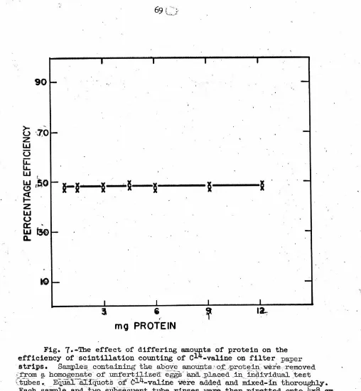
open [image:14.547.47.501.65.594.2]normal value. Each sample tested had its own control which was placed
in the reference beam of the spectrophotometer and contained the
reac-tion mixture minus the substrate. Non-enzymatic hydrolysis of the
· substrate at this pH during the reaction ti.me was found to be
insignifi-cant as was .also the case for the activity in boiled homogenates of
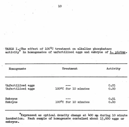
unfertilized eggs or larvae (Table I).
Preparation:and assay for study of ion effects
Entire cultures of 72-hour plutei were used as a source of alkaline
phosphatase activity. The embryos were collected by cooling the, cultures
with carbon:dioxide gas falling from pieces of "dry" ice suspended in
wire baskets above them. Under these conditions the embryos settled to
the bottom of the culture vessel very qui~, allowing for rapid
concen-tration and washing. The embryos were homogenized in a Potter-Elvehjem
tissue grinder with Teflon pestle. The debris from~ 15-minute settling
period was discarded, and the remainder pelleted by a centrifugation at
105
,ooo
X g for 120 minutes. This pellet was washed twice in distilledwater or 0.55 molar KCl. Adjustment of the ionic strength to the test
values was done in a 0.05 molar veronal-HCl buffer-substrate solution
(pH 8.5) by addition of calculated amounts of chloride salts. The value
of ionic strength as given in Figs. 5 and
6
refers to the added chloride10
· TABLE I.-Tb.e ef'f'ect of' ioo0c treatment on alkaline phosphatase
activity* in homogenates of unfertilized eggs and embryos of' L. pictus.
Homogenate
Unfertilized eggs Unfertilized eggs
Embryos Embryos
Treatment
ioooc f'or 10 minutes
ioo0c f'or 10 minutes
Activity
0.05 o.oo
0.91 o.oo
[image:16.548.38.490.20.465.2]Preparation and assay of homogenate fractions
5
For preparation of the homogenate fractions approximately 5 X 10
~mbryos were collected as above and washed twice by settling in a solution
containing 0.55 molar KCl and 0.005 molar MgC12 • They were finally sus-pended in a solution containing 0.24 molar KCl, 0.24 molar sucrose, 0.005
molar MgCl2 and 0.05 molar veronal-HCl buffer at pH 8.5. The embryos were
ground up in a Potter-Elvehjem homogenizer only until no intact cells were
observed. The various fractions were separated in 0.328 x 1-15/16 inch
centrif'uge tubes using an International refrigerated centrifuge with high
speed attachment and a Spinco Model L ultracentrif'uge. The centrifugal
forces listed were calculated with reference to rave. The various
frac-tions were assayed at pH 10.15 in 0.1 molar carbonate-veronal-HC1 buffer
12
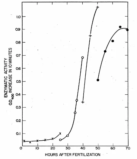
RESULTS
Alkaline phosphatase activity during . development
ReeuJ.ta of
a
aet of determ1nat1ona ofal.ka.J.1ne
phosphataseact1vi-ties in homogenates of' L. pictus embryos at various stages of' development
are plotted in Fig.
3.
The general pattern of the changes resembles thatfound by Mazia, Blumenthal and Benson (3) for A.punctulata and by
Gustaf's on and Ha.sselberg ( 4) for P. miliaris.
· The values of' enzyme activity have been expressed in Fig. 3 as moles
of' p-nitrophenol formed per embryo per minute~ The relationship of' values expressed as activity per embryo to activity per mg of protein present has
not been determined.. There is only a relatively small decrease in total
protein in sea urchin embryos up to the pluteus stage; namely about
16.
5'/o
decrease in S. pur:puratus (22) and about la{o decrease in P. lividus (23).
I f it can be assumed that this is common to the development of sea urchins,
one might expect that during development the changes in activity based on
protein content will be similar to changes in activity per embryo.
Further-more, based upon approximate total cell numbers given by Ma.zia (24), the
change in activity per cell would be one-third to one-half' the magnitude
of the change· in activity per embryo during the period from 10 to
4o
hours after fertilization (see Appendix, p. 149 ). However, since the enzymeappears to be localized in newly formed areas which represent only a
frac-tion of' the total number of' cells, this type of comparison is not likely
to be valid. Thus i~ remains a possibility that the activity per cell does not change in the gut and skeleton-forming eel.ls and that the increase
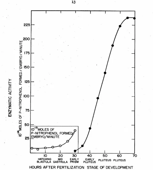
w
200
I-:::>z
~ 175 ...~
0::: CD ~ 150 w ... Cl w ~>-
0::: 125 t-~
>
t- ..J 0
(.)
z
<l: w (.) I Cl.. 100
-
~
00:::
:E
I-.
>-
z
75
~
ICl.. L&J ~
-<n
w 50 ..J 0
I0-15MOLES OF
s~
'Q
P-NITROPHENOL FORME!¥25
EMBRYO/Ml NUTE10 20 30
40
50 60 70HATCHING MID EARLY
BLASTULA GASTRULA PRISM PC~~0s PLUTEus PLUTEus
HOURS AFTER FERTILIZATION STAGE OF DEVELOPMENT
Fig. 3.-Al.kaline phosphatase activity in homogenates of L. pictus eggs at dif'f'erent stages of development at
2l.5°c. All
points are :froma single cultu;re, .and each point represents five separate samplings of the culture. After combining ·the five samplings, each resulting sample
was
asseyed at30°c
in0.1
molar carbonate - veronal - HCl buffer (pH10.15),
0.002 molar MgC12 and 0.0002 molar p-nitrophenyl phosphate. In add+tion, sea water salts were present at one-sixth the concentration in regular
[image:19.548.27.526.39.596.2]14
the increase in activity per embryo which begins at about 10 hours af'ter
fertilization, the activity per cell decreases approximately one
thousand-fold, though the activity per unit volume does not change.
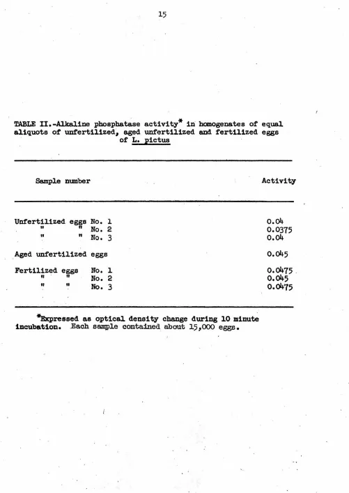
Samples containing equal aliquots of unfertilized, aged unfertilized,
and newly fertilized eggs (Table II) indicate that alkaline phosphatase
activity is not affected by fertilization. The unfertilized eggs were
given the same prefertilization treatment as the other two classes but
were put on ice at the time that fertilization took place. The aged
unfertilized eggs received the same treatment as the fertilized eggs,
and both were placed on ice after the fertilized eggs had developed for
one and·one-half hours. All sperm were removed from the fertilized eggs
by washing several times. The slight difference in activity between
unfertilized eggs and fertilized eggs cannot be attributed to
fertiliza-tion since the aged unfertilized eggs also showed a slightly increased
activity.
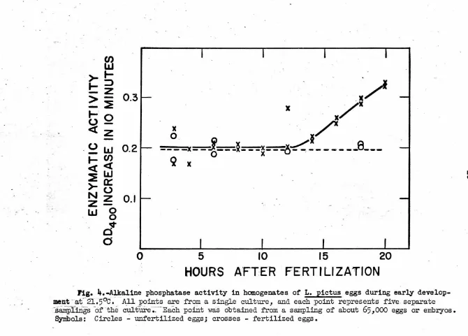
A detailed study showed that alkaline phosphatase activities
remained constant .,during cleavage stages and began to increase shortly
after hatching (Fig.
4).
The exact time that this increase began afterfertilization in the three cultures which were grown under reasonably
·identical conditions appeared to vary nearly four hours. The cause of
this variation was not studied though the simplest explanation is that
some difference in culture conditions was responsible. The unfertilized
eggs did not show any increase in activity after as long as 18 hours
·under culture conditions.
Cultures of embryos which were fed could be maintained several days
longer under crowded conditions than those which were not. The activities
TABLE II.-Alkaline
pbospha~se
activity* iii hanogenates of equal aliquots of unfertilized~ aged unfertilized and fertilized eggsof
L.
pictusSample n1J111ber
Unfertilized eggs
No.
l"
"
No.
2 It"
No.
3
Aged unfertilized eggsFertilized eggs
No.
lfl II
No.
2II It
No.
3
*Expressed as optical density change during
10
minute incubation. .Each sampl.e contained about 15,ooo
eggs.Activity
o.04
0.0375
o.04
o.045
[image:21.550.33.534.24.731.2]f3
)-
f-f-
:::>
>
~
0.3
-
:E
1-o
2
<(z
-.
0
I.LI
0.2
- fl)
I - < ( <(
I.LI
~
a::
)- 0
~ ~
0.1
UJ
0
0
~
0
0
0
.
.
/
x
x/x
.
~
. ·.,/'
----x-
_.j;_ __
x ___ x __
~x.~--
_ -- __
9 __ _
q
x
0
·X X -U .5
HOURS
10
15
20
A
'.
FT
:
ER
FERTILIZATION
Fig. 4.~aline phosphatase activity in homogenates of L. pictus eggs during early
develop-.
ment
-
a£
·
21.5°c.
fl:ll points are from a single culture, and each point represents five. separate~.88.Ilrpli.rigs
of
·the culture-;.:-Each pofutwas
obtained from a sa.irrpling of about65,000
eggs or embryos. Symbols: Circles ..:. unfertilized eggs; crosses - fertilized eggs.....
[image:22.740.19.689.20.501.2]was the case with non-fed cultures, but continued to increase. '!he
activity at the 80-hour stage was 5a{o higher than the activity in the
60-hour embryos.
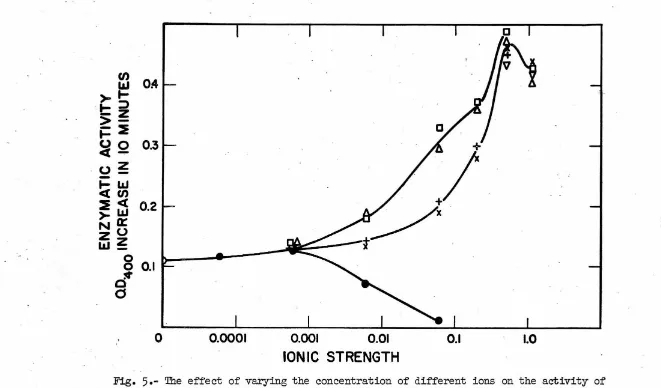
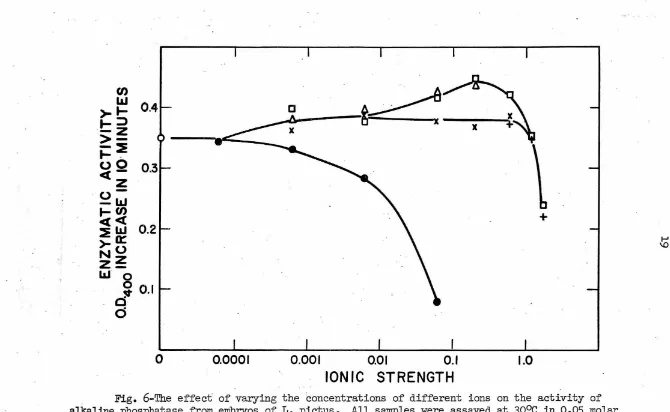
Ionic requirements for optimum alkaline phosphatase activity
'I'b.e ionic strength optimum for alkaline phosphatase activity was
studied in solutions containing several developmentally significant
cations. In order to view the relative importance of ionic strength
and of specific ions, one set of tests was run by adding only the salts
to the bui'fer-substrate solution. Tb.is set gave a range of total ionic
strengths of approximately 0.05 to 1.25 (Fig.
5).
In the other set thebui'fer-substrate contained o.6 molar KCl in addition to the added salts
and represented an approximate total ionic strength range of only 0.65
to 1.85 (Fig. 6). From these it can be seen that optimum activity is
obtained at a total ionic strength in the range of 0.6 to 1.0 and that
it is relatively insensitive to specific ion effects. On the other hand,
the two divalent ions, magnesium and calcium, showed a more specific
maxi-mal activation in the region of 0.02 to 0.07 molar under both experimental·
conditions.
EDTA inhibits alkaline phosphatase activity at concentrations of
0.0001 molar and higher. For purposes of comparison with the effects
of other salts at similar ionic strengths the EDTA was treated as a
trivalent ion. This could be done because the pH of the bui'fer which
was used fell midway between the :pK3 and :pK4 of EDTA at a point where
D
fl)
OA
l&J
>~
.
~::>
- z
>-t:=
:i
~Q
0.3
<..>
~
-~w <( fl)
:i
~
0.2
>a:
I
~
_ /
I
N
<..>
Zz
l&.I_
8
0.1
,,.
00
0
0.0001
0.001
0.01
0.1
1.0
ION IC STRENGTH
Fig.
5
.
-
The eff'ect of varying the concentration of different ions on the activity ofalkaline phosphatase from embryos of L. ;pictus. All samples were assayed at 3ooc in 0.05 molar
veronal - HCl buf'fer (;pH
8.5)
at a substrate concentration of 0.0002 molar. The ionic strengthsrefer to the salts which were added and do not include the contributions by the buffer or substrate.
Symbols: Squares -
Mg;
triangles - Ca; crosses - K; ;pluses - Li; inverted triangles - Na; closedcircles - NaEDTA; o;pen circles - buffer and substrate only.
[image:24.736.30.691.32.420.2]o
.
+
>.._
.._~
- z
>--:e
._
,oO
0
.
3
<{
-z
u--W
.... en
I <{ <{:E
w
0
.
2
>-a:
I
NO
z~
w
·
o
0
""0
.
1
d
d
0
0
.
000
1
&~
x
x
. '\
\
0
.
001
0.01
0.1
STRENGTH
IONI
C
+
.
I
1.0
Fig. 6-'l'he effect of varying the concentrations of different ions on the activity of
alkaline phosphatase from embryos of L. pictus. All samples were assayed at 30°c · in 0 .05 molar veronal - HCl buffer (pH
8
.
5
)
,
o.6 molar KCl and 0.0002 molar substrate. The ionic strengths refer to the salts which were added and do not include the contributions by the buffer, substrateor 0.6 molar KCl. Symbols: Squares -
Mg;
triangles - Ca; crosses - K; pluses - Li; closedcircles - NaEDTA; open circles - buffer, substrate and o.6 molar KCl.
[image:25.736.31.701.14.426.2]20
EDTA depressed the pH of the reaction mixture through chelation of
the divalent ions and corresponding liberation of protons. However,
at the concentration where inhibition of activities was :first noticed
the pH depression was insigriificant.
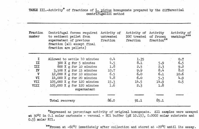
In vitro identification o:f alkaline phosphatase activity in cell :fractions
Alkaline phosphatase activity of the 48-hour embryo was spread
throughout most of the fractions when subcellular particles were
iso-lated by differential centrifugation (Table III). Each fraction was
washed twice with suspending medium and divided so that it could be
assayed before and after DOC treatment. A third portion of the
frac-tion was frozen at
-6o
0c
immediately after collection.and used as anactivity control for the other two samples which necessarily remained
at
o
0c
for at least eight hours. The washings from each fraction werenot combined with the supernatants but assayed separately. Tn this
experiment, as in the one that follows, the activity of each fraction
is expressed as a percentage of the total activity of the initial
homogenate.
Light microscope examination showed that the first fraction contained
mostly skeletal fragments. Fraction II (note Table III for sedimenting
forces) contained nuclei and a few whole cells. The number of whole cells
is probably indicated by the increase in activity in this fraction after
DOC treatment. The third fraction contained a few nuclei and some smaller
particles. Fractions IV and V fall into the range of centrifugal forces
Fraction number
Centrif'ugal forces required Activity of
to sediment pellet from untreated
supernatant of previous fraction
fraction (all except final
fraction are pellets)
I Allowed to settle
10
minuteso.4
II
300
X g for5
minutes4.5
III
6oo
X g for10
minutes1.8
'IV
3,500
X g for15
minutes10.7
V
12,000
X g for10
minutes6.5
VI
14,ooo
X g for20
minutes4.8
VII
105,000
X g for120
minutes15-3
VIII
105,000
X g for120
minutes1.6
supernatant
Total recovery
86.o
Activity of Activity DOC treated of frozen
fraction fraction**
1.35
8.1
5.9
2.5
2.3
9.9
8.4
6.o
6.1
6.o
5.3
14.5
14.9
2.3
1.8
91.1
85.1
Activity of
washings***
0.7
6.5
9.2
8.o
10.6
4.9
0.5
*Expressed as percentage activity of original homogenate. ~11 samples were assayed
at
30°c
in0.1
molar carbonate - veronal - HCl buffer (pH10.15), 0.0002
molar substrate and0.55
molar KCl.**Frozen at
-6o
0c
immediately after collection and stored at-20°c
until its assay.***The washings from each fraction were assayed separately. The wash solution
contained
0.24
molar KCl,0.24
molar sucrose,0.005
molarMgC12
and0.05
molar veronal - HClbuffer (pH
8.5).
'
I\)
[image:27.738.49.688.26.439.2]Fraction DI was quite large and may have been the major mitochondrial fraction. The main function of Fraction VI was to rid the supernatant of any remaining mitochondria-sized particles, and its actual composi-tion is unknown. Fraction VII can be considered as the major microsomal. pellet (27, 28), and Fraction VIII contains the supernatant materials. It is indicated from these data that the major portion of activity is concentrated in the mitochondrial and microsomal fractions with very little in the nuclear and cell debris fractions and virtually none in the supernatant fraction. Assay of the quickly frozen samples confirmed the two initial values for each fraction.
The above conclusions are also supported by the following experiment during which a different fractionation scheme was used (Table DI). A homogenate made from 72-hour plutei was freed of skeletal fragments by settling and centrif'uged at 14,000 X g for 20 minutes to pellet the nuclei and mitochondria. This pellet was then resuspended and washed eight sepa-rate times with the suspending medium. The activity was checked in the initial supernatant, the washing fluids and the final pellet. The pellet was then fractionated in the following manner. The nuclear fraction pre-pared by centrif'ugation at 600 X g for 10 minutes was washed twice, and the washings combined with the 600 X g supernatant. This was then
centrif'uged at 12,000 X g for 10 minutes to bring down the mitochondria. This fraction was washed once, and the wash was combined with the 12,000 X g supernatant. Assay of these fractions after DOC treatment showed
most of the activity to be associated with the mitochondria as opposed to
. the nuclei. Two separate experiments of this type were run, and each gave
*
TABLE. IV .-Activity of three major fractions of L. pictus homogenate prepared by the differential centrif'ugation method
Fractionation scheme Activity
14,ooo
x
g for 20 minutes supernatant 19.5II II
1st wash 9.6
II II
2nd II
7.3
II II
3rd II
3.4
II II
4th
"
3.0II II
5th
"
2.5II II
6th II
2.0
II II
7th II
1.6
II II
8th II
1.5
II
pellet
46
600 X g for 10 minutes pellet 3.3
12,000
x
g for 10 minutes pellet 34.612,000
x
g for 10 minutes supernatant 8.2Total recovery
*Expressed as percentage activity of original homogenate. All samples were assayed at 30°c in 0.1 molar carbonate - veronal - HCl buffer
24
exactly the same pattern of activity distribution with very similar
quantitative results.
Alkaline phosphatase activity was found in both the mitochondrial
and microsomal fractions of two earlier stages in development, earls"
blastula (6 hour) and gastrula (24 hour). However, in contrast to the
later stages, al.most all of the activity was in the microsomal fraction
instead of being approximately distributed equally between the
mitochon-drial and microsomal fractions.
A nearly complete solubilization of microsomal alkaline phosphatase
results from a lO-minute agitation of the fraction in a l~ DOC solution
(Table V). On the other hand, the untreated sample retains most of the
bound enzyme throughout the four washings. The activity associated with
TABI.iE V.-The effect of deoxycholate treatment on L •. pictus microsomal
fraction alkaline phosphatase
*
Fractionation scheme Activity Activity after DOC treatment
.105,000 X g for 60 minutes supernatant 0.07 0.77
II II
1st wash 0.02 0.15
.. II II
2nd II
0.03 0.02
II II
3rd II
0.01 0.02
II II
4th II
o.oi
0.01
II
pellet 0.82 0.02
'*'.Expressed as optical density change dur:,l.ng 10 minutes incubation. Wash solution and conditions of assay the same as
26
DISCUSSION
The control of alkaline phosphatase activity during development
The observation that there is a change in alkaline phosphatase activity at fertilization (29) was not confirmed, and the present
measurements show that it first begins to increase shortzy after
hatching of the blastula. Since the leveling of activities of the
embryos at 65 hours after fertilization could be delayed by adding food to the cultures, this leveling is not likezy to be an indica-tion of the sort of functional maturation of an organ or tissue which has been suggested by Rogers (30,31) for alkaline phosphatase activity
plateaus in developing avian nervous systems. In the cultures to which
food had been added an activity plateau was reached as the embryos began
to die off. These results also indicate that for feeding stages (after
4o
hours) the data presented here may have very little value inrefer-ence to activities of embryos in natural environments except perhaps to suggest strongzy that their activity continues to increase for some· time
beyond 80 hours after fertilization.
From experiments with echinoid hybrids in which paternal influence
upon the activity during development was observed, Flickinger (32)
con-eluded that there is evidence of a nuclear control of alkaline phosphatase activity. It is, of course, general:cy- assumed that all cellular enzymes are direct or indirect products of nuclear activity, and there does not
the pertinent questions relate to the time and place at which specific
genes become active. Conceivably. the enzyme for alkaline phosphatase
activity could be present in the unfertilized egg or synthesized soon
after fert111zat1on in masked form and become unmasked (rather tha.n
newly synthesized) during development. In addition, from the studies
.which will be presented later, it can be postulated that cytoplasmic
elements instead of the nucleus might be directly responsible for the
onset and continued synthesis of the enzyme.
The increase in alkaline phosphatase activity which is begun during
the late blastula stage is the only point in the early development of the
sea urchin embryo at which a change in activity so obviously implies that
a controlling mechanism has been switched from off to on. Though the
nature of this control mechanism has not yet been extensively studied,
a considerable a.mount of evidence can be presented which suggests that
the genetic material contained within the nucleus begins actively direct
-ing development at this time. Prior to this stage the phenomena which go
to make up cleavage appear to originate at a cytoplasmic level (cf. p. 50 ).
The rate of cleavage is always maternal even when non-nucleate fragments
are fertilized with heterologous spermatozoa
(33, 34, 3
5
).
On the otherhand, development which follows gastrulation has been observed to show both
maternal and paternal.characteristics, including a compromise in the rate
of development
(34,
.
36).
It is also notable that approximately 8afo of theknown lethal hybrid combinations of amphibians pass through the cleavage
and blastula stages before arresting (cf.
37).
Sea urchin embryos, rearedin sea water containing 5-iododeoxyuridine and 5-bromodeo:xyuridine,
28
through the blastula stage (
38, 39) •
Further development was abnormaland death soon resulted :for al.most all of the embryos. The :few sUJ1Vivors
were in every case abnormal. In addition, as reviewed elsewhere,
artif'i-.. cially activated non-nucleate :fragments (p. 50 ) and actinomycin D treated
embryos (p.
46)
never develop beyond the blastula stage.The above is not to imply that the nuclear genetic material is
necessarily inactive during the cleavage stages. There is a high positive
correlation which exists between respiratory rate and DNA content in
:fertilized sea urchin eggs
(35,
40). Moreover, nuclear activity may beindicated by the RNA synthesis which is going on during the cleavage
stages (p. 44 :for review; p. 133 for discussion).
The view that the increase in alkaline phosphatase activity represents
a turning-on o:f the appropriate genes at the gastrula stage is consistent
with the customary findings that gastrulation represents the earliest stage
at which nuclear influences on embryonic development are observed. Whether
or not this increase represents synthesis of' enzyme at that time or a gene~
tically directed activation of' a pre-existing ·inactive form is not known.
Borrowing f'rom·other systems, at least three possibilities along this
.latter line can be put forth. The first suggests that if .active sea urchin
alkaline phosphatase is composed of dimers as is the case for E. coli
alka-line phosphatase (41, 42, 43),·the activity would ultimately depend upon
the ability of the two monomers to combine. Thus they might be present in
the egg and restricted from combining. A second possibility is that the
active enzyme might require an activator molecule which is not supplied
until the observed time of increase. The phenomenon, though not known to
enzyme systems (44), and its importance in the maintenance of normal cel1
functions is not dif'.ficult to surn:iise. Finally, it has been shown that
alkaline phosphatase activity increases two to three times in the 14~to
·21-da;y lllouse embryo.duodenum as a result of the adl!linistration of
pti.ro-mycin and actinomyqin D (45). These findings strongly suggest that the
enzyme itsel'f' is already present in an inactive· form; probably inhibited
by a labile protein. These possibilities cannot, however, be seriously
considered until the question of synthesis or activation at the time of
increase in activity has been resolved.
The synthesis of specific proteins at the gastrula stage is strongly
in4icated by studies using the Oucll,terlony method of immunological analysis
(46,
47). In these studies the appearance of new antigens in normal embryosand hybrids was first detected at this stage. Furthermore, paternal antigens
could not be detected until this time. Autoradiogram studies (48, 49, 50)
also give support to the idea of a synth~sis of enzyme which begins at the late blastula stage. Mesenchyme blastulae and other later stages which had.
been allowed to. incorporate radioactive protein and RNA precursors showed that
both protein and RNA synthesis were increasing rapidly in the regions where
histochemical studies had indicated that the alkaline phosphatase activity
was localized. Since, however, more recent studies were in direct
contra-diction to the above observation with respect to amino acid incorporation
(5i),
final judgment· on even this matter must await f'urther developments.The alkaline phosphatase of the developing chick embryo has been found
to be "adaptive" in the sense that it responded to the addition of substrate
30
embryos with ~xo~e~ous substrates have met with no success (32).
Ionic effects on a:ikaline phosphatase activity
In these studies magnesium and calcium ions show negligible
differences of effect on in vitro alkaline phosphatase activity,
whereas a considerable difference was noted in their effects on
morphological development in sea tirchin embryos (12, 13). There
remains, however, a serious limitation to this type of comparison;
name~, that one does not know the effective concentrations of the
responsible ions in viv;o. . In the absence of such information, it
cannot be said that these two ions do not specifica~ affect the
in vivo activity in bringing about the observed developmental effects.
However, the similarity of the magnesium and calcium effects on the
in vitro activity appears to favor this conclusion. Other studies
indicate that exogenous ion changes can affect protein synthesis as
well as development
(53).
This suggests the possibility that thesynthesis of either alkaline phosphatase or the organic matrix of the
skeleton might be inhibited under the conditions which alter
morpho-·logical development in these later stages.
Several observations may be made concerning the response of
alkaline phosphatase to the different ions and ionic strengths. The
loss of alkaline phosphatase activity from sea urchin embryo
prepare.-tions after the addition of EDTA indicates that a cation or cations
chelated by this agent is required to activate the molecule. The
two prime suspects (11) have relatively iittle effect on the activity,
especially at Jl.igher salt concentrations. The data presented here suggest
two possibilities; either that there was not a significant amount of the
cofactor washed from the enzyme by the methods used or that an ion
differ-ent f'r0m these two was the activator. Perhaps a situation exists similar
to th~t found for purified alkaline phosphatase prepared from swine kidney
(54). In this case zinc appeared to be intimately associated with the
enzyme molecule. In addition, a magnesium ion optimum existed although
at a much higher concentration (5 x lo-3M) than the estimated
concentra-tion of enzyme (10-Si..1). This led to the proposiconcentra-tion that the magnesium
salt of the phosphate ester is the preferred substrate for the enzyme
while zinc is the actual cofactor. Although the requirements shown here
for sea urchin alkaline phosphatase could conform to the above activation
pattern,there is no direct evidence in its support as yet.
There are a number of possible ways in which changes in ionic
strength may affect alkaline phosphatase activity. As sunnnarized by
Webb (55.)
ati
increase in enzyme activity with an increase in ionicstrength can, in general, be attributed (a) to an increased efficiency
· in the formation of the enzyme-substrate complex, (b) to an increased
rate of release of the product by the enzyme, (c) to direct influences
on the structure of the enzyme molecule, or (d) to an effect of ionic
strength upon the substrate itself especially when the pH of' the reaction
system is close to a dissociation constant (acidic or basic) of' the
sub-strate. These experiments on sea urchin alkaline phosphatase provide
another exariiple of the susceptibility of ail enzymatic activity to changes
32
A final observation which concludes this part of the discussion
is that chloride ions have been shown to activate .some·enzymes
(56).
Since no discrimination was made with regard to this possibility, it
remains as a possible activator.
Alkaline phosphatase activity in different cell fractions
The two different procedures by which subcellular fractions were
prepared accomplished different purposes. The first procedure, as
outlined in Table III, minimized contamination of the later fractions
by particles damaged during the washings and at the same time still
clearly showed the lower limit of activity in all of the fractions.
On
the other hand, the second scheme (Table IV) gave a quantitativeresult while minimizing contamination of the faster sedimenting
parti-cles with the microsomal fraction. By these two methods one feels
reasonably certain that both qualitative and quantitative results have
been obtained.
The· activity which could be attributed to nuclei was never over
5%
of the total activity. It does not seem likely that this small· amount woul,d account for the observations that the histochemical
alka-line phosphatase reactions always developed first in the nuclei of the
larval cells and also that the increase in activities seen during the
larval stages appeared to take place almost entirely in the nucleus
(7,
14).The first question which might be asked is if this difference represents
a leaching of the native enzyme from the nuclei during the isolation
procedures. However, keeping in mind that there was almost no activity
unlikely. In view of this apparent inconsistency it can be suggested
that the histochemical methods used for this type of localization were
subject to diffusion artifacts (cf.
57).
A clear example of this hasrecently been shown. Whole-tissue histochemical studies on the nervous
system of the developing chick embryo have consistently indicated that
most of the activity of the cells was associated with the nuclei
(58, 59).
However, fractionation of homogenates of these tissues by differential
centrifugation has shown most of the activity to be located in the
cyto-plasmic fractions (60). Histochemical studies on these fractions have
gone further to indicate that the amount of residual activity associated
with these isolated nuclei was directly proportional to the amount of
cytoplasm remaining with the nuclei. In agreement with this latter
find-ing, it has been suggested by Duve (61) that especially when the enzyme
is also localized in the microsomal fraction, one might expect to find
small strands of endoplasmic reticulum still attached to the nucleus
after homogenization.. Another possibility is that the outer membrane
of the nuclear envelope has enzymes associated with it. This association
could be similar to that between enzymes and the endoplasmic reticulum
. since the latter appears to be structurally identical with the outer
nuclear membrane
(62-6
5
).
There is also a possibility that the activity shown here for the nuclear fraction results from mitochondrialcontamina-tion. However, this does not seem likely since the combined wash fluids
from this fraction do not show a great deal more activity than the
frac-tion itself (Table
IV).
In
these experiments the mitochondrial fractions containedcorrespond roughly to the mitochondrial and lysosomal fractions of rat
liver homogenates
(66)
it suggests that perhaps sea urchin embryoalkaline phosphatase may itself be .contained within these particles.
This is, on the other hand, not likely in view of its lack of need for
hypotonic or detergent activation which seems to be characteristic of
lysosomal and some of the mitochondrial enzymes
(67, 68).
The evidencefor the existence of special particles for each individual lysosomal
enzyme was very inconclusive, though it was highly suggestive in the
case of uricase
(69,
70). Alkaline phosphatase may have a particle ofits own as suggested above or it just as plausibly may be found on the
outside of particles in these fractions as has been suggested for some
other mitochondrial enzymes (71). The possibility also remains that the
activity in these fractions comes from contamination by broken nuclei.
This, however, does not appear to be a reasonable hypothesis since the
nuclear fraction itself contains such a small amount of the activity.
Approximately
5afo
of the total activity of the homogenate wasfound in the microsomal fraction. Nearly all of the enzyme could be
solubilized by use of a detergent with no apparent gain or loss of acti-·
vity. The detergent used, sodium deoxycholate, is known to disrupt
mitochondria (72) and to free ribosomes from the endoplasmic reticulum
(73) presumably by dissolution of the lipid portions of the membranes.
These observations suggest that sea urchin alkaline phosphatase is probably
structurally associated with the lipid portions of membranes in both the
mitochondrial and micr.osomal fractions.
The mechanism by which the enzyme becomes distributed in these
phenoloxidase system of Calliphora erythrocephalas (74) suggests a
possible model. Inactive proenzyme·circulated in the hemolymph and
became activated only when brought in contact with a factor localized
in the cuticle. If activated i.n the mitochondria-free supernatant of a cuticle preparation the active unit remained in the supernatant even
after additional centrifugation. If, on the other hand, the activation
took place in the total homogenate the active unit then became associated with the mitochondrial fraction. It was also found that the specificity
of the enzyme varied depending upon whether or not it was particulately
associated. These findings suggest that there can be specificity in the
assoGiation between an enzyme and a subcellular fraction and that the particle containing the enzyme does not necessarily represent the site of synthesis of the enzyme •. They suggest fUrther that in a system where
there is no non-particulately associated activity the enzyme might be
·transported from the site of synthesis to the particle in an inactive
form and become active only on associating with the particle. ·Finally, the above findings showing that enzymes may become associated with
parti-cles in homogenates also signal that cell fractionation studies may possibly contain artifacts of preparation. Though this type of artifact
cannot be ruled out, ther.e is as y:et no reason to suspect it in the data
presented here •
As visualized by Moog (75) the location or change of location of an enzyme within a cell may be of ultimate importance as a factor governing the function and perhaps developmental role that it will have. The experiments reported here are. inherently restricted in their functional
activities which were found in the two intracellular fractions, i.e.
mitochondrial and microsomal, were from different enzymes and that
the alkaline phosphatase-containing particles in these two fractions
ca.me respectively from cells of either of the two alkaline phosphatase· -rich organs. In this regard electrophoretic studies have demonstrated
two separate bands containing alkaline phosphatase activity in extracts
of later stage embryos
(76).
Furthermore, on the basis of differencesin pH and temperature optima for reaction rates, it has been suggested
that two different alkaline phosphatases exist separately in adult sea
urchin organs (TT) which correspond roughly to the alkaline
phosphatase-rich embryonic organs •
The functions of alkaline phosphatase within the cell are most
likely related to substrate hydrolysis rather than synthesis or transfer
reactions
(61).
In this capacity, it has been proposed that alkalinephosphatase has a role of providing inorganic phosphate to the cell
(78).
Furthermore, in view of the ability of the enzyme to hydrolyze
monophos-phate esters of a wide variety of compounds, including primary and
secondary B.liphatic alcohols, sugar alcohols, cyclic alcohols, phenols
·and mononucleotides at the 21
,
3'
or5'
position (cf. 10), it may also be useful in degrading these compounds to where they can be more widelyused in synthetic processes. The enzyme in sea urchins is associated
with organs which commonly contain alkaline phosphatase in other· inverte-brates and in verteinverte-brates (cf.
79).
Judging from the. organs in which it is found, the enzyme appears to function somehow in calcification of theskeleton and resorption of food from the intestine. Though very little
SUMMARY AND CONCLUSIONS
Alkaline phosphatase activity in the sea urchin L. pictus remains
constant during the cleavage stages and begins to increase at the beginning
of gastrulation; eventually increasing at least 300 times. On the basis
of several lines of evidence it seems likely that this increase is due to
a synthesis of new enzyme.
The enzyme activity showed an ionic strength requirement which was
optimum somewhere between
o.6
to l.O. Calcium and magnesium showed amore specific additional optimum concentration at 0.02 to 0.07 molar.
Since these two ions affected the in vitro activity in a similar manner
it does not seem likely that defects in skeleton development caused by
these two ions in abnormal amounts in sea water were due to ion effects
at the enzyme level.
Finally, even though activity was distributed throughout all of the homogenate fractions which were collected it seems very unlikely on the
basis of the sma.11 amount of activity consistently found in the nuclear
:fraction, that the enzyme becomes a nuclear enzyme in later development
II. STUDIES ON THE ACTIVATION OF PROTEIN
4o
INTRODUCTION
Protein synthesis in sea urchin eggs
It appears that in sea urchin eggs (as in :inany others) there is
no increase in total protein (22, 23) or total nitrogen per embryo (82)
during development. Nevertheless it has been shown that numerous enzymes
increase in activity during development (83) and also that new proteins
appear
(46,
47). This implies that protein synthesis is taking placein these eggs and that it does so at the expense of materials already
present within them.
It is well known that many biochemical activities alter upon
ferti-lization; generally in the direction of increased activities (83). · The
penetration of labelled protein precursors into the intact sea urchin
egg was first noted by Hultin (84) to increase at this time. Paralleling
this increased uptake of Nl5-NH4Cl was also an increased labelling of the
proteins of the egg. The interpretation of these experiments was very
difficult because the change in amount of incorporation into proteins upon
fertilization correlated very closely with the observed change in
permea-bility. In similar experiments with N15 labelled glycine and DL-alanine
the phenomenon of increased incorporation into the protein fraction after
fertilization was confirmed (85) though the possibility of a permeability
change was not investigated.
A striking increase in the labelling of proteins coincident to the
formation of the blastula was found in embryos reared in heavy water (86).
Based upon several lines of reasc:ming which are now innnaterial, the authors
acid incorporation. Since, however, there was very little difference
in the amounts of deuterium incorporated into protein by the unfertilized
and fertilized eggs after as long as three and one-half hours of
develop-ment, nothing could be concluded with respect to the innnediate effect of
fertilization. Even if there had been some indication of an increased
activity the interpretation would have had to remain ambiguous largely
due to reports that the sea urchin egg became more permeable to water
(and presumably D20) as well as other substances innnediately after
ferti-lization (87-91).
An increase in incorporation of labelled amino acid into protein
was unequivocally shown to occur upon fertilization of sea urchin eggs
(27) only after a method was developed for preloading the ovarian eggs
in vivo so that effects caused by permeability changes in the egg would
be eliminated (89). This method consisted mainly of injecting the labelled
precursor, s35_methionine, into the body cavity of the adult female sea
urchin and allowing approximately four hours for its penetration into the
eggs. Eggs treated in this manner contained the label primarily in the
acid soluble fraction. Fertilization or artificial activation by butyric
acid treatment resulted in an increase in the labelling of the acid ins
olu-ble fraction (93)~ Fractionation by centrifugation of homogenates from
eggs at different times after fertilization showed that the label was
incorporated into the microsomal and supernatant fractions almost
exclu-sively for the first four or five hours (26, 94). Thereafter a considerabl.e