Original Article
Analysis of BMP4 expression during development
of the striated muscle complex in rat embryos
with anorectal malformations
Yuan-Yuan Geng1, Hong Gao2, Hui-Min Jia1, Wei-Lin Wang1
1Department of Pediatric Surgery, 2Key Laboratory of Health Ministry for Congenital Malformation, Shengjing Hospital of China Medical University, Shenyang 110004, PR China
Received December 14, 2016; Accepted January 20, 2017; Epub March 1, 2017; Published March 15, 2017
Abstract: This study aimed to investigate the spatiotemporal expression pattern of BMP4 in the striated muscle complex (SMC) in ethylenethiourea (ETU)-exposed rat embryos with anorectal malformations (ARMs), and to explore the possible role of BMP4 during morphogenesis of the SMC. The ETU-induced ARM model- on embryonic day 10 (E10)- was used to investigate the expression pattern of BMP4 during development of the SMC. Immunohistochem-istry staining, western blotting and quantitative real-time PCR (qRT-PCR) were applied to confirm the expression lev-els of protein and mRNA of BMP4 between normal rat embryos and embryos with ARMs. Immunostaining revealed that BMP4 expression showed space-time dependent changes in the developing SMC of embryos with ARMs. With the growth of rat embryos, the BMP4 positive stained cells gradually increased from E17. The same changes in ex-pression of BMP4 were detected in embryos with ARMs. However, compared to embryos with ARMs, BMP4 expres-sion was significantly higher in normal tissues. In western blotting and qRT-PCR, BMP4 expresexpres-sion in the SMC of rat embryos with ARMs was lower at both the mRNA level and the protein level compared with normal rat embryos. This study demonstrated that BMP4 expression in embryos with ARMs was remarkably reduced, which indicated that BMP4 could play an important role in the pathogenesis of the SMC in embryos with ARMs.
Keywords: Anorectal malformations, BMP4, gene expression, rat embryos, striated muscle complex, development
Introduction
Anorectal malformations (ARMs) are very com-mon surgical problems, with a clinical spectrum that ranges from anal stenosis to imperforate anus to persistent cloaca, and affects approxi-mately 1/5000 to 1/1500 live births [1]. In spite of numerous technical advances in the surgical treatment of ARMs, some patients con-tinued to suffer from fecal incontinence and constipation after procedures [1-5]. The etiolo-gy of postoperative defecation problems are multifactorial, and maldeveloped pelvic floor musculature (PFM) has been identified as one of the most important underlying factors [6-10]. The striated muscle complex (SMC) in rats resembles the gross anatomy of the puborecta-lis muscle in humans, and contributes substan-tially to micturition, defecation, continence, and support of the pelvic organs [11].
Previous studies have demonstrated that bone morphogenetic proteins (BMPs) are important
myo-genic differentiation [19]. Furthermore, Ono Y
et al. found that the stimulation of BMP signal-ing- by adding BMP4- resulted in an increased number of activated satellite cells and a decreased number of differentiating myoblasts [13]. In addition, it was revealed that BMP4 played an important role in the development of the hindgut, and downregulation of BMP4 could be partly related to the maldevelopment of the terminal hindgut in ARMs [20, 21]. However, it is unknown whether BMP4 continues to play a role in the development of the SMC after the occurrence of ARMs. To gain an insight into the pattern of BMP4 expression and the possible role of BMP4 during development of the SMC in embryos with ARMs, we conducted a system-atic study on the spatiotemporal expression of BMP4 in normal rat embryos compared to rat embryos with ARMs.
Materials and methods
Animal model and tissue collection
Ethical approval was obtained from the China Medical University Animal Ethics Committee before the study. A total of 80 time-mated preg-nant Wistar rats were divided into normal and ARMs groups. In the ARMs group, pregnant rats were gavage fed a single dose of 125 mg/kg of 1% ethylenethiourea (ETU; 2-imidazolidinethi-one, Aldrich Chemical Co., Germany); In the nor-mal group, pregnant rats received an equal dose of saline on E10 (E0-sperm in vaginal smear after overnight mating). Embryos were collected via cesarean delivery on E17, E19 and E21. For immunohistochemical studies, normal embryos and embryos with ARMs were fixed in 4% paraformaldehyde for 12 to 24 h depending on their size. Embryos were then embedded in paraffin in a routine manner and sectioned sagittally at a thickness of 4 μm. For western blot and qRT-PCR analysis, the SMC tissues were collected under magnification and immediately stored at -80°C until required for use. Only male fetuses were used in this study, because the SMCs are thinner in female fetal rats. We were able to observe the gonads under the light microscope to determine the sex of the rats; Specifically, the testis has a characteristic “striped” appearance, while the ovary has a characteristic “spotty” appearance.
Immunohistochemical staining
Immunohistochemical staining was performed according to the previously described method
[22]. For antigen retrieval, slides were incubat-ed in boiling 0.01 mol/L citrate buffer (pH 6.0) for 10 min, followed by cooling at room temper-ature, and endogenous peroxidase activity was then blocked with 3% H2O2. After incubation in 10% normal goat serum for 30 min to block nonspecific binding sites, sections were incu-bated with BMP4 primary antibody at a dilution of 1:200 (rabbit polyclonal, ab39973, Abcam, Cambridge, UK) overnight at 4°C. Sections were then incubated with biotinylated goat anti-rabbit IgG (Maixin, Fuzhou, China) for 30 min at room temperature after the primary antibody was washed off. Immunoreactions were visual-ized by DAB (Maixin, Fuzhou, China) and sec-tions were counterstained with hematoxylin. Specimens were photographed using a digi-tized microscope camera (Nikon E800, Japan). In all experiments, negative controls were per-formed by either omitting the primary or sec-ondary antibody step.
Protein preparation and western blotting
SMC tissue collected from the normal and ARMs groups were pooled and sonicated in ddH2O containing protease inhibitors. Protein extracts (50 μg) were heated at 100°C for 5 min and size fractionated on Bis-Tris SDS-PAGE gels (Invitrogen, Carlsbad, CA, USA). Protein samples were denatured, separated by SDS/ PAGE, and transferred to polyvinylidene fluoride (PVDF) membranes (Millipore, Billerica, MA, USA); This was followed by blocking with 5% fat-free milk in Tris-buffered saline (1.5 h, room temperature). PVDF membranes with proteins were incubated overnight at 4°C in primary antibody against BMP4 (dilution 1:1000), and then incubated with secondary antibody (dilu-tion 1:2000) for 2.5 h at room temperature. The immunostained bands were detected with a Proto Blot II AP System using a stabilized sub-strate (Promega Biological Products, Ltd., Shanghai, China). GAPDH was used as an inter-nal standard to normalize protein levels in each lane.
RNA extraction and quantitative real-time PCR (qRT-PCR)
diluted to a concentration of 1 μg/μl and stored at -80°C. The total RNA was reverse-tran-scribed into cDNA using a SYBR Prime Script RT-PCR kit (Takara, Dalian, China), according to the manufacturers’ instructions. The BMP4 primers used for qRT-PCR were as follows: For- ward, 5’-TGCCATTGTGCAGACCCT-3’ and rever- se, 5’-CACCACCTTGTCGTACTCGTC-3’. The hou- sekeeping gene β-actin (Takara, code D3783) was used as an internal control. qRT-PCR was performed with a 20 μl reaction system in tripli-cate for each specimen in the presence of SYBR® Green PCR Master Mix (TaKaRa Biotechnology Co., Dalian, China) in a Light- cycler® (Roche Molecular Biochemicals, Co., Mannheim, Germany). The reaction program
was: 3 min pre-denaturation at 95°C and 45 cycles of 10 sec of denaturation at 95°C and 30 sec of annealing at 60°C. After the termina-tion of qRT-PCR, the productermina-tion was automati-cally analyzed by the Lightcycler system. A dis-sociation procedure was performed to generate a melting curve to confirm amplification speci-ficity. The relative levels of gene expression were determined as ΔCt=Ct gene -Ct reference, and the fold change in gene expression was cal-culated using the 2-ΔΔCt method.
Statistical analysis
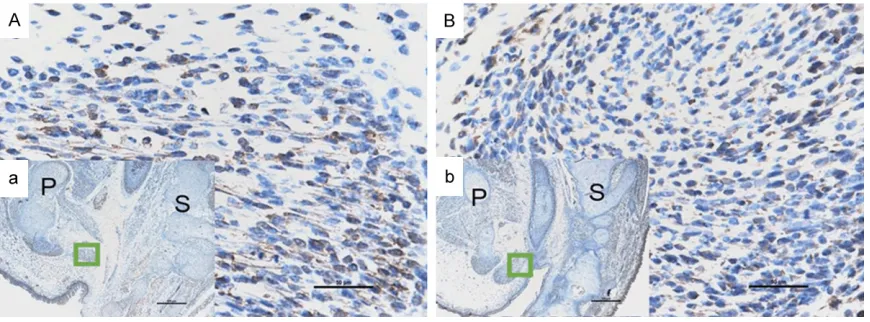
[image:3.612.91.528.71.230.2]The Statistical Program for Social Sciences, version 13.0 (SPSS, Chicago, IL), was used for Figure 1. Immunohistochemical staining of BMP4 on E17. (A and a) Represent the normal group; On E17, sporadic BMP4 positive stained cells were detected in the SMC. (B and b) Represent the ARMs group; No obvious immuno-reactivity specific to BMP4 was detected in the SMC on E17. (A and B: Original magnification ×400; a and b: original magnification ×40). P, pubis; S, sacrum. The region marked with a square in (a and b) is magnified in (A and B).
[image:3.612.90.525.301.462.2]statistical analysis. We used the student t-test to compare the BMP4 levels between the nor-mal and ARMs groups. P values of less than 0.05 were considered statistically significant. Results
General observation
In our study, no malformations were observed in the 156 male normal rat embryos. We
was very distinct, and increasingly more BMP4 positive stained cells were detected in the SMC and bulbocavernosus muscle (Figures 2A and 3A).
[image:4.612.91.524.73.231.2]However, in embryos with ARMs, on E17, no obvious immunoreactivity specific to BMP4 was detected in the SMC (Figure 1B). From E19, there was an increasing number of cells with BMP4 immunoreactivity in the SMC, but this Figure 3. Immunohistochemical staining of BMP4 on E21. (A and a) Represent the normal group; On E21, increas-ingly more BMP4 positive stained cells were detected in the SMC and the bulbocavernosus muscle. (B and b) Represent the ARMs group; cells showing BMP4 immunoreactivity increased gradually on E21. (A and B: Original magnification ×400; a and b: Original magnification ×40). P, pubis; S, sacrum. The region marked with a square in (a and b) is magnified in (A and B).
Figure 4. BMP4 protein expression relative to GAPDH expression (y-ax-is) in the developing SMC of normal rat embryos and rat embryos with ARMs on E17, 19 and 21 (x-axis). The immunoblot shows a remarkable BMP4 expression in the normal group, but a weak signal in the ARMs group. The trends of expression at each time point are shown. N, normal group; A, ARM group.
obtained a total of 196 embryos with ARMs from 413 ETU-treated male rat embryos. The incidence of ARMs in embryos of the ETU-treated group on E17 to E21 was 66.4%. However, externally visible spinal bifida and/or meningocele were also observed, and 17 embryos died in utero. As denerva-tion could affect the development of SMC, specimens with neurolog-ic defects were excluded [23]. In this study, all specimens with ARMs were determined by means of observing the fistula between the rectum and the urethra in sag-ittal planes under the light mi- croscope.
Immunohistochemical analysis
[image:4.612.95.353.311.522.2]was significantly lower compared to normal embryos (Figures 2B and 3B).
Western blot analysis
Western blot analysis specific for BMP4 was performed to quantify protein expression in the development of the SMC in the normal and ARMs groups. Accordingly, BMP4 was approxi-mately detected as a 47 kDa band on western blots of protein extracts. The expression level of BMP4 was normalized to the protein level of GAPDH from the same specimens of embryos at E17, 19, and 21, which are the key periods of SMC development; BMP4 expression gradually increased in the normal group, while in the ARMs group, BMP4 protein expression was weak. BMP4 protein expression significantly decreased in ARMs SMC compared with the normal SMC at each time point (0.29±0.04 ver-sus 0.69±0.06; 0.45±0.09 verver-sus 0.82±0.09; 0.60±0.05 versus 0.91±0.13; Respectively; P<0.05; Figure 4).
QRT-PCR analysis
The OD value of total RNA calculated by A260/ A280 was in the range of 1.8 to 2.0. In our study, a significant increase in the expression pattern of BMP4 was detected in the normal group compared to the ARMs group at each
[image:5.612.90.356.87.149.2]found that dysregulation of apoptosis was implicated as one of the fundamental factors in the pathogenesis of SMC maldevelopment in rats with ARMs [26]. Mi J et al. demonstrated that spatiotemporal expression of Wnt5a was imbalanced during SMC development in embry-os with ARMs [27]. Numerous efforts have been made to understand the mechanisms of SMC development in embryos, but our current understanding of normal and abnormal devel-opment of the SMC remains incomplete. The development of skeletal muscle proceeds through five phases, as follows [28]: Phase 1 (specification), the development of muscle pro-genitor cells is specified toward muscle cells in somites; Phase 2 (migration), muscle progeni-tor cells migrate to the presumptive places at which muscles are formed; Phase 3 (prolifera-tion), progenitor cells proliferate and increase in number; Phase 4 (differentiation), progenitor cells finally differentiate to become myoblasts, which in turn fuse to form multinucleated myo-tubes; Phase 5 (maturation), the multinucleat-ed myotubes mature into myofibers, including slow-twitch myofibers or fast-twitch myofibers. Zhang SW et al. found that the fibroid structure of the SMC in normal embryos and embryos with ARMs could not be identified until E16 [25]. In other words, the muscle progenitor cells Table 1. The relative quantity of BMP4 mRNA on E17
[image:5.612.90.354.186.246.2]Group BMP4 average Ct value β-actin average Ct value ∆Ct ∆∆Ct Times of gene (compared to ARMs group) A17 26.08±1.35 18.90±1.21 7.18 0 1 N17 20.52±1.17 14.21±1.62 6.31 -0.87 1.83
Table 2. The relative quantity of BMP4 mRNA on E19
Group BMP4 average Ct value β-actin average Ct value ∆Ct ∆∆Ct Times of gene (compared to ARMs group) A19 22.39±1.99 15.38±1.51 7.01 0 1 N19 21.64±1.39 16.07±1.43 5.57 -1.44 2.71
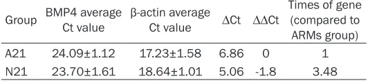
Table 3. The relative quantity of BMP4 mRNA on E21
Group BMP4 average Ct value β-actin average Ct value ∆Ct ∆∆Ct Times of gene (compared to ARMs group) A21 24.09±1.12 17.23±1.58 6.86 0 1 N21 23.70±1.61 18.64±1.01 5.06 -1.8 3.48
time point, which was consistent with the results obtained from we-stern blot analysis. Results showed that in the normal group, the mRNA levels of BMP4 (1.83-fold, 2.71-fold and 3.48-fold) were higher than those in the ARMs group on E17, E19 and E21 (P<0.05), respectively (Tables 1-3).
Discussion
[image:5.612.89.355.283.343.2]of the SMC start to migrate to the inherent loca-tion on E16, and they then participate in prolif-eration and differentiation processes from E17 to E21.
During development of the skeletal muscle, many signaling pathways participate in the embryogenesis and development of the SMC, namely Shh, BMP, Wnt and Notch. Previous studies have demonstrated that BMP4 plays a critical role in muscle development and regen-eration. TakenaoUmemoto et al. reported that BMP4 expressed in myoblasts plays a positive role in myotube formation/maturation through myogenin expression, and the presence of myo-tubes inhibits BMP4 expression in proliferating myoblasts through transcriptional regulation, although the expression is intrinsically increa- sed with the time of culture [29]. In this study, BMP4 expression gradually increased from E17 to E21, both in normal embryos and in embryos with ARMs, which is consistent with the previ-ous reports mentioned. Recent studies have shown that BMPs are important in preventing premature myogenic differentiation [13]. In the current study, we found that at the same stage, the expression level of BMP4 was reduced in embryos with ARMs compared with normal embryos. These results implythat this downreg-ulation of BMP4 expression in embryos with ARMs may accelerate premature myogenic dif-ferentiation. Considerable connective tissue infiltrated into intermuscular bundles, which resulted in the malformation of SMC in ARMs, further suggesting that optimal BMP4 levels are critical for the development of the SMC. Furthermore, the genetics of ARMs is an ex- tremely complex event. Morphological changes of the SMC take place after the occurrence of abnormal anorectum in rats with ARMs [25]. We suspectedthat rectum maldevelopment in- fluenced the development of the SMC to a cer-tain extent. Bourdelat et al. emphasized the importance of the rectum in the development of the sphincter, which corroborated this pres-ent study [30]. We found that spatiotemporal expression of BMP4 was imbalanced during development of the SMC in embryos with ARMs, and this trend was consistent with that in termi-nal hindgut development [20, 21], suggesting that the rectum could play a key role in the development of the SMC.
In conclusion, BMP4 is extremely important for the development of the terminal hindgut and
the SMC in embryos with ARMs. However, our study was unable to confirm whether BMP4 was the initial event leading to malformation of the SMC, and it has been shown that numerous signaling molecules are involved in the differ-ent phases of the developmdiffer-ent of the SMC. For a precise understanding of the developmental processes and the pathogenesis in ARMs, fur-ther analysis of the molecular mechanisms should be carried out.
Acknowledgements
This work was supported by grants from the National Natural Science Foundation of China (Grant Nos. 81270436 and 81170334). Disclosure of conflict of interest
None.
Address correspondence to: Dr. Wei-Lin Wang, Department of Pediatric Surgery, Shengjing Hospital of China Medical University, No. 36 Sanhao Street, Heping District, Shenyang 110004, PR China. Tel: +86 24 96615 57111; Fax: +86 24 96615 57111; E-mail: [email protected]
References
[1] Pena A, Guardino K, Tovilla JM, Levitt MA, Ro-driguez G and Torres R. Bowel management for fecal incontinence in patients with anorectal malformations. J Pediatr Surg 1998; 33: 133-137.
[2] Levitt MA and Pena A. Outcomes from the cor-rection of anorectal malformations. Curr Opin Pediatr 2005; 17: 394-401.
[3] Sonnino RE, Reinberg O, Bensoussan AL, La-berge JM and Blanchard H. Gracilis muscle transposition for anal incontinence in children: Long-term follow-up. J Pediatr Surg 1991; 26: 1219-1223.
[4] Bai Y, Yuan Z, Wang W, Zhao Y, Wang H and Wang W. Quality of life for children with fecal incontinence after surgically corrected anorec-tal malformation. J Pediatr Surg 2000; 35: 462-464.
[5] Rintala RJ and Lindahl H. Is normal bowel func-tion possible after repair of intermediate and high anorectal malformations? J Pediatr Surg 1995; 30: 491-494.
[6] Li L, Li Z, Wang LY and Xiao FD. Anorectal anomaly: neuropathological changes in the sacral spinal cord. J Pediatr Surg 1993; 28: 880-885.
ex-ternal anal sphincter in children with anorectal malformation. J Pediatr Surg 2000; 35: 1052-1057.
[8] Fernandez-Fraga X, Azpiroz F and Malagelada JR. Significance of pelvic floor muscles in anal incontinence. Gastroenterology 2002; 123: 1441-1450.
[9] Yuan ZW, Lui VC and Tam PK. Deficient motor innervation of the sphincter mechanism in fe-tal rats with anorecfe-tal malformation: a quanti-tative study by fluorogold retrograde tracing. J Pediatr Surg 2003; 38: 1383-1388.
[10] Jia H, Zhang K, Zhang S, Yuan Z, Bai Y and Wang W. Quantitative analysis of sacral para-sympathetic nucleus innervating the rectum in rats with anorectal malformation. J Pediatr Surg 2007; 42: 1544-1548.
[11] Poortmans A and Wyndaele JJ. M. levator ani in the rat: does it really lift the anus? Anat Rec 1998; 251: 20-27.
[12] Frank NY, Kho AT, Schatton T, Murphy GF, Mol-loy MJ, Zhan Q, Ramoni MF, Frank MH, Kohane IS and Gussoni E. Regulation of myogenic pro-genitor proliferation in human fetal skeletal muscle by BMP4 and its antagonist Gremlin. J Cell Biol 2006; 175: 99-110.
[13] Ono Y, Calhabeu F, Morgan JE, Katagiri T, Amthor H and Zammit PS. BMP signalling per-mits population expansion by preventing pre-mature myogenic differentiation in muscle sa-tellite cells. Cell Death Differ 2011; 18: 222-234.
[14] Cobourne MT and Sharpe PT. Tooth and jaw: molecular mechanisms of patterning in the first branchial arch. Arch Oral Biol 2003; 48: 1-14.
[15] Chen D, Zhao M and Mundy GR. Bone morpho-genetic proteins. Growth Factors 2004; 22: 233-241.
[16] Wan M and Cao X. BMP signaling in skeletal development. Biochem Biophys Res Commun 2005; 328: 651-657.
[17] Massague J and Chen YG. Controlling TGF-beta signaling. Genes Dev 2000; 14: 627-644. [18] Miyazono K, Maeda S and Imamura T. BMP
re-ceptor signaling: transcriptional targets, regu-lation of signals, and signaling cross-talk. Cyto-kine Growth Factor Rev 2005; 16: 251-263. [19] Clever JL, Sakai Y, Wang RA and Schneider DB.
Inefficient skeletal muscle repair in inhibitor of differentiation knockout mice suggests a cru-cial role for BMP signaling during adult muscle regeneration. Am J Physiol Cell Physiol 2010; 298: C1087-1099.
[20] Zhang J, Zhang ZB, Gao H, Zhang D and Wang WL. Down-regulation of SHH/BMP4 signalling in human anorectal malformations. J Int Med Res 2009; 37: 1842-1850.
[21] Mandhan P, Quan QB, Beasley S and Sullivan M. Sonic hedgehog, BMP4, and Hox genes in the development of anorectal malformations in Ethylenethiourea-exposed fetal rats. J Pedi-atr Surg 2006; 41: 2041-2045.
[22] Pluznick JL, Wei P, Grimm PR and Sansom SC. BK-{beta}1 subunit: immunolocalization in the mammalian connecting tubule and its role in the kaliuretic response to volume expansion. Am J Physiol Renal Physiol 2005; 288: F846-854.
[23] Adhihetty PJ, O‘Leary MF, Chabi B, Wicks KL and Hood DA. Effect of denervation on mito-chondrially mediated apoptosis in skeletal muscle. J Appl Physiol (1985) 2007; 102: 1143-1151.
[24] Valasek P, Evans DJ, Maina F, Grim M and Patel K. A dual fate of the hindlimb muscle mass: cloacal/perineal musculature develops from leg muscle cells. Development 2005; 132: 447-458.
[25] Zhang SW, Bai YZ, Zhang SC, Wang DJ, Zhang T, Zhang D and Wang WL. Embryonic develop-ment of the striated muscle complex in rats with anorectal malformations. J Pediatr Surg 2008; 43: 1452-1458.
[26] Chen QJ, Jia HM, Zhang SW, Zhang SC, Bai YZ, Yuan ZW and Wang WL. Apoptosis during the development of pelvic floor muscle in anorec-tal malformation rats. J Pediatr Surg 2009; 44: 1884-1891.
[27] Mi J, Chen D, Ren X, Jia H, Gao H and Wang W. Spatiotemporal expression of Wnt5a during the development of the striated muscle com-plex in rats with anorectal malformations. Int J Clin Exp Pathol 2014; 7: 1997-2005.
[28] Buckingham M, Bajard L, Chang T, Daubas P, Hadchouel J, Meilhac S, Montarras D, Rocan-court D and Relaix F. The formation of skeletal muscle: from somite to limb. J Anat 2003; 202: 59-68.
[29] Umemoto T, Furutani Y, Murakami M, Matsui T and Funaba M. Endogenous Bmp4 in myo-blasts is required for myotube formation in C2C12 cells. Biochim Biophys Acta 2011; 1810: 1127-1135.