Original Article
Lactose induced redox-dependent senescence
and activated Nrf2 pathway
Shuli Xing1, Lanxin Zhang1, Huiling Lin1, Zhifan Mao1, Keting Bao1, Peng Hao3, Zhe Pei3, Jian Li2, Zelan Hu1 1Shanghai Key Laboratory of New Drug Design, School of Pharmacy, East China University of Science and
Technology, Shanghai, China; 2State Key Laboratory of Bioreactor Engineering, East China University of Science
and Technology, Shanghai, China; 3Duke University Medical School, Durham, NC, USA
Received March 5, 2019; Accepted March 28, 2019; Epub June 1, 2019; Published June 15, 2019
Abstract: Lactose is a disaccharide found in milk and thus a part of our daily food intake. Upon ingestion, it is hydro-lyzed to glucose and galactose by the enzyme lactase and absorbed in the small intestine. People who suffer from
lactose intolerance are unable to completely digest it due to deficiency of lactase, leading to intestinal problems
such as diarrhoea, and bloating. Various studies have focused on treating these symptoms. However, the effects of lactose that diffuses passively into cells, on cellular senescence have largely remained unknown. Thus, the pres-ent study investigated the effects and mechanisms of lactose on senescence both in vitro and in vivo. The study was conducted in MRC-5 cells. The cellular senescence was estimated by determining the expression of SA-β-gal
and p16ink4a. The cell viability of MRC-5 cells was determined by the CCK-8 Assay. Activity of intracellular reactive
oxygen species was estimated by measuring the levels of superoxide dismutase (SOD), glutathione (GHS), and reac-tive oxygen species (ROS). The mechanism of lactose on cellular senescence was explored by western blotting. We also studied the effect of lactose on the lifespan of Caenorhabditis elegans. Increased activities of SA-β-gal and
p16ink4a revealed the ability of lactose to induce senescence in MRC-5 cells. The elevated intracellular ROS level
and decreased GSH and SOD levels in these cells were indicative of cellular oxidative stress induced by lactose. Furthermore, western blotting analysis of Nrf2 and mRNA expression of its downstream genes suggested the Nrf2/ ARE pathway was involved in the oxidative stress induced by lactose. These results were further validated by the shortened lifespan of C. elegans after lactose supplement. Moreover, the lactose-induced senescence could be al-leviated by an antioxidant, N-Acetyl-L-cysteine (NAC), both in vitro and in vivo. The present study observed a positive correlation between lactose and cellular oxidative stress, suggesting the latter to be an underlying mechanism of lactose-induced senescence.
Keywords: Lactose, ROS, oxidative stress, cellular senescence, Nrf2
Introduction
Aging is defined as an inescapable and irrevers -ible phenomenon in mammals characterized by slow deterioration and degeneration of various physiologic functions [1]. Cellular senescence, a special state of irreversible cell cycle arrest, plays an important role in aging and is respon-sible for causing age-related diseases [2]. Many experimental studies have revealed that cellu-lar senescence can induce functional degener-ation of tissues and organs. For example, Baker and colleagues found that removal of senes-cent cells could prevent or delay dysfunction of tissues and thus extend the lifespan [3]. The ability of senescent cells to induce permanent
proliferative arrest is now being exploited as a therapeutic target for major diseases [2]. This
phenomenon of cellular senescence was first reported by Hayflick in serially cultured human fibroblasts in 1961 [4]. The cellular senescence is identified as a limited proliferative phase of
lifespan caused by various types of stress in which cells stop proliferating and undergo vari-ous alterations. These include, but are not lim-ited to, changes in morphology, failure of DNA replication, senescence-associated secretory phenotype (SASP), accumulation of senesce- nce-associated heterochromatin foci (SAHF), high level of p16ink4a and increase of
senes-cence-associated β-galactosidase (SA-β-gal)
Advancements in medical sciences have made it evident to extend mammals’ lifespan by ge- netic, dietary, and drug interventions. However, genetic intervention or dietary restriction is
dif-ficult to realize in humans, and drug treatment
is associated with some unacceptable side ef- fects [6]. Therefore, recently the effects of nu- trients taken as a part of our daily food intake on aging have drawn much attention of the
sci-entific community [7, 8]. Moreover, various
nu-trients have been reported to possess anti-aging properties. For example, Zwighaft and colleagues demonstrated the administration of spermidine to the diet, mice could extend their lifespan by regulating their circadian period [9]. On the other hand, nutrients such as galactose and so on can induce cellular senescence thereby shortening the lifespan in many ani-mals [10-12].
Lactose is a disaccharide that is composed of galactose and glucose and is originally found in milk and milk products. It is therefore one of the most common nutrients taken by human beings as daily dietary intake. Upon absorption, it is hydrolyzed by lactase to glucose and galac-tose on the surface of the small intestine [13].
However, a deficiency of lactase would prevent
its metabolism, leading to lactose intolerance. Approximately 15% of people in Northern Eur- opean are reported as lactose intolerant, this percentage is even higher in other races, up to 80% in Latinos and nearly 100% in Chinese [14]. People with lactose intolerance are unable to digest it completely, leading to accumulation
of lactose in the small intestine that is
reflect-ed by an array of intestinal distress [13]. How- ever, effects of accumulated lactose on cellular senescence and aging remain largely unknown. In the present study, we have studied the ef- fects of lactose on the senescence of human
fetal lung fibroblast cell lines (MRC-5 cell lines),
a model for research on aging. We found la-
ctose can increase the expression of SA-β-gal
[15] and cyclin-dependent kinase (CDK) inhi- bitor p16ink4a, which are the most widely used biomarkers of cellular senescence [5, 16]. Tre- atment of MRC-5 cells with lactose also incre- ased the intracellular level of oxidative stress accompanied by activation of an antioxidant signaling pathway, nuclear factor erythroid 2- related factor 2 (Nrf2) pathway. In addition, lac-tose treatment could shorten the lifespan of C.
elegans, which could be rescued by treatment
with antioxidants.
Material and methods
Cell culture and drug treatment
The normal human fetal lung fibroblast line,
MRC-5, was obtained from the Cell Bank of the Chinese Academy of Sciences at passage 18 (Shanghai, China). The cell line was used for
experiments at passage 23 to 28. All fibro -blasts were cultured in MEM (Life Technolo- gies, Grand Island, United States) supplement-ed with 10% fetal bovine serum (FBS) (Gibco, Grand Island, United States), sodium pyruvate, non-essential amino acids and antibiotics (pen-icillin and streptomycin; Biological Industries, Connecticut, United States) at 37°C with 5%
CO2. Lactose (Macklin, Shanghai, China) was dissolved in sterile water and added to the
growth medium at final concentration of 10
mmol/L, 25 mmol/L and 50 mmol/L, respec-tively. Rapamycin (Selleckchem, Houston, Unit- ed States), doxorubicin (Selleckchem), N-Ace- tyl-L-cysteine (Selleckchem) were dissolved in sterile water and added to the growth medium
at final concentration of 1 nmol/L, 50 μmol/L
and 1 mmol/L, respectively. The drugs were
replaced one day in between for 7 days,
exce-pt where noted. The cultures were passaged
1:2 at 90% confluence and the medium was
renewed thrice a week.
Senescence associated β-galactosidase activ-ity assay
The SA-β-gal activity assay was performed
according to the manufacturer’s protocol (Be-
yotime, Shanghai, China). In brief, fibroblast cells grown on a six-well plate were fixed in a fixative solution for 15 minutes, then rinsed
with phosphate buffered saline (PBS) thrice,
following a mixture containing 1% staining fluid A, 1% staining fluid B, 93% staining fluid C (flu -ids A, B and C were provided with the kit), and 5% X-gal was added. The cells were allowed to
get stained overnight (12 hours) at 37°C in the
dark. Images were captured using the Nikon Ni microscope.
Detection of intracellular lactose concentration by ELISA
detec-tion of lactose was performed as described.
Briefly, 96-well flat-bottomed microtiter ELISA
plates were coated with the streptavidin-HRP
and the specific antibody, followed by incuba
-tion for 60 min at 37°C. After repeated wash -ings, agents A and B were added. The plate was
then incubated for 10 min at 37°C in dark-ness. Finally, 50 μL stop buffer was added to
stop the reaction. The absorbance was detect-ed using a microplate reader (Biotek, Vermont, United States) at 450 nm.
Determination of cell viability by the CCK-8 as-say
The cell counting kit-8 (CCK-8) assay was per-formed according to the manufacturer’s proto-col (Yeasen, Shanghai, China). A 100-μL cell
suspension containing various concentrations of lactose was dispensed into a 96-well plate
and incubated for 24 hours at 37°C with 5%
CO2. After incubation, 10 μL of CCK-8 solution
was added to each well of the plate and further incubated for 2 hours. The absorbance was then measured at 450 nm using a microplate reader (Biotek).
Measurement of anti-oxidative activities
After treatment with lactose for 7 days, activi -ties of superoxide dismutase (SOD) and gluta-thione (GSH) were measured using the ap- propriate kits (Jiancheng, Nanjing, China). The absorbance was detected using a microplate reader (Biotek) at corresponding wavelengths.
Determination of cellular glutathione level
The formation of GSH was determined by a thiol probe, monochloromobimanem (mBCI), follow-ing the protocol described (Thermo, New York, United States). The thiol-reactive reagents of mBCI react with the thiol groups of living cells
to form fluorescent products. For this, a 100
mmol/L stock solution of the thiol probe in di- methylsulfoxide was prepared before use. After
exposure to lactose for 7 days, MRC-5 cells
were rinsed with Hank’s balanced salt solution (HBSS) twice, and then mBCI was added to
HBSS at a final concentration of 100 μmol/L. Pluronic F-127, a cell-permeable agent, was added at a final concentration of 0.04% to dis-perse the thiol dye. The cells were then
incu-bated for 20 minutes at 37°C in the dark be-fore fluorometric detection (excitation at 394
nm, emission at 490 nm) with a Nikon fluores -cence microscope.
Determination of intracellular reactive oxygen species
After treatment with lactose for 7 days, levels
of reactive oxygen species (ROS) were deter-mined by an indicator, DCFH-DA dye, (Yeasen, Shanghai, China). The medium was replaced
with 10 μmol/L of DCFH-DA dye (diluted in
FBS-free medium) and incubated for 20 minutes at
37°C in the dark before fluorometric detection
(excitation at 488 nm, emission at 525 nm)
with a Nikon fluorescence microscope.
RNA purification, reverse transcription, and real-time PCR
The cells were harvested after being exposed
to lactose for 7 days. Total RNA was extracted
by the Eastep Super Total RNA extraction kit following manufacturer’s instruction (Promega, Wisconsin, United States). The cDNA was sy- nthesized using an oligo (dT) primer, random primer and reverse transcriptase. All primers were designed and synthesized using Primer Premier Software 5.0 (PREMIER Biosoft In- ternational) by Sangon Biotech (Shanghai) Co., Ltd. The sequences were: GCLC FW: 5’-CAAG- AGAAGGGGGAAAGGAC-3’, RV: 5’-GACCTCGGG- CAGTGTGAAC-3’; GCLM FW: 5’-TCAGGGAGTTT- CCAGATGTC-3’, RV: 5’-CAATAGGAGGTGAAGCA- ATG-3’; GPx7 FW:
5’-ACTTCAAGGCGGTCAACA-TC-3’, RV: 5’-GGCAAAGCTCTCAATCTCC-3’; GSR FW: 5’-CCCAAGCCCACAATAGAGG-3’, RV: 5’-AC- CTGCACCAACAATGACG-3’; GSTM1 FW: 5’-GCA- TGATCTGCTACAATCC-3’, RV: 5’-CTTGGGCTCAA- ATATACGG-3’; GSTM4 FW: 5’-CCTTGCTCCCTGA- ACACTC-3’, RV: 5’-GTCGTCACTTCCAACCAAC-3’; HO-1 FW: 5’-AAGACTGCGTTCCTGCTCAAC-3’, RV: 5’-AAAGCCCTACAGCAACTGTCG-3’; NQO-1 FW: 5’-GGCATTCTGCATTTCTGTG-3’, RV: 5’-GG- CGTTTCTTCCATCCTTC-3’; TALDA FW: 5’-GGGC- CGAGTATCCACAGAAG-3’, RV: 5’-GGCGAAGGA- GAAGAGTAACG-3’; TBP FW: 5’-CGCCAGCTTCG- GAGAGTTC-3’, RV: 5’-ACAACCAAGATTCACTGTG- GATACA-3’; β-actin FW:
5’-CTGGAACGGTGAAG-GTGACA-3’, RV: 5’-AAGGGACTTCCTGTAACAATG- CA-3’. β-actin was used as the housekeeping
gene. The real-time PCR was performed using 4
μL of cDNA, 0.4 μL of each primer, and 10 μL SYBR Green PCR master mix in a 20 μL reac -tion volume. PCR runned for a 2 min activa-tion
15 s, 60°C for 60 s, and finally at 60°C to 95°C
for dissociation. Every sample was run in
tripli-cate. The -ΔΔCt method was applied for quanti
-fication of RNA relative expression.
Western blotting analysis of p16ink4a and Nrf2
After incubation with 0, 10, 25, and 100
mmol/L of lactose for 7 days, the proteins were
extracted using the lysis buffer (Yeasen) and
quantified by the bicinchoninic assay (Yeasen). Protein samples (15 μg) were electrophoresed
on a 12% SDS-polyacrylamide gel and
trans-ferred to polyvinyldiflouride membranes. After
blocking for 1 hour in 5% (wt/vol) skim milk in TBST, membranes were labeled with
corre-sponding antibodies. β-actin was used as the
loading control. The molecular sizes of the
immunoreactive proteins were confirmed by
molecular weight markers separated, and all the bands were visualized by enhanced
chemi-luminescence using hyperfilm and enhanced
chemiluminescence reagent (Yeasen) accord-ing to the manufacturer’s instruction.
C. elegans lifespan assay
The N2 C. elegans were obtained from Caen- orhabditis Genetics Center. These were
culti-vated at 20°C on standard NGM plates with
OP50 Escherichia coli and developmentally
synchronized by hypochlorite treatment. E. coli OP50 was used as the food for worms. 250 to 300 synchronized L4 hermaphrodites were tra- nsferred to a fresh Petri dish containing 12.5
mg/L 5-fluoro-2’-deoxyuridine to eliminate
self-progeny. Then, 200 mg/mL streptomycin was added to the plates to prevent any bacterial
contamination. The first day of adulthood was
counted as day 1 in survival curves, following which the hermaphrodites were transferred to a new Petri dish every 3 days during the
repro-ductive period (approximately the first 7 days).
Each hermaphrodite was scored as alive, dead, or lost every day. Worms were judged to be dead when they no longer responded to gentle prodding. Plates containing experimental treat-ments were prepared from the same batch of NGM agar as the control plates except that the
respective chemical was added to a final con -centration of 10, 25, 50 and 100 mmol/L from a sterile 500 mmol/L stock solution of lactose
and to a final concentration of 5 mmol/L from
a 500 mmol/L aqueous stock of NAC (all from Selleckchem). 15 worms were placed on NGM
plates with lactose for 10 days, and then re- moved on NGM plates with or without NAC for 10 days. Worms viability was counted every day. A total of 90 worms were used per condi-tion in three independent experiments.
Statistical analysis
All results were reported as average of three biological replicates, each consisting at least three technical replicates, unless otherwise stated, and analyzed by ANOVA in Graphpad Prism 7.0.
Results
Lactose could induce senescence signs in nor-mal fibroblast cells
To examine whether lactose could induce the senescence in normal middle-age diploid hu-
man fibroblast cells, MRC-5 cell line was
in-cubated with lactose at different
concentra-tions for 7 consecutive days. Dairy products
tend to contain dozens, or even hundreds of millimoles per liter of lactose [17]. Meanwhile,
considering the concentration-dependent in- volvement of other monosaccharide [18, 19] in the research of cellular senescence, 0 to 80 mmol/L lactose was administrated to MRC-5
cells. Then β-galactosidase staining was per -formed to evaluate the senescent characteris-tics in MRC-5 cells treated with lactose. The
percent of SA-β-gal+ cells was observed to increase with the increasing concentration and incubation time of lactose (Figure 1A-C). At a lactose concentration of 20 mmol/L, the
per-centage of SA-β-gal+ cells was significantly ele -vated compared with control group (P<0.05;
Figure 1B). Moreover, the percent of SA-β-gal+
cells was significantly elevated as of the third
day compared with the control group (P<0.05;
Figure 1C). Further, the expression of p16ink4a
was assessed, which is indicative of irrevers-ible cell cycle exit. It is therefore used as a bio-marker of cellular senescence in combination
with SA-β-gal expression [16]. The protein
ex-pression of p16ink4a increased in a dose-depen-dent manner (Figure 1D), which was in
agree-ment with the results of SA-β-gal staining.
Do-xorubicin, as a positive control, caused
senes-cence in diploid human fibroblasts, as reported
previously [20]. In conclusion, lactose could induce senescence in MRC-5 cells, as evident
2038 Int J Clin Exp Pathol 2019;12(6):2034-2045 Figure 1. Lactose induced senescence signs in normal fibroblast cells. A. Senescence-associated β-galactosidase (SA-β-gal) staining in normal, middle-age diploid human cells (MRC-5 cell lines, Passage 23-25) treated with vehicle, lactose, and doxorubicin for 7 days (10×). Scale bar =100 μm. B. The cells incubated with lac
-tose (7 days) and cellular senescence examined by SA-β-gal staining. C. The cells incubated (from 0-7 days) with 25 mM lac-tose and cellular senescence examined by SA-β-gal staining. D. MRC-5 cells treated with lactose and doxorubicin for 7 days. Whole cell lysates analyzed by western blotting with antibodies p16ink4a. β-actin
was used as a loading control. Results (mean ± SD) obtained from three independent experiments. E. Concentration of lactose in cells (104 cell equivalent per ELISA
well) with varying concentrations of lactose added to the cells for 7 days. F. Concentration of lactose in cells (104 cell equivalent per ELISA well) incubated with 25
expression, two widely used senescent bioma- rkers. The intracellular concentration of lacto- se was examined using ELISA. It was found to increase with the exogenous lactose in time- and concentration-dependent manners (Fig- ure 1E, 1F) indicating the ability of lactose to reach the cytoplasm. To exclude the effect of cell proliferative activity, lactose with concen-trations ranging from 0 to 80 mmol/L was added to cells for 24 hours followed by mea- suring cell proliferation. The data showed that lactose had no effects on the survival of MRC-5 cells (Figure 1G).
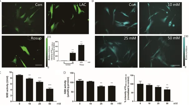
Lactose induced cellular oxidative load in MRC-5
To examine whether the lactose-induced sen- escence was triggered by intracellular redox state, we investigated the effects of lactose on ROS (Figure 2A). The cells were treated with 25
mmol/L lactose for 7 days, and the intracellular
ROS levels were indicated by DCFH-DA dye. The
fold change of fluorescent brightness revealed that lactose could significantly increase the
ROS levels in MRC-5 cells (P<0.01). Moreover, GSH (Figure 2B, 2D) and SOD (Figure 2C), the key anti-oxidative enzymes in redox metabo-lism, reported a dose-dependent decrease, as detected by microscope analysis and micro-plate reader. All assays suggested that lactose induces ROS generation in a dose-dependent manner.
NAC can rescue the lactose-induced oxidative stress and cellular senescence
We next investigated whether the oxidative damage induced by lactose was the main rea-son of premature senescence. For this, NAC, a widely used ROS scavenger, was supplemented
to the culture medium at a final concentration
of 1 mmol/L to eliminate lactose-induced ROS. We initially detected the ROS levels using the DCFH-DA dye, and found that the addition of
NAC resulted in a significant reduction in ROS
levels (P<0.01 versus high lactose; Figure 3A,
3B). In parallel, NAC could also rescue cellular senescence induced by lactose. Also,
accumu-lation of SA-β-gal decreased in the presence
of NAC (Figure 3C). Similarly, western blotting analysis showed that p16ink4a expression signi-
ficantly decreased to the basal level as com -pared with stable high lactose after incubation of NAC (P<0.01; Figure 3D).
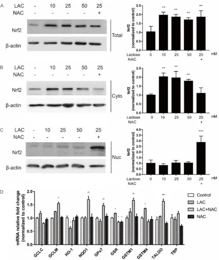
Nrf2/ARE pathway is involved in oxidative stress induced by lactose in MRC-5
The Nrf2 pathway is known to regulate the total antioxidant system through modulating the expression of antioxidant-associated ge- nes involved in repairing oxidative damage. Once released, it migrates to the cell nucleus and binds the DNA to activate antioxidant re- sponse elements (AREs), which works as anti-oxidant and detoxicant. Western blotting analy-sis showed the levels of total and cytoplasmic
Nrf2 significantly increase in the cells exposed
to 10 mmol/L, 25 mmol/L and 50 mmol/L of lactose (Figure 4A, 4B). The addition of NAC did not affect the total Nrf2 level but decreased its cytoplasmic level, whereas increased its nuclear level (Figure 4A-C). These results indi-cated that NAC might function by activating the Nrf2 translocation to the nucleus.
We also examined the mRNA expression of
Nrf2 downstream ARE-containing genes in
fib-roblasts. Phase II enzymes and endogenous antioxidants had been well reported to be Nrf2-dependent [21]. The regulation of phase II enzymes, such as glutamate-cysteine ligase catalytic subunit (GCLC), glutamate-cysteine
ligase modifier subunit (GCLM), heme oxygen -ase 1 (HO-1), NAD(P)H quinone dehydrogen-ase 1 (NQO1), would deactivate and detoxify foreign
compounds and were referred to as detoxifica -tion enzymes [22]. Moreover, endogenous
anti-oxidants, such as glutathione peroxidase 7 (GPx7), glutathione-disulfide reductase (GSR),
glutathione S-transferase Mu (GSTM), and tra-
nsaldolase (TALDO) play a significant role in detoxification by catalyzing radicals into
non-toxic substances and scavenging ROS. RT-PCR analysis revealed lactose to have no effects on the mRNA expression of many Nrf2 targeting genes. However, addition of NAC induced a
sig-nificant increase in the mRNA expression of
these genes, including NQO1, TALDO, GCLM,
GPx7 and GSTM1, which are members of anti -oxidative proteins and protect the cells from oxidative stress (Figure 4D). In brief, lactose could affect the intracellular redox state by acti-vating Nrf2 signaling, which was rescued by NAC.
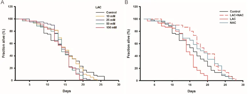
Lactose supplement shortens the lifespan of C. elegans
Lactose treatment significantly induced ROS
atten-2040 Int J Clin Exp Pathol 2019;12(6):2034-2045 Figure 2. Lactose induced intracellular oxidative stress. A. The cells treated with 25 mM lactose for 7 days. 100 μM Rosup treated as positive control. ROS stained by DCFH-DA dye and imaged by inversion fluorescence microscope. Scale bar =100 μm. B. The GSH level of living cells treated with lactose and imaged by inversion fluorescence microscope. Fluorescent image of GSH stained with mBCI dye. Scale bar =100 μm. C. The levels of SOD tested by SOD assay kit. Results obtained from
Figure 3. The oxidative stress induced by lactose could be abrogated by anti-oxidant. A. The cells incubated with lactose (7 days), and then treated with 1 mM NAC for 24 hours. Fluorescent image of DCFH-DA dye examined by inversion fluorescence microscope. Scale bar =100 μm. B. The optical density analysis of fluorescence imaging. C. The cells treated with 1 mM NAC, 1 nM Rapamycin, 1 mM NAC and 25 mM lactose. The rate of senescence stained by SA-β-gal staining and analyzed by SA-β-gal positive cells (%). D. The expression of p16ink4a analyzed by western blotting. Values represented average ± SD from at least 3 experiments. *P<0.05,
uated the phenomenon. We observed similar results in C. elegans. All worms were adminis-tered with various concentrations of lactose from middle age onward to 10 days. Lactose at 10, 25, 50 and 100 mmol/L of concentrations decreased the mean lifespan of worms by
[image:9.612.93.523.68.578.2]10.4%, 17.4% (P<0.05), 22.8% (P<0.01), 16.5% (P<0.05) respectively (Figure 5A). However, after co-administration of NAC and lactose, the mean lifespan of worms increased by 28.2% (P<0.01) as compared with 50 mmol/L of lac-tose treatment. It was consistent with our in
vitro data which showed that only NAC treat-ment had no effect on the lifespan of worms
(Figure 5B). Taken together, the presence of
NAC could completely rescue the decreased longevity induced by the high concentration of lactose, indicating redox stress to be, at least in part, responsible for the lactose-induced senescence.
Discussion
The present study reported that treatment of
lung fibroblasts (MRC-5 cells) with lactose
in-duced cellular senescence through an increase in the generation of intracellular ROS. Lactose has been known to reduce the lifespan of C. elegans. Indeed, lactose seemed to increase dose-related ROS accumulation both in vitro
and in vivo, which promoted imbalance of redox state and affected the Nrf2 pathway. This also suggested that activation of Nrf2 could resist aging induced by oxidants, and could thus act as a potential therapeutic target.
Lactose is a distinctive carbohydrate in milk and dairy products. It is an essential marker for the evaluation of dairy quality. Studies have focused on its physiological function in facilitat-ing assimiliation of nitrogen, calcium and phos-phorus markedly, as well as promoting the de- velopment of brain tissue [23, 24]. Dairy prod-ucts contain a varying amount of lactose, such
as 4 to 5% lactose in human milk, 2 to 7% lac -tose in yogurt and 3 to 8% lac-tose in ice cream.
The concentration of lactose in skimmed milk has been reported to be considerably high, up
to 47 g/L [17, 25] (equivalent to 137 mmol/L
lactose, greater than what we reported). Thus,
lactose can realistically exert a significant
ef-fect on the cells.
Upon ingestion, lactose is metabolized and dis-tributed through passive diffusion. Normally lactose is hydrolyzed into glucose and galac-tose by lactase and gets absorbed in the
small intestine. Unfortunately, lactase deficien -cy is associated with race and age worldwide. Pochart and colleagues studied the extent of lactose absorption in people suffering from lac-tose intolerant to exclude the distribution of lactase. They analyzed the absorption of lac-tose after administration of 18 g of laclac-tose meals (without lactase in food), and collected the lactose from terminal ileum. The results
showed that only 1.74 to 2.83 g of lactose was left [26], which suggested that passive diffu-sion was another important way to transport lactose, besides catabolism.
We also studied the long-term effect of lactose on cells and C. elegans. It was noted that
lac-tose exposure would significantly induce, in a
[image:10.612.92.525.75.239.2]aging cells, proteins, and DNA [27]. This might
serve as one of the mechanisms by which lac-tose induces senescence by increasing ROS generation. The results obtained in the present study indicated cellular dysfunction, which, in turn, speculated the involvement of
redox-relat-ed pathways. We then triredox-relat-ed to study the influ -ence of lactose on Nrf2-ARE pathway, a key pathway for oxidation resistance, which can induce several cytoprotective proteins against toxicities and chronic diseases linked with oxi-dative stress [28]. According to the results of our research, lactose, as a chemical inducer, activated the expression of Nrf2 protein and ARE-containing genes, such as HO-1, GSR, and GSTM4. We speculated it could disrupt the combination of Nrf2 and Keap1, thereby acti-vating the Nrf2-ARE signaling. We found that the effects of lactose on the Nrf2 pathway could be rescued by the antioxidant, NAC. So, it was reasonable to hypothesize that intracellu-lar redox state regulated by NAC might stimu-late uncoupling of Nrf2 from Keap1. Nrf2 then translocated to the nucleus, where it activated an array of antioxidant response element-regu-lated genes expression, including endogenous
antioxidative system (TALDO, GPx7, GSR and
GSTM1) and phase II detoxifying enzymes (GCLM, NQO-1). These results further support-ed the fact that lactose acceleratsupport-ed the onset of senescence and the protective effect of NAC
in vitro and in vivo.
In conclusion, we confirmed that high lactose
for an extended period would result in oxidative
stress in fibroblasts and model animals. The
present study also emphasized that Nrf2 could serve as an essential therapeutic target to resist aging induced by oxidants.
Acknowledgements
This work was supported by the Exploratory Research Foundation of ECUST (Grant number:
222201714058) and National Natural Science Foundation of China (Grant number:
8177-2689).
Disclosure of conflict of interest
None.
Address correspondence to: Dr. Zelan Hu, Shanghai Key Laboratory of New Drug Design, School of Pharmacy, East China University of Science and
Technology, 130 Mei Long Road, Shanghai 200237,
China. E-mail: [email protected]; Dr. Jian Li, State Key Laboratory of Bioreactor Engineering, East China University of Science and Technology,
130 Mei Long Road, Shanghai 200237, China. E-mail: [email protected]
References
[1] Campisi J. Aging, cellular senescence, and cancer. Annu Rev Physiol 2013; 75:685-705.
[2] van Deursen JM. The role of senescent cells in ageing. Nature 2014; 509: 439-446.
[3] Baker DJ, Wijshake T, Tchkonia T, LeBrasseur NK, Childs BG, van de Sluis B, Kirkland JL and van Deursen JM. Clearance of p16Ink4a-posi-tive senescent cells delays ageing-associated
disorders. Nature 2011; 479: 232-236.
[4] Hayflick L and Moorhead PS. The serial cultiva -tion of human diploid cell strains. Exp Cell Res 1961; 25: 585-621.
[5] Sharpless NE and Sherr CJ. Forging a signature of in vivo senescence. Nat Rev Cancer 2015;
15: 397-408.
[6] Longo VD, Antebi A, Bartke A, Barzilai N, Brown-Borg HM, Caruso C, Curiel TJ, Cabo R, Frances-chi C, Gems D, Ingram DK, Johnson TE, Ken-nedy BK, Kenyon C, Klein S, Kopchick JJ, Lepperdinger G, Madeo F, Mirisola MG, Mitch-ell JR, Passarino G, Rudolph KL, Sedivy JM, Shadel GS, Sinclair DA, Spindler SR, Suh Y, Vijg J, Vinciguerra M and Fontana L. Interventions to slow aging in humans: are we ready? Aging
Cell 2015; 14: 497-510.
[7] Simpson SJ, Le Couteur DG and Raubenheimer D. Putting the balance back in diet. Cell 2015; 161: 18-23.
[8] Levine ME, Suarez JA, Brandhorst S, Balasub-ramanian P, Cheng CW, Madia F, Fontana L, Mirisola MG, Guevara-Aguirre J, Wan J, Passa-rino G, Kennedy BK, Wei M, Cohen P, Crimmins EM and Longo VD. Low protein intake is associ-ated with a major reduction in IGF-1, cancer, and overall mortality in the 65 and younger but not older population. Cell Metab 2014; 19:
407-417.
[9] Zwighaft Z, Aviram R, Shalev M, Rousso-Noori L, Kraut-Cohen J, Golik M, Brandis A, Reinke H, Aharoni A, Kahana C and Asher G. Circadian clock control by polyamine levels through a mechanism that declines with age. Cell Metab
2015; 22: 874-885.
[10] Elzi DJ, Song M and Shiio Y. Role of galactose
in cellular senescence. Exp Gerontol 2016; 73:
1-4.
domesti-ca is associated with oxidative stress.
Bio-gerontology 2004; 5: 317-325.
[12] Parameshwaran K, Irwin MH, Steliou K and Pinkert CA. D-galactose effectiveness in mod-eling aging and therapeutic antioxidant treat-ment in mice. Rejuvenation Res 2010; 13:
729-735.
[13] Deng Y, Misselwitz B, Dai N and Fox M. Lactose intolerance in adults: biological mechanism
and dietary management. Nutrients 2015; 7:
8020-8035.
[14] Swagerty DL Jr, Walling AD and Klein RM. Lactose intolerance. Am Fam Physician 2002; 65: 1845-50.
[15] Maier AB, Westendorp RG, VAN Heemst D. Beta-galactosidase activity as a biomarker of replicative senescence during the course of
human fibroblast cultures. Ann N Y Acad Sci 2007; 1100: 323-332.
[16] Xia X, Chen W, McDermott J and Han JJ. Mo-lecular and phenotypic biomarkers of aging.
F1000Res 2017; 6: 860.
[17] Conzuelo F, Gamella M, Campuzano S, Ruiz MA, Reviejo AJ and Pingarron JM. An integrated amperometric biosensor for the determination of lactose in milk and dairy products. J Agric Food Chem 2010; 58: 7141-8.
[18] Cui X, Li W and Zhang B. Studies on cell senes-cence induced by D-galactose in cultured
neu-rons and fibroblasts. Zhongguo Ying Yong Sheng Li Xue Za Zhi 1997; 13: 131-133.
[19] He X, Kan H, Cai L and Ma Q. Nrf2 is critical in defense against high glucose-induced oxida-tive damage in cardiomyocytes. J Mol Cell Cardiol 2009; 46: 47-58.
[20] Baar MP, Brandt RM, Putavet DA, Klein JD, Derks KW, Bourgeois BR, Stryeck S, Rijksen Y, van Willigenburg H, Feijtel DA, van der Pluijm I, Essers J, van Cappellen WA, van IWF, Hout-smuller AB, Pothof J, de Bruin RW, Madl T, Hoeijmakers JH, Campisi J and de Keizer PL. Targeted apoptosis of senescent cells restores tissue homeostasis in response to
chemotoxic-ity and aging. Cell 2017; 169: 132-147, e116.
[21] Zhang DD. Mechanistic studies of the Nrf2-Keap1 signaling pathway. Drug Metab Rev
2006; 38: 769-789.
[22] Shen G and Kong AN. Nrf2 plays an important role in coordinated regulation of Phase II drug metabolism enzymes and Phase III drug trans-porters. Biopharm Drug Dispos 2009; 30: 345-355.
[23] Talbot FB and Hill LW. The influence of lactose
on the metabolism of an infant: With special reference to fat, nitrogen and ash. American Journal of Diseases of Children 1914; VIII:
218-227.
[24] Jarvis B. Milk sugar in infant feeding: a study of the effects of the routine use of milk sugar in infant feeding. American Journal of Diseases of Children 1930; 40: 993-999.
[25] F. Fox P and McSweeney P. Advanced Dairy Chemistry. 2009.
[26] Marteau P, Flourie B, Pochart P, Chastang C, Desjeux JF and Rambaud JC. Effect of the mi-crobial lactase (EC 3.2.1.23) activity in yoghurt on the intestinal absorption of lactose: an in
vivo study in lactase-deficient humans. Br J
Nutr 1990; 64: 71-9.
[27] Ameziane-El-Hassani R and Dupuy C. Detection of reactive oxygen species in cells undergoing oncogene-induced senescence. Methods Mol
Biol 2017; 1534: 139-145.