Original Article
Scanning all chromosomal abnormalities with
microarray-based comparative genomic hybridization
in differential diagnosis of pediatric cancers
Hulya Tosun Yildirim1, Safiye Aktas2, Gulden Diniz3, Tekincan Cagri Aktas2, Burcin Baran2, Serdar Bayrak2,
Zekiye Altun2, Yasemin Cakir2, Nur Olgun2
1Department of Pathology, Antalya Education and Research Hospital, Health Sciences University, Antalya,
Turkey; 2Oncology Institute, Department of Basic Oncology, Dokuz Eylul University, Izmir, Turkey; 3Department of
Pathology, Izmir Democracy University, Izmir, Turkey
Received May 28, 2019; Accepted July 22, 2019; Epub August 1, 2019; Published August 15, 2019
Abstract: Objective: Despite conventional histopathological and immunohistochemical methods, difficulties may be experienced in the differential diagnosis of pediatric cancers, especially in small round-cell undifferentiated tumors. In these cases, the determination of chromosomal abnormalities may be helpful. The aim of this study was to evalu-ate the place of the whole genome array comparative genomic hybridization method in pediatric cancers where difficulty is experienced in differential diagnosis. Method: In Comparative Genomic Hybridization (CGH), 135,000 probes were scanned as 3 probes per gene in all genomes. It was possible to analyze paraffin block tissues obtained from the archive of the Pathology Laboratory of Dr. Behcet Uz Children’s Hospital. DNA extraction was made from the paraffin blocks of 24 cases where difficulty had been experienced in making the differential diagnosis and in each case, comparisons with the control samples were made for all anomalies in all chromosomes using microarray technology. Results: Together with the typically observed chromosomal anomalies, additional derangements with debatable importance were determined. Conclusion: The whole genome CGH method may be useful in pediatric cancers where difficulties are experienced in making differential diagnoses. Since technical difficulties are experi-enced in the examination of paraffin-embedded tissue samples, storing fresh tissue samples from each tumor will be helpful for genetic and molecular examinations.
Keywords: Comparative genomic hybridization (CGH), pediatric cancers, small round cell malignant tumors, dif-ferential diagnosis
Introduction
Malignancies are one of the important causes of death in children. In developed countries, less than one percent of cancers occur in chil-dren under 15 years of age. The overall cancer prevalence is between 70-160 per million peo-ple under the age of 15 years [1]. In developing countries such as our country, malignancies are the fourth most common cause of death. Differential diagnosis is more important in childhood malignancies because of the longer life expectancy of children [1].
Primitive small round cell tumors observed in childhood belong to heterogeneous malignant tumor groups that constitute 0.1% of childhood
malignant tumors. These tumors mainly seen in childhood may be listed as neuroblastoma, sar-comas, including rhabdomyosarsar-comas, non-rhabdomyosarcoma soft tissue sarcomas, Ew- ing sarcoma (ES)/primitive neuroectodermal tumor (PNET), desmoplastic small round cell tumor (DSRCT) and malignant rhabdoid/tera-toid tumor (RT) [1-3].
techniques can be identified more reliably, allowing both the comparison of at least two genomes with each other and the analysis of the whole genome in a single experiment [4-6]. The aim of this study is to investigate the role of whole genome-comparative genomic hybridiza-tion in pediatric cancers posing difficulties in differential diagnosis.
Materials and methods
In the Pathology Laboratory of Dr. Behçet Uz. Children’s Hospital, DNA samples were extract-ed from the paraffin blocks of 24 patients wh- ere difficulty was experienced in the differential diagnosis, and a CGH analysis was performed. The anticipated chromosomal anomalies were compared with the clinicopathological findings. In the comparative genomic hybridization anal-ysis, the differences in the number of copies in the whole genome or in a region on the genome with a referenced genome were determined using NimbleGen CGH arrays. The DNA of 24 patients (test DNA) and normal DNA (control DNA) to be analyzed for CGH were labeled with different fluorochromes and hybridized with metaphase chromosomes obtained from nor-mal cells. Differences in fluorescence intensi-ties across chromosomes in the reference me- taphase domain refer to the differences in the number of copies (amplifications or deletions) in the tumor DNA. If DNAs showing different fluorescent phenotypes overlap with normal metaphase chromosomes, they are have a blue-orange color.
If there is a deletion in the test DNA, hybridiza-tion does not occur in this region, so this region appears red. In contrast, if there is an amplifi-cation in the DNA, the region is appears green and is brighter than the other regions because hybridization is more intense in this region. Chromosome hybridization profiles were calcu-lated according to hybridization rates using a special image analysis software program. Results
Our study population consisted of patients with Ewing sarcomas (n = 10), neuroblastomas (n = 5), and undifferentiated malignant tumors (n = 9) (Table 1). Previous diagnoses of RT (n = 2), DSRCT (n = 2), and RMS (n = 1) were consid-ered in 5 cases with diagnoses of
undifferenti-ated malignant tumors. The ages of the patients ranged from 2 months to 18 years. The mean age of the patients was 8.7 ± 5.3 years. The series consisted of 10 boys (41.7%) and 14 girls (58.3%). The tumors were localized in the kidneys and adrenal glands (n = 6), the abdomi-nal cavity (n = 4), the chest wall (n = 4), the soft tissue of the hip or of an extremity (n = 3), the lungs or pleura (n = 2), the pelvic region (n = 2), and the liver (n = 1). The location of the tumor was unknown in 2 cases sent for the consulta-tion (Table 1).
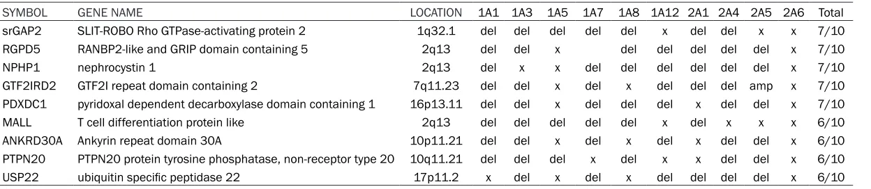
In all, 3 male and 7 female Ewing sarcoma patients were analyzed (Figure 1). The median age of the patients was 8. 6 years. The oldest and the youngest patients were 14 years and 1 year old, respectively. The tumors were located in different body parts. These tumors were localized in the extremities (n = 2), the lungs (n = 1), and in the soft tissue of the trunk (n = 7). The CGH analysis frequently detected 8 gene domains (commonly shared gene domains found in 6 and 7 cases with Ewing sarcoma) (Table 2).
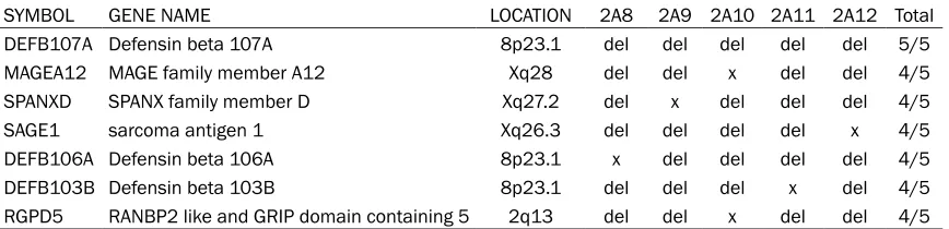
In our series, 1 male, and 4 female patients with a median age of 7.2 years (range, 2-16 years) received the diagnosis of undifferentiat-ed neuroblastoma. The tumors were localizundifferentiat-ed in the adrenal glands in 3 cases. In the CGH analysis, 8 gene regions (commonly shared in 4 or 5 of 9 cases) were often detected (Table 3). In addition, 9 cases were diagnosed as undif-ferentiated malignant tumors. Among the cases diagnosed as undifferentiated tumors, the pre-vious diagnoses of RT (n = 2), DSCRT (n = 2), and RMS (n = 1) were considered. In the CGH analysis, 8 gene regions (commonly shared in 4 or 5 of 9 cases) were often detected (Table 4). Discussion
can be found, or, it may be necessary to look at
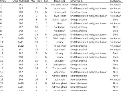
multiple sites for the differential diagnosis. In interacts with Crk-associated substrates and controls cell division, cell-cell, and cell-matrix Table 1. Patient demographic data
CASE CASE NUMBER AGE (year) SEX LOCATION DIAGNOSIS SURVIVAL
1 1A1 4 F Iliac bone region Ewing sarcoma Not known
2 1A2 4 M Abdomen Undifferentiated malignant tumor Not known
3 1A3 11 M Thoracic wall Ewing sarcoma Not known
4 1A4 9 M Pelvic region Undifferentiated malignant tumor Not known
5 1A5 8 M Sacral region Ewing sarcoma Died
6 1A6 0 F Liver Undifferentiated malignant tumor Not known
7 1A7 18 F Not known Ewing sarcoma Alive
8 1A8 17 F Not known Ewing sarcoma Died
9 1A9 13 M Lung/pleura Undifferentiated malignant tumor Died 10 1A10 2,5 F Pelvic region Undifferentiated malignant tumor Not known 11 1A11 10 F Kidney Undifferentiated malignant tumor Not known
12 1A12 7 F Thoracic wall Ewing sarcoma Not known
13 2A1 13 F Abdomen Ewing sarcoma Not known
14 2A2 3 M Kidney Undifferentiated malignant tumor Died
15 2A3 3 M Kidney Undifferentiated malignant tumor Died
16 2A4 15 M Shoulder Ewing sarcoma Alive
17 2A5 15 F Lung/pleura Ewing sarcoma Died
18 2A6 14 F Thoracic wall Ewing sarcoma Alive
19 2A7 5 M Abdomen Undifferentiated malignant tumor Alive
20 2A8 7 F Adrenal gland Neuroblastoma Alive
21 2A9 16 F Abdomen Neuroblastoma Not known
22 2A10 2 M Adrenal gland Neuroblastoma Alive
23 2A11 7 F Adrenal gland Neuroblastoma Alive
[image:3.612.92.522.89.409.2]24 2A12 5 F Thoracic wall Neuroblastoma Alive
Figure 1. (A) General appearence of ES/PNET tumor cells (HE × 200). Im-munohistochemical stains in the same tumor: (B) Ki67 proliferation index, (C) Expression of CD99 (DAB × 200), and (D) Expression of Synaptophysin (DAB × 200).
these cases, CGH may help detect chromosomal anoma-lies [6].
Table 2. Copy number altered genes among patients with ES/PNET
SYMBOL GENE NAME LOCATION 1A1 1A3 1A5 1A7 1A8 1A12 2A1 2A4 2A5 2A6 Total
srGAP2 SLIT-ROBO Rho GTPase-activating protein 2 1q32.1 del del del del del x del del x x 7/10
RGPD5 RANBP2-like and GRIP domain containing 5 2q13 del del x del del del del del x 7/10
NPHP1 nephrocystin 1 2q13 del x x del del del del del del x 7/10
GTF2IRD2 GTF2I repeat domain containing 2 7q11.23 del del x del x del del del amp x 7/10
PDXDC1 pyridoxal dependent decarboxylase domain containing 1 16p13.11 del del x del del del x del del x 7/10
MALL T cell differentiation protein like 2q13 del del del del del x del x x x 6/10
ANKRD30A Ankyrin repeat domain 30A 10p11.21 del del x del x del x del del x 6/10
PTPN20 PTPN20 protein tyrosine phosphatase, non-receptor type 20 10q11.21 del del del x del x x del del x 6/10
adhesion signaling. GTF2IRD2 is a transcrip-tional factor under the control of the retinoblas-toma protein.
MALL participates in raft-mediated trafficking in endothelial cells. MAL1 interacts with caveo-lin and is localized in glycolipid or cholesterol-enriched membrane rafts. PTPN20 is a mem-ber of the protein tyrosine phosphatases and is involved in actin polymerization. USP22 is an interesting gene that regulates various cellular and organismal processes such as cell prolif-eration, apoptosis, and malignant transforma-tion. It also deubiquitinates histones H2A and H2B, thereby acting as a coactivator. USP22 participates in the activation of several onco-genes such as MYC and contributes to stem-ness in colorectal carcinogenesis as well as the epithelial mesenchymal transition in osteosar-coma cell lines. Altered levels of ANKRD30A expressed in epithelial cells and in the testes have been found to be associated with breast cancer. TPTE is a PTEN-related tyrosine phos-phatase and plays a role in the endocrine sig-nal transduction pathways and in the sper-matogenic function of the testes. CTSE encodes peptidase involved in antigen processing and the maturation of secretory proteins [1, 8]. Even with the availability of advanced molecu-lar and immunohistochemical diagnostic meth-ods, the diagnosis of poorly differentiated and undifferentiated neuroblastomas may be espe-cially difficult. The defensin beta 107A gene, where copy number variations were most fre-quently detected in the CGH analysis per-formed in our patient series, and the defensin family produced by neutrophils, are antimicro-bial, and cytotoxic peptides are effective on the immune system. There is evidence that abnor-mal defensin expression is related to a suscep-tibility to infectious diseases and inflammatory disorders. The loss/deletion of defensin beta
107A has been especially observed in breast, cervix, colon, and bladder cancers [10-12]. The MAGE family member A12 gene is closely related to several other genes clustered on the X chromosome. It has been reported that these genes may be overexpressed in melanoma and breast cancer tumors [13, 14]. However, in our study, a deletion was detected in the MAGE family member A12 gene in neuroblastoma cases. The SPANX family member D sarcoma antigen 1 plays a role in the formation of matu- re spermatozoa. It is necessary to initiate the sequence of molecular and morphological changes in the germ cells. This gene is a mem-ber of the gene family associated with the SPA- NX cancer/testis localized in a cluster on the X chromosome. SPANX genes encode diverse- ly differentiated testis-specific proteins that are localized in different subcellular compart-ments. Although the specific function of this gene has not yet been determined, polymor-phisms in this gene may be associated with a susceptibility to prostate and breast cancer [15, 16].
[image:5.612.91.525.85.190.2]Defensin beta 106A is an interesting gene that regulates the processes of stimulating the immune system, especially in the acquired immune response [17]. Defensin beta 103B is also effective in the immune system. A beta-defensin 3, expressed in the epithelium in re- sponse to various stimulations, including hu- man papilloma virus infection, has recently been found to be overexpressed in head and neck cancers and cervical cancers and has been shown to exhibit tumorigenic activities [17, 18]. However, the RGPD5 gene is a small GTP binding protein of the RAS superfamily, thought to control various cellular functions associated with nuclear membranes and inter-actions with other proteins. Its relationship with cancerogenesis has not been detected yet [19]. Table 3. Copy number changes among patients with neuroblastoma
SYMBOL GENE NAME LOCATION 2A8 2A9 2A10 2A11 2A12 Total
DEFB107A Defensin beta 107A 8p23.1 del del del del del 5/5
MAGEA12 MAGE family member A12 Xq28 del del x del del 4/5
SPANXD SPANX family member D Xq27.2 del x del del del 4/5
SAGE1 sarcoma antigen 1 Xq26.3 del del del del x 4/5
DEFB106A Defensin beta 106A 8p23.1 x del del del del 4/5
DEFB103B Defensin beta 103B 8p23.1 del del del x del 4/5
Table 4. Copy number changes among patients with undifferentiated malignant tumors
SYMBOL GENE NAME LOCATION 1A2 1A4 1A6 1A9 1A10 1A11 2A2 2A3 2A7 Total
ANKRD30B ankyrin repeat domain 30B 18p11.21 del X X del del del del x x 5/9
SLC6A4 solute carrier family 6 member 4 17q11.2 del del x x del del del x x 5/9
PPIB peptidylprolyl isomerase B 15q22.31 del del x del x del del x x 5/9
HERC2 HECT and RLD domain containing E3 ubiquitin protein ligase 2 15q13.1 del del x del del del x x x 5/9
TDO2 tryptophan 2,3-dioxygenase 4q32.1 amp amp x del del del x x x 5/9
IGSF21 immunoglobin superfamily member 21 1p36.13 x x x del del x del del x 4/9
CAMK2B calcium/calmodulin dependent protein kinase II beta 7p13 x x x del del x del del x 4/9
Difficulties encountered in making the differen-tial diagnoses of undifferentiated malignant tumors, especially in the group of small round cell malignant tumors should be expected. Ev- en immunohistochemical and molecular tests may not show the cases’ degrees of differentia-tion. In such cases, the CGH analysis can be used both to detect DNA copy number varia-tions and to make a differential diagnosis. In 9 cases of our patient series, we could not arrive at a definite differential diagnosis. The immuno-globulin superfamily member 21 gene, which is one of the genes most frequently demonstrat-ing copy number variations in the CGH analysis, encodes a protein that is a member of the immunoglobulin superfamily. These proteins are usually found in cell membranes and act as receptors in immune response pathways. Its association with carcinogenesis has not been determined yet [20].
The product of the calcium/calmodulin-depen-dent protein kinase II beta (CAMK2B) gene belongs to the serine/threonine protein kinase family and the Ca (2+)/calmodulin-dependent protein kinase subfamily. Calcium signaling is very important for many different aspects of plasticity in glutamatergic synapse. In one stu- dy, the dysfunction of CAMK2B was associated with osteoblastic bone metastases of the pros-tate, and it is closely related to osteoblasto- genesis of human mesenchymal stem cells [21]. POM121 transmembrane nucleoporin is a component of the nuclear pore complex (NPC). The NPC functions in the regulation of nucleus-cytoplasmic transport, transcription, and the stability of the genome. However, their function-al roles in cancer have been poorly understood. There is evidence in the literature that NPC may play a role in the development of primary meta-static prostate cancer [22].
Ankyrin repeat domain 30B gene expression is contained in the testes. The solute carrier fam-ily 6 member gene expresses the integral mem-brane protein that transports serotonin from synaptic domains to presynaptic neurons. It encodes a transport protein that mediates the uptake of long-chain fatty acids in the cell. The function of these proteins is mainly related to lipid biosynthesis, the fatty acid metabolic pro-cess, and fatty acid transport. It has been shown to play a role in depression sensitivity in people with aggressive behavior and emotion- al trauma, especially in Alzheimer’s patients. In
addition, the deletion of this gene may contrib-ute to the development of breast and thyroid cancer and may be effective in reducing sur-vival in patients with colorectal cancer [23-25]. Peptidylprolyl isomerase B, a gene product cy- clophilin B (CypB), is an endoplasmic reticulum protein that binds to cyclophilin A, which is a member of the cyclophilin family [26, 27]. CypB has peptidyl-prolyl cis-trans isomerase (PPIa- se) activity. PPIases catalyze the cis-trans isom-erization of proline and peptide bonds in oligo-peptides and accelerate the folding of proteins. The encoded protein is a cyclophilin A (CypA) binding protein and may play a role in cyclophil- in mediated immunosuppression. CypA, the protein produced by this gene, has been report-ed to be upregulatreport-ed in breast, stomach, cervi-cal, and pancreatic cancers [28-31]. In particu-lar, it has been reported to regulate the growth and survival of gastric cancer cells and may be related to the recovery of tumor growth and anti-apoptotic functions, and it has been noted as a very important link in the pathogenesis of cancer [26]. In addition, the overexpression of CypB inhibits the activation of antioxidant en- zymes in the cell and the resulting reduction of oxidative stress, especially the neurogenic cy- totoxicity by mitogen-activated protein kinase through the c-Jun N-terminal kinase pathway [27].
In conclusion, the CGH array may be a useful method for the detection of DNA copy number alterations among pediatric patients with undif-ferentiated malignant tumors. In our ES/PNET dataset, the copy number alterations were mo- stly found in genes related to cytoskeletal ele-ments, migration, and protein trafficking in our cohort.
Changes in the number of copies in our neuro-blastoma series were mostly found in genes that regulate the stimulation of the immune system [11, 12, 17, 18]. Changes in the number of copies of undifferentiated malignant tumors were found in genes that may be important in mesenchymal stem cell differentiation and in genes that may have played a role in tumor development through the suppression of im- mune responses and that may cause oxidative stress [21, 26-31]. In order to better under-stand the role of these genes in tumorigenesis and their contribution to the differential diagno-sis, it is necessary to examine the functions of genes in larger series.
Acknowledgements
This text has been edited by Gurkan Kazanci, a professional translator from Logos Publishing. The study was supported by Turkish Pediatric Oncology Group (TPOG).
Disclosure of conflict of interest
None.
Address correspondence to: Dr. Gulden Diniz, De- partment of Pathology, Izmir Democracy University Medical School, 13. sok. No: 2 Guzelyali, 35290, Izmir, Turkey. Tel: +90 542 2431309; Fax: +90 232 3625522; E-mail: [email protected]; agdiniz@ gmail.com
References
[1] Rajwanshi A, Srinivas R, Upasana G. Malignant small round cell tumors. J Cytol 2009; 26: 1-10.
[2] Merchant MS, Mackall CL. Current approach to pediatric soft tissue sarcomas. Oncologist 2009; 14: 1139-53.
[3] Kapoor G, Das K. Soft tissue sarcomas in chil-dren. Indian J Pediatr 2012; 79: 936-42. [4] King W, Proffitt J, Morrison L, Piper J, Lane D,
Seelig S. The role of fluorescence in situ hy-bridization technologies in molecular
diagnos-tics and disease management. Mol Diagn 2000; 5: 309-19.
[5] Lau CY. Molecular pathology in cancer re-search. In: Lakhani SR, Fox SB, editors. New York, U.S.A.: Springer; 2016.
[6] Bryndorf T, Kirchhoff M, Rose H, Maahr J, Gerdes T, Karhu R, Kallioniemi A, Christensen B, Lundsteen C, Philip J. Comparative genomic hybridization in clinical cytogenetics. Am J Hum Genet 1995; 57: 1211-20.
[7] Chang CC, Shidham VB. Molecular genetics of pediatric soft tissue tumors: clinical applica-tion. J Mol Diagn 2003; 5: 143-54.
[8] Savola S, Klami A, Tripathi A, Niini T, Serra M, Picci P, Kaski S, Zambelli D, Scotlandi K, Knuu-tila S. Combined use of expression and CGH arrays pinpoints novel candidate genes in Ew-ing sarcoma family of tumors. BMC Cancer 2009; 9: 1-17.
[9] Delattre O, Zucman J, Melot T, Garau XS, Zuck-er JM, Lenoir GM, Ambros PF, SheZuck-er D, Turc-Carel C, Triche TJ, et al. The Ewing family of tu-mors--a subgroup of small-round-cell tumors defined by specific chimeric transcripts. N Engl J Med 1994; 331: 294-299.
[10] Zhang D, Jiang F, Wang X, Li G. Downregulation of ubiquitin-specific protease 22 ınhibits prolif-eration, invasion, and epithelial-mesenchymal transition in osteosarcoma cells. Oncol Res 2017; 25: 763-771.
[11] Jones EA, Kananurak A, Bevins CL, Hollox EJ, Bakaletz LO. Copy number variation of the beta defensin gene cluster on chromosome 8p in-fluences the bacterial microbiota within the nasopharynx of otitis-prone children. PLoS One 2014; 9: e98269.
[12] Hollox EJ, Armour JA, Barber JC. Extensive nor-mal copy number variation of a beta-defensin antimicrobial-gene cluster. Am J Hum Genet 2003; 73: 591-600.
[13] Zhao G, Bae JY, Zheng Z, Park HS, Chung KY, Roh MR, Jin Z. Overexpression and ımplications of melanoma-associated antigen A12 in patho-genesis of human cutaneous squamous cell carcinoma. Anticancer Res 2019; 39: 1849-1857.
[14] Wu J, Wang J, Shen W. Identification of MA-GEA12 as a prognostic outlier gene in gastric cancers. Neoplasma 2017; 64: 238-243. [15] Kouprina N, Noskov VN, Solomon G, Otstot J,
Isaacs W, Xu J, Schleutker J, Larionov V. Muta-tional analysis of SPANX genes in families with X-linked prostate cancer. Prostate 2007; 67: 820-8.
[17] De Paula VS, Gomes NS, Lima LG, Miyamoto CA, Monteiro RQ, Almeida FC, Valente AP. Structural basis for the interaction of human β-defensin 6 and its putative chemokine re-ceptor CCR2 and breast cancer microvesicles. J Mol Biol 2013; 425: 4479-95.
[18] Xu D, Zhang B, Liao C, Zhang W, Wang W, Chang Y, Shao Y. Human beta-defensin 3 con-tributes to the carcinogenesis of cervical can-cer via activation of NF-κB signaling. Oncotar-get 2016; 7: 75902-75913.
[19] Nothwang HG, Rensing C, Kübler M, Denich D, Brandl B, Stubanus M, Haaf T, Kurnit D, Hildeb-randt F. Identification of a novel Ran binding protein 2 related gene (RANBP2L1) and detec-tion of a gene cluster on human chromosome 2q11-q12. Genomics 1998; 47: 383-92. [20] Lin X, Wang J, Yun L, Jiang S, Li L, Chen X, Li Z,
Lu Q, Zhang Y, Ma X. Association between LE-KR1-CCNL1 and IGSF21-KLHDC7A gene poly-morphisms and diabetic retinopathy of type 2 diabetes mellitus in the Chinese Han popula-tion. J Gene Med 2016; 18: 282-287.
[21] Mamaeva OA, Kim J, Feng G, McDonald JM. Calcium/calmodulin-dependent kinase II regu-lates notch-1 signaling in prostate cancer cells. J Cell Biochem 2009; 106: 25-32.
[22] Rodriguez-Bravo V, Pippa R, Song WM, Carceles-Cordon M, Dominguez-Andres A, Fuji-wara N, Woo J, Koh AP, Ertel A, Lokareddy RK, Cuesta-Dominguez A, Kim RS, Rodriguez-Fer-nandez I, Li P, Gordon R, Hirschfield H, Prats JM, Reddy EP, Fatatis A, Petrylak DP, Gomella L, Kelly WK, Lowe SW, Knudsen KE, Galsky MD, Cingolani G, Lujambio A, Hoshida Y, Do-mingo-Domenech J. Nuclear pores promote le-thal prostate cancer by increasing POM121-Driven E2F1, MYC, and AR nuclear import. Cell 2018; 174: 1200-1215.
[23] Savas S, Hyde A, Stuckless SN, Parfrey P, Youn-ghusband HB, Green R. Serotonin transporter gene (SLC6A4) variations are associated with poor survival in colorectal cancer patients. PLoS One 2012; 7: e38953.
[24] De Summa S, Graziano F, Pilato B, Pinto R, Danza K, Lacalamita R, Serratì S, Sambiasi D, Grassi M, Tommasi S. Six low-penetrance SNPs for the estimation of breast cancer heri-tability: a family-based study in caucasian Ital-ian patients. Oncol Lett 2017; 14: 4384-4390. [25] Suh S, Kim YH, Goh TS, Jeong DC, Lee CS, Jang JY, Cha W, Han ME, Kim SJ, Kim IJ, Pak K. mRNA expression of SLC5A5 and SLC2A family genes in papillary thyroid cancer: an analysis of the cancer genome Atlas. Yonsei Med J 2018; 59: 746-753.
[26] Nakano N, Sakashita S, Matsuoka R, Murata Y, Shiba-Ishii A, Kobayashi N, Sato Y, Noguchi M. Cyclophilin A expression and its prognostic sig-nificance in lung adenocarcinoma. Pathol Int 2017; 67: 555-563.
[27] Oh Y, Jeong K, Kim K, Lee YS, Jeong S, Kim SS, Yoon KS, Ha J, Kang I, Choe W. Cyclophilin B protects SH-SY5Y human neuroblastoma cells against MPP(+)-induced neurotoxicity via JNK pathway. Biophys Res Commun 2016; 478: 1396-402.
[28] Meng DQ, Li PL, Xie M. Expression and role of cyclophilin B in stomach cancer. Genet Mol Res 2015; 14: 5346-54.
[29] Ray P, Sullenger BA, White RR. Further charac-terization of the target of a potential aptamer biomarker for pancreatic cancer: cyclophilin B and its posttranslational modifications. Nucle-ic Acid Ther 2013; 23: 435-42.
[30] Fang F, Zheng J, Galbaugh TL, Fiorillo AA, Hjort EE, Zeng X, Clevenger CV. Cyclophilin B as a co-regulator of prolactin-induced gene expres-sion and function in breast cancer cells. J Mol Endocrinol 2010; 44: 319-29.
[31] Lee J, Kim SS. An overview of cyclophilins in human cancers. J Int Med Res 2010; 38: 1561-74.
[32] Bonanno L, Costa C, Majem M, Sanchez JJ, Ro-driguez I, Gimenez-Capitan A, Molina-Vila MA, Vergnenegre A, Massuti B, Favaretto A, Rugge M, Pallares C, Taron M, Rosell R. Combinatory effect of BRCA1 and HERC2 expression on out-come in advanced non-small-cell lung cancer. BMC Cancer 2016; 16: 312.
[33] Pham QT, Oue N, Sekino Y, Yamamoto Y, Shige-matsu Y, Sakamoto N, Sentani K, Uraoka N, Yasui W. TDO2 Overexpression ıs associated with cancer stem cells and poor prognosis in esophageal squamous cell carcinoma. Oncol-ogy 2018; 95: 297-308.