Original Article
Elevated GSK3β expression predicts good prognosis in
hepatocellular carcinoma
Yingying Hu
1,2*, Xian Lin
1*, Shi Zuo
3*, Rongcheng Luo
1, Weiyi Fang
11Cancer Center, Traditional Chinese Medicine-Integrated Hospital, Southern Medical University, Guangzhou, Guangdong, People’s Republic China; 2Dongguan Health School of Guangdong Province, Dongguan, Guangdong, People’s Republic China; 3Department of Hepatobiliary Surgery, Affiliated Hospital of Guizhou Medical University, China. *Equal contributors.
Received January 2, 2018; Accepted February 7, 2018; Epub May 1, 2018; Published May 15, 2018
Abstract: Hepatocellular carcinoma (HCC) is the second most common cause of cancer-related death worldwide.
The role of GSK-3β in cancer progression is considered critical. However, the prognostic value of total GSK-3β pro
-tein levels in HCC remains undetermined. In this study, the expression and biologic significance of total GSK-3β in HCC were evaluated at mRNA and protein levels. We showed that GSK-3β mRNA levels were significantly upregu -lated in HCC tissues relative to the levels in the adjacent non-tumor tissues as recorded on the TCGA database (P
< 0.001). Notably, GSK-3β protein levels were significantly downregulated in HCC tissues relative to those in the
adjacent non-tumor tissues by immunohistochemistry (P < 0.001). We found that GSK-3β was negatively associated
with the American Joint Committee on Cancer (AJCC) stage (P = 0.030) and positively correlated with good progno-sis for HCC patients (P = 0.036). The data further indicated that GSK3β expression tended to be an independent
prognostic marker for HCC after surgical resection (HR = 1.658, 95% CI 0.945-2.909, P = 0.078) and can potentially serve as a biomarker for the clinical diagnosis and prognosis of HCC.
Keywords: GSK3β, bioinformatic analysis, hepatocellular carcinoma, prognosis, immunohistochemistry
Introduction
Hepatocellular carcinoma (HCC) is currently the
second most common cause of cancer-related
death worldwide [1] and the third leading cause
of cancer deaths among both men and women
in China [2]. Although several strategies have
been applied for the management of HCC,
unsatisfactory clinical outcomes emphasize
the need for novel indicators of survival and
reliable therapeutic targets.
Numerous studies on GSK-3β in various
can-cers have been conducted, and most of them
focus on the phosphorylated form of GSK-3β.
Two phosphorylation sites were identified in
GSK-3β, which is activated by phosphoryla-
tion at Tyr-216 and inactivated by phospho-
rylation at Ser-9. Accumulating evidence
sug-gests that pGSK-3β (ser9) regulates Wnt sig-
naling and is responsible for HCC progres-
sion [3-5]. Total GSK-3β has received increas-
ed interest from the research community in
recent years. Although some reports
demon-strated no relationship between total GSK-
3β and pGSK-3β (ser9) levels [6, 7], other
stud-ies indeed suggested a negative correla-
tion between total GSK-3β and pGSK-3β (ser9)
levels [8, 9], indicating a potential progno-
stic value of total GSK-3β in HCC patients.
However, the role of total GSK-3β protein levels
in HCC is yet to be investigated and remains
unclear.
Materials and methods
Bioinformatics analysis
RNA-seq data on liver hepatocellular carci-
noma (LIHC) and stomach adenocarcino-
ma (STAD) were downloaded from the Cancer
Genome Atlas (TCGA) Web site
(http://can-cergenome.nih.gov/). The data included 424
patients with HCC. Among these patients, 50
came with paired non-cancerous tissues, and
407 had gastric cancer, 32 of which had paired
non-cancerous tissues.
Patients and tissues
Overall, 93 HCC and 87 non-tumor subjects
with available clinical information and
paraffin-the slides were incubated with secondary
anti-body for 30 min and counterstained using 3,
3’-diaminobenzidine and hematoxylin.
Evaluation of immunohistochemical staining
[image:2.612.92.354.99.519.2]The IHC sections were scored by 2 experienc-
ed pathologists who were blinded to the clini-
copathological data. Immunohistochemical st-
aining was scored according to the intensity
and the percentage of positively-stained cells.
Staining intensity was estimated on a scale of 0
to 4, as follows: 0 (no staining), 1 (weakly
posi-tive staining), 2 (moderately posiposi-tive staining),
and 3 (strong staining). The percentages of
cells were scored as 0 (0%), 1 (≤ 25%), 2
(26%-50%), 3 (51%-75%), and 4 (76%-100%). The
Table 1. Correlations between GSK3β expression and the
clin-icopathologic features of hepatocellular carcinoma patients
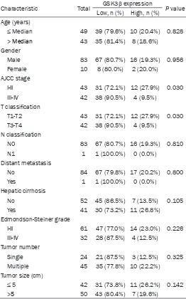
Characteristic Total GSK3β expression P value Low, n (%) High, n (%)
Age (years)
≤ Median 49 39 (79.6%) 10 (20.4%) 0.828 > Median 43 35 (81.4%) 8 (18.6%)
Gender
Male 83 67 (80.7%) 16 (19.3%) 0.956 Female 10 8 (80.0%) 2 (20.0%)
AJCC stage
I-II 43 31 (72.1%) 12 (27.9%) 0.030 III-IV 42 38 (90.5%) 4 (9.5%)
T classification
T1-T2 43 31 (72.1%) 12 (27.9%) 0.030 T3-T4 42 38 (90.5%) 4 (9.5%)
N classification
N0 83 67 (80.7%) 16 (19.3%) 0.810
N1 1 1 (100.0%) 0 (0.0%)
Distant metastasis
No 84 67 (79.8%) 17 (20.2%) 0.800
Yes 1 1 (100.0%) 0 (0.0%)
Hepatic cirrhosis
No 52 45 (86.5%) 7 (13.5%) 0.105
Yes 41 30 (73.2%) 11 (26.8%) Edmondson-Steiner grade
I-II 61 47 (77.0%) 14 (23.0%) 0.226 III-IV 32 28 (87.5%) 4 (12.5%)
Tumor number
Single 24 21 (87.5%) 3 (12.5%) 0.325 Multiple 45 35 (77.8%) 10 (22.2%) Tumor size (cm)
≤ 5 42 31 (73.8%) 11 (26.2%) 0.142 >5 50 43 (80.4%) 7 (19.6%)
embedded blocks participated in
this study. All patients had und-
ergone surgical resection as ini-
tial treatment. Cancer and
adja-cent non-cancerous tissues were
collected from the patients dur-
ing surgery. Each patient had
been pathologically diagnosed
with HCC. The protocols were
approved by the Ethical Commit-
tee and Institutional Review Board
of Traditional Chinese
Medicine-Integrated Hospital of Southern
Medical University, and written
informed consent was obtained
from each patient. All
clinicopatho-logical information was
retrospec-tively collected from the medical
records of the patients. The study
was performed in accordance with
the approved protocols.
Immunohistochemistry (IHC)
final staining scores were calculated by
[image:3.612.92.373.73.189.2]multi-plying the 2 scores.
characteristics of 93 patients with HCC are
listed in Table 1.
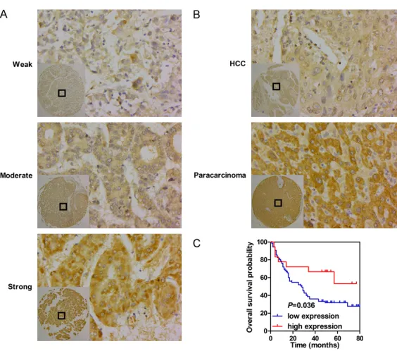
Figure 1. GSK-3β is downregulated in HCC tissues and confers good progno-sis for HCC patients. A. Representative images of GSK-3β staining in HCC. B. Representative images of GSK-3β staining in paired cancerous tissues and
[image:3.612.92.374.256.505.2]non-cancerous tissues.
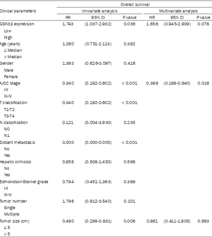
Figure 2. The bioinformatics analysis of GSK-3β expression in HCC and gas
-tric cancer tissues, and peritumoral tissues. A. The comparison of GSK-3β
expression between HCC tissues and non-cancerous tissues. B. Comparison
of GSK-3β expression between gastric tissues and non-cancerous tissues. C. Kaplan-Meier survival analysis based on GSK-3β expression.
Statistical analysis
All data were analyzed with
SPSS ver. 21.0 (SPSS Inc.,
USA). For comparison, diff-
erential expression analysis
was performed using the
Wilcoxon rank sum test. Re-
lationships between gene
expression and
clinicopatho-logical characteristics were
investigated using the
Chi-square test and Fisher’s ex-
act test. Log-rank tests were
performed on Kaplan-Meier
survival curves to elucidate
any significant correlation be-
tween gene expression and
overall patient survival. Uni-
variate and multivariate
sur-vival analyses were
conduct-ed using the Cox
proportional-hazards regression model.
The hazard ratio (HR) and
cor-responding 95% confidence
intervals (95% CI) were
calcu-lated for each factor. All tests
were 2-sided and
considered
statistically significant when
P
< 0.05.
Results
Clinicopathological
charac-teristics
The median age of 93 patients
with HCC was 54 y (range:
25-73 y). Follow-up ranged
from 4 y to 6.7 y. According to
the criteria set by the AJCC
Cancer Staging Manual, 8th
Edition, 12 patients with HCC
were in stage I, 31 in stage II,
40 in stage III, and only 2 in
stage IV. The available
medi-cal records of 93 patients
showed that only 1 patient
had lymph node metastasis,
and 1 case had distant
metas-tasis. The clinicopathological
Table 2. GSK3β expression in HCC tissues and adjacent non-tumor
tissues
Group Cases (n) GSK3β expression P value
Low High
[image:3.612.90.375.613.667.2]Bioinformatics analysis and
immunohisto-chemistry of GSK3β in HCC and non-cancer
-ous tissues
To explore the role of GSK3β in HCC, we initially
investigated its mRNA expression in HCC on
the basis of the TCGA LIHC dataset. RNA-seq
data from HCC tissues and para-carcinoma
tissues showed that GSK3β expression in
HCC tissues was significantly elevated relative
to that in matched non-tumor tissues (
P
<
[image:4.612.94.519.95.571.2]0.001,
Figure 1A). We then detected GSK3β
expression in 93 HCC tissues and 87 adja-
cent non-tumor tissues by
immunohistochem-istry. The analysis indicated that patients ex-
hibited different GSK3β levels, as determin-
ed by the strength of staining from weak to
strong (Figure 2A). Notably, GSK3β protein
levels were significantly downregulated in
HCC samples relative to those in matched
para-carcinoma tissues (
P
< 0.001, Table 2;
Figure 2B).
Table 3. Univariate and multivariate survival analysis of clinicpathologic variables of hepatocellular
carcinoma patients
Clinical parameters
Overall survival
Univariate analysis Multivariate analysis
HR 95% CI P value HR 95% CI P value
GSK3β expression 1.743 (1.037-2.932) 0.036 1.658 (0.945-2.909) 0.078 Low
High
Age (years) 1.260 (0.751-2.113) 0.382
≤ Median
> Median
Gender 1.393 (0.626-3.097) 0.416
Male Female
AJCC stage 0.340 (0.192-0.602) < 0.001 0.399 (0.189-0.840) 0.016 I-II
III-IV
T classification 0.340 (0.192-0.602) < 0.001 T1-T2
T3-T4
N classification 0.121 (0.004-3.930) 0.235 N0
N1
Distant metastasis 0.000 (0.000-0.000) < 0.001 No
Yes
Hepatic cirrhosis 0.858 (0.508-1.450) 0.568 No
Yes
Edmondson-Steiner grade 0.784 (0.451-1.363) 0.388 I-II
III-IV
Tumor number 1.796 (0.912-3.540) 0.101 Single
Multiple
Tumor size (cm) 0.490 (0.289-0.831) 0.008 0.861 (0.411-1.805) 0.693
≤ 5
Correlations between GSK3β expression and
the clinicopathological characteristics of
pa-tients with HCC
The correlations between the GSK3β levels
and the clinicopathological features of pa-
tients with HCC are presented in Table 1. High
GSK3β expression was negatively correlated
with AJCC staging (I-II
vs
. III-IV) and T
classifica-tion (T1-2
vs.
T3-4) (
P
= 0.030, Table 1);
how-ever, no relationship was found between GSK3β
expression and other parameters, such as age,
gender, N classification, distant metastasis,
hepatic cirrhosis, Edmondson-Steiner grade,
number of tumors, and size of tumors.
High GSK3β as an indicator of good prognosis
in HCC tissues
The relationship between GSK3β levels and the
overall survival of patients with HCC was
elucidated by survival analysis. As shown in
Figure 2C, high GSK3β expression correlated
with good prognosis in patients with HCC
(medi-an survival 50.5 months vs. 28 months)
(Log-rank,
P
= 0.036). Moreover, univariate Cox
pro-portional-hazard analysis was performed to
evaluate the potential value of GSK3β in the
prediction of HCC prognosis. High GSK3β
expression, AJCC staging (I-II), T classification
(T1-2), absence of distant metastasis, and
small tumor size (≤ 5 cm in diameter) conferred
longer overall survival time in patients with HCC
(Table 3).
Subsequently, multivariate Cox
proportional-hazard analysis was conducted to investigate
meaningful parameters identified by univariate
Cox analysis. T classification was consistent
with the AJCC stage; only 1 patient had lymph
node metastasis, and 1 case had distant
metastasis; thus, we included the
characteris-tics of GSK3β expression, AJCC stage, and
tumor size in the multivariate Cox analysis for
patients with HCC. As summarized in Table 3,
only the AJCC stage (HR = 0.424, 95% CI
0.206-0.873,
P
= 0.020) was an independent
prog-nostic factor for patients with HCC. However,
GSK3β expression tended to be an
indepen-dent prognostic marker for patients with HCC
(HR = 1.658, 95% CI 0.945-2.909,
P
= 0.078).
Discussion
GSK3β participates in the regulation of
tumori-genesis and cancer progression and may
func-tion as a “tumor suppressor” or “tumor
promot-er” for certain types of tumors [10]. In the
present study, GSK3β expression and its
bio-logic significance in HCC were investigated.
GSK-3β mRNA levels were upregulated;
howev-er, GSK3β protein levels in HCC were
downregu-lated. High GSK3β expression, which was
nega-tively correlated with the AJCC stage, was
indicative of good prognosis for patients with
HCC. Moreover, we showed that GSK3β
expres-sion tended to be an independent prognostic
marker for patients with HCC.
deter-mined that GSK-3β functions as a negative
regulator of HCC progression. GSK-3β mRNA
levels were increased in HCC and stomach
can-cer tissues relative to those in non-tumor
tis-sues, according to the TCGA database (Figure
1). Interestingly, GSK-3β protein levels were
decreased in HCC and gastric cancer [26]
tis-sues relative to those in non-tumor tistis-sues. In
the current study, we consider that
post-tran-scriptional modification of GSK3β may partly
explain the obtained results. We also
demon-strated that high GSK3β expression was
nega-tively correlated with the AJCC stage, and the
findings might partly explain its role in HCC. The
previous study suggested that reduced
expres-sion of GSK-3β was associated with good
clini-copathological prognostic markers at the
mRNA level in HCC [27]; however, low GSK3β
protein level was identified as a poor prognostic
factor for HCC in the study. Similarly, reduced
GSK3β confers poor prognosis in squamous
cell carcinoma of the tongue and malignant
glioma [28, 29]. Although further analysis
revealed that GSK3β expression tended to be
an independent prognostic marker for patients
with HCC, we believe that significant results
could be obtained by increasing the sample
volume.
In addition, the locations of GSK3β also have
distinct functions. Nuclear GSK3β was
corre-lated with shorter overall survival in colon
carci-noma [30]; conversely, another study
suggest-ed that GSK3β formsuggest-ed a complex with β-catenin
in the nucleus to inhibit the canonical Wnt
sig-naling pathway [31]. In human bladder cancer,
nuclear accumulation of GSK-3β was also
iden-tified as a novel prognostic marker contributing
to urothelial cancer cell proliferation and
sur-vival [32]. By contrast, we demonstrated in the
present study that GSK-3β is mainly located in
the cytoplasm of HCC cells.
In conclusion, our results aid in supporting a
tumor suppressor role of GSK-3β in HCC. We
also show that GSK-3β can potentially serve
as a biomarker for the clinical diagnosis and
prognosis of HCC. Lastly, the targeted inhibition
of GSK-3β might be an alternative strategy for
the treatment of HCC.
Acknowledgements
This study was funded by the National Natural
Science Foundation of China (NSFC) (No.
81773151), and the Guangzhou Science and
Technology Plan Projects (No. 201604020009).
Disclosure of conflict of interest
None.
Address correspondence to: Weiyi Fang and Rong- cheng Luo, Cancer Center, Traditional Chinese Medicine-Integrated Hospital of Southern Medical University, 13 Shiliugang Road, Haizhu District, Guangzhou, Guangdong, People’s Republic China. Tel: 86-20-61650036; E-mail: fangweiyi1975@163. com (WYF); luorc02@vip.163.com (RCL); Shi Zuo,
Department of Hepatobiliary Surgery, Affiliated
Hospital of Guizhou Medical University, China. E-mail: drzuoshi@qq.com
References
[1] Omata M, Cheng AL, Kokudo N, Kudo M, Lee JM, Jia J, Tateishi R, Han KH, Chawla YK, Shiina S, Jafri W, Payawal DA, Ohki T, Ogasawara S, Chen PJ, Lesmana CRA, Lesmana LA, Gani RA,
Obi S, Dokmeci AK and Sarin SK. Asia-Pacific
clinical practice guidelines on the manage-ment of hepatocellular carcinoma: a 2017 up-date. Hepatol Int 2017; 11: 317-370.
[2] Chen W, Zheng R, Baade PD, Zhang S, Zeng H, Bray F, Jemal A, Yu XQ and He J. Cancer statis-tics in China, 2015. CA Cancer J Clin 2016; 66: 115-132.
[3] Yang WJ, Chang CJ, Yeh SH, Lin WH, Wang SH, Tsai TF, Chen DS and Chen PJ. Hepatitis B virus X protein enhances the transcriptional activity of the androgen receptor through c-Src and gly-cogen synthase kinase-3beta kinase path-ways. Hepatology 2009; 49: 1515-1524. [4] Clevers H. Axin and hepatocellular carcinomas.
Nat Genet 2000; 24: 206-208.
[5] Ban KC, Singh H, Krishnan R and Seow HF. GSK-3beta phosphorylation and alteration of beta-catenin in hepatocellular carcinoma. Can-cer Lett 2003; 199: 201-208.
[6] McDonnell SR, Hwang SR, Basrur V, Conlon KP, Fermin D, Wey E, Murga-Zamalloa C, Zeng Z, Zu Y, Elenitoba-Johnson KS and Lim MS. NPM-ALK signals through glycogen synthase kinase 3beta to promote oncogenesis. Oncogene 2012; 31: 3733-3740.
[7] Zhao P, Li Q, Shi Z, Li C, Wang L, Liu X, Jiang C, Qian X, You Y, Liu N, Liu LZ, Ding L and Jiang BH. GSK-3beta regulates tumor growth and an-giogenesis in human glioma cells. Oncotarget 2015; 6: 31901-31915.
en-hances GSK-3beta to attenuate beta-catenin via phosphatase 2A to block metastatic ef-fects of HA22T cells and hepatocellular carci-noma xenografted nude mice. Environ Toxicol 2017; 32: 2133-2143.
[9] Fishman P, Madi L, Bar-Yehuda S, Barer F, Del Valle L and Khalili K. Evidence for involvement of Wnt signaling pathway in IB-MECA mediated suppression of melanoma cells. Oncogene 2002; 21: 4060-4064.
[10] Luo J. Glycogen synthase kinase 3beta (GSK-3beta) in tumorigenesis and cancer chemo-therapy. Cancer Lett 2009; 273: 194-200. [11] Lee H and Ro JY. Differential expression of
GSK3beta and pS9GSK3beta in normal hu-man tissues: can pS9GSK3beta be an epithe-lial marker? Int J Clin Exp Pathol 2015; 8: 4064-4073.
[12] Grimes CA and Jope RS. The multifaceted roles of glycogen synthase kinase 3beta in cellular signaling. Prog Neurobiol 2001; 65: 391-426. [13] Wang XH, Meng XW, Xing H, Qu B, Han MZ,
Chen J, Fan YJ, Lu CQ and Lu ZW. STAT3 and beta-catenin signaling pathway may affect GSK-3beta expression in hepatocellular carci-noma. Hepatogastroenterology 2011; 58: 487-491.
[14] Gao Y, Liu Z, Zhang X, He J, Pan Y, Hao F, Xie L, Li Q, Qiu X and Wang E. Inhibition of cytoplas-mic GSK-3beta increases cisplatin resistance through activation of Wnt/beta-catenin signal-ing in A549/DDP cells. Cancer Lett 2013; 336: 231-239.
[15] Watson RL, Spalding AC, Zielske SP, Morgan M, Kim AC, Bommer GT, Eldar-Finkelman H, Giordano T, Fearon ER, Hammer GD, Lawrence TS and Ben-Josef E. GSK3beta and beta-catenin modulate radiation cytotoxicity in pan-creatic cancer. Neoplasia 2010; 12: 357-365. [16] Vincent T, Kukalev A, Andang M, Pettersson R
and Percipalle P. The glycogen synthase ki-nase (GSK) 3beta represses RNA polymerase I transcription. Oncogene 2008; 27: 5254-5259.
[17] Yang Y, Li Z, Chen G, Li J, Li H, Yu M, Zhang W, Guo W and Tian W. GSK3beta regulates ame-loblast differentiation via Wnt and TGF-beta pathways. J Cell Physiol 2018; 233: 5322-5333.
[18] Zhou W, Wang L, Gou SM, Wang TL, Zhang M, Liu T and Wang CY. ShRNA silencing glycogen synthase kinase-3 beta inhibits tumor growth and angiogenesis in pancreatic cancer. Cancer Lett 2012; 316: 178-186.
[19] Zeng J, Liu D, Qiu Z, Huang Y, Chen B, Wang L, Xu H, Huang N, Liu L and Li W. GSK3beta over-expression indicates poor prognosis and its inhibition reduces cell proliferation and surviv-al of non-smsurviv-all cell lung cancer cells. PLoS One 2014; 9: e91231.
[20] Ghosh JC and Altieri DC. Activation of p53-de-pendent apoptosis by acute ablation of glyco-gen synthase kinase-3beta in colorectal can-cer cells. Clin Cancan-cer Res 2005; 11: 4580-4588.
[21] Zhang N, Liu L, Dou Y, Song D and Deng H. Gly-cogen synthase kinase-3beta antagonizes ROS-induced hepatocellular carcinoma cell death through suppression of the apoptosis signal-regulating kinase 1. Med Oncol 2016; 33: 60.
[22] Zhang Y, Shu YM, Wang SF, Da BH, Wang ZH and Li HB. Stabilization of mismatch repair gene PMS2 by glycogen synthase kinase 3beta is implicated in the treatment of cervical carci-noma. BMC Cancer 2010; 10: 58.
[23] Fu Y, Hu D, Qiu J, Xie X, Ye F and Lu WG. Over-expression of glycogen synthase kinase-3 in ovarian carcinoma cells with acquired pacli-taxel resistance. Int J Gynecol Cancer 2011; 21: 439-444.
[24] Li R, Erdamar S, Dai H, Sayeeduddin M, Frolov A, Wheeler TM and Ayala GE. Cytoplasmic ac-cumulation of glycogen synthase kinase-3beta is associated with aggressive clinicopathologi-cal features in human prostate cancer. Anti-cancer Res 2009; 29: 2077-2081.
[25] Chen S, Sun KX, Liu BL, Zong ZH and Zhao Y. The role of glycogen synthase kinase-3beta (GSK-3beta) in endometrial carcinoma: a carci-nogenesis, progression, prognosis, and target therapy marker. Oncotarget 2016; 7: 27538-27551.
[26] Tang X, Zheng D, Hu P, Zeng Z, Li M, Tucker L, Monahan R, Resnick MB, Liu M and Ramrat-nam B. Glycogen synthase kinase 3 beta inhib-its microRNA-183-96-182 cluster via the beta-Catenin/TCF/LEF-1 pathway in gastric cancer cells. Nucleic Acids Res 2014; 42: 2988-2998. [27] Zekri AR, Bahnassy AA, Abdel-Wahab SA, Khaf-agy MM, Loutfy SA, Radwan H and Shaarawy
SM. Expression of pro- and anti-inflammatory
cytokines in relation to apoptotic genes in Egyptian liver disease patients associated with HCV-genotype-4. J Gastroenterol Hepatol 2009; 24: 416-428.
[28] Goto H, Kawano K, Kobayashi I, Sakai H and Yanagisawa S. Expression of cyclin D1 and GSK-3beta and their predictive value of prog-nosis in squamous cell carcinomas of the tongue. Oral Oncol 2002; 38: 549-556. [29] Li Y, Lu H, Huang Y, Xiao R, Cai X, He S and Yan
G. Glycogen synthase kinases-3beta controls differentiation of malignant glioma cells. Int J Cancer 2010; 127: 1271-1282.
[31] Caspi M, Zilberberg A, Eldar-Finkelman H and Rosin-Arbesfeld R. Nuclear GSK-3beta inhibits the canonical Wnt signalling pathway in a beta-catenin phosphorylation-independent manner. Oncogene 2008; 27: 3546-3555.
[32] Naito S, Bilim V, Yuuki K, Ugolkov A, Motoyama T, Nagaoka A, Kato T and Tomita Y. Glycogen