Original Article
TLR4 signaling is activated in ventilator-induced
diaphragm dysfunction in rats
Pei Liu1, Hongmei Zhang2, Ke Hu1, Hongmei Zheng3
1Division of Respiratory Disease, Renmin Hospital of Wuhan University, Wuhan 430060, Hubei Province, P. R. C.; 2Department of Respiratory Disease, Taihe Hospital, Hubei University of Medicine, Hubei, P. R. C.; 3Department of
Skill Training Center, Taihe Hospital, Hubei University of Medicine, Hubei, P. R. C.
Received February 16, 2017; Accepted July 20, 2017; Epub August 1, 2017; Published August 15, 2017
Abstract: Inflammation is involved in ventilator-induced diaphragm dysfunction. Toll-like receptor 4 (TLR4) is an important inflammatory factor, but it remains unclear whether TLR4 contributes to ventilator-induced diaphragm dysfunction. This study aimed to investigate the role of TLR4 signaling in ventilator-induced diaphragm dysfunc
-tion. Total 30 adult male SD rats were randomly divided into control group, low tidal volume and high tidal volume
group (n = 10). Control group received tracheotomy and endotracheal intubation but no mechanical ventilation;
low tidal volume group and high tidal volume group received tracheotomy, mechanical ventilation after intubation, and then received tidal volume 6 ml/kg and 20 ml/kg, respectively. Ventilation rate was 60 beats/min, inspiratory
to expiratory ratio was 1:3, FiO2 was 21%, ventilation was 24 h. Diaphragmatic muscle contraction, tumor necrosis factor (TNF-α) and TLR4 expression, and malondialdehyde (MDA) and superoxide dismutase (SOD) contents in the diaphragm tissues were detected. TLR4 and TNF-α expression in diaphragm tissues of high tidal volume group were significantly increased and diaphragm muscle contraction was significantly decreased, compared to other groups. In conclusion, high tidal volume ventilation may activate TLR4 signaling and cause pathological changes in dia
-phragm tissues. TLR4 is a promising target for the prevention and treatment of ventilator-associated dia-phragmatic
injury.
Keywords: Ventilator-induced diaphragm dysfunction, TLR4 signal, TNF-α, malondialdehyde, superoxide dis -mutase
Introduction
Mechanical ventilation has been increasingly used in the clinic for the treatment of various causes of respiratory failure, but improper use of mechanical ventilation will lead to ventilator-associated diaphragmatic injury [1]. Toll-like
receptors are transmembrane receptor mainly
expressed in innate immune cells and function
as pathogen pattern recognition receptors [2].
Recent studies have shown that TLR4 plays important role in early inflammation of acute
lung injury and thus is involved in acute lung
injury occurrence and development [3, 4]. However, no study has reported the role of TLR4 in ventilator-associated diaphragmatic injury. Therefore, this study attempts to investi
-gate the role of TLR4 signaling in mechanical
ventilation associated diaphragmatic injury.
Materials and methods
Animals and groups
Thirty specific-pathogen-free (SPF) adult male SD rats (weight 250-300 g) were provided by the Animal Experimental Center of Hubei Medical College, and randomly divided into con -trol group, low tidal volume group and high tidal
volume group (n = 10 for each). Control group, SD rats underwent intraperitoneal anesthetiz
-ing us-ing 10% chloral hydrate (5 ml/kg body
weight), and underwent tracheotomy,
intuba-tion and the right carotid artery intubaintuba-tion after successful anesthesia, without mechanical
ve-ntilation. Low tidal volume group and high tidal volume group, rats underwent intraperitoneal
intubation and the right carotid artery
intuba-tion after successful anesthesia, then under -went mechanical ventilation using the animal
ventilator with the tidal volume of 6 ml/kg body weight and 20 ml/kg body weight in low tidal
volume group and high tidal volume group,
respectively. The mechanical ventilation fre -quency was 60 beats/min, inspiratory to expira-tory ratio was 1:3, FiO2 was 21%, and
ventila-tion lasted 24 h [5]. At the end of ventilaventila-tion, all rats were sacrificed by air embolism and right diaphragm specimens were taken for patho -logical and immunohistochemically analysis,
left diaphragm specimens were taken to
de-tect diaphragmatic contractility. Animal experi-ments were approved by animal ethics
commit-tee at the Renmin Hospital of Wuhan University.
Pathological examination
Paraffin sections were stained with HE, patho
-logical changes of the diaphragm was observed
by a pathologist under light microscope. Immunohistochemical analysis
Lung tissues were fixed in paraformaldehyde and paraffin embedded, and then cut into 4 μm thick serial sections. The sections were washed with phosphate buffered saline (PBS) three times and then blocked by incubation with 2% goat serum in PBS containing 0.3% Triton X-100 (PBS-X) for 1 h at room temperature.
Next the sections were incubated with anti-rat
TLR4 or anti-TNF-α primary antibody (Boster, Wuhan, China) at 4°C overnight, and then incu
-bated with secondary antibody and DAB chro
-mogen. The section was counterstained with
Hematoxylin and eosin and observed under
optical microscope. The staining was analyzed by Image-Pro 6.0 software.
Detection of oxidative stress
Malondialdehyde (MDA) content in the
diaph-ragm was detected by using thiobarbituric acid
(TBA) kit (Jiancheng Bioengineering Institute,
Nanjing, China) and Superoxide dismutase
(SOD) activity was detected by using Xanthine oxidase assay kit (Jiancheng Bioengineering Institute, Nanjing, China) following the manu
-facturer’s manuals.
Diaphragm muscle contraction force detection
After the animals were sacrificed, the left rib diaphragm was taken comprising a central ten
-don and ribs, and connected to 95% O2 and 5% CO2 gas mixture Ringer bath (pH 7.4) at 37°C. The diaphragm was cut into 5 mm × 15 mm muscle strips. Muscle strips were fixed to the bottom end of the bath, central tendon end was connected to a standardized and calibrat -ed tension transducer, which was connect-ed
to a polygraph recorder. The muscle strips
we-re placed between two platinum electrodes, a stimulator was used to give 0.5 ms single square wave pulse stimulation (stimulation pa-
rameters preload 2 g, time constant DC, high frequency filter 30 Hz, gain 10 g), each stimulus had an interval of 2 min. Initial intensity of the
stimulus was 0.1 mA, the stimulus intensity
was gradually increased with increments of 0.05 mA, when the contractile force of muscle
contraction was not increased we obtained the
maximum contraction force. Diaphragm mus
-cle contraction force = maximum contraction force/muscle strip cross-sectional area, and muscle cross-sectional area = weight of the bar (g)/[muscle strip length × muscle density
(1.056 g/cm3)] [6]. Statistical analysis
Data were expressed as x ± s and analyzed by SPSS13.0 software. The differences between
groups were compared by using one-way
analy-sis of variance. P<0.05 was considered as sig
-nificant difference.
Results
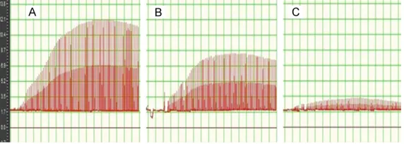
Diaphragmatic biopsy in each group
Diaphragmatic structure in the control group
was normal (Figure 1A). In the diaphragm in low tidal volume group, mild edema and a small
amount of inflammatory cell infiltration were
observed (Figure 1B). In the diaphragm in high tidal volume group, obvious edema and some
inflammatory cell infiltration were observed
(Figure 1C).
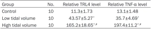
TLR4 and TNF-α expression in the diaphragm
in each group
We performed immunohistochemically staining to detect TLR4 and TNF-α expression in dia
-phragm tissues. The results showed that TLR4 expression was very weak in control group,
Similarly, TNF-α expression was very weak in
control group, strong in low tidal volume group, and very strong in high tidal volume group
(Figure 3). Quantitative analysis showed that
both TLR4 and TNF-α expression levels were significantly higher in low tidal volume group
and high tidal volume group than in control
group. Furthermore, both TLR4 and TNF-α
expression levels in the diaphragm in high tidal
volume group were significantly higher than in
low tidal volume group control (Table 1). In
addition, TLR4 expression level was positively correlated with TNF-α expression level, with a correlation coefficient of 0.904.
ty in rats decreased significantly, especially in rats with high tidal volume ventilation. TNF-α
expression in diaphragm in high tidal volume
group increased significantly, and we observed diaphragm edema and inflammatory cell infil
-tration, which indicated that inflammatory
re-sponse can play an important role in
ventilator-induced diaphragm dysfunction.
Toll-like receptors are frequently expressed in the surface of innate immune cells where they recognize pathogens to activate innate immune system to produce pro-inflammatory cytokines [10]. Toll-like receptors are distributed in vari
-Table 1. TRL4 and TNF-α expression in diaphragm in each group
Group No. Relative TRL4 level Relative TNF-α level
Control 10 11.3±1.73 13.1±1.48
Low tidal volume 10 43.57±5.27* 35.7±4.69*
High tidal volume 10 165.2±18.65*,# 197.4±11.2*,#
[image:3.612.90.376.73.142.2]*P<0.05 compared to control group, #P<0.05 compared to low tidal volume group.
Figure 1. Histopathology of diaphragm in each group. A. Control group. B.
Low tidal volume group. C. High tidal volume group. Shown were
[image:3.612.89.377.203.275.2]representa-tive images of HE staining. Scale bar: 100 μm.
Figure 2. TLR4 expression in the diaphragm in each group. A. Control group. B. Low tidal volume group. C. High tidal volume group. Scale bar: 100 μm.
Figure 3. TNF-α expression in the diaphragm in each group. A. Control group. B. Low tidal volume group. C. High tidal volume group. Scale bar: 100 μm.
Oxidative stress parameters and diaphragmatic contractil-ity in each group
As shown in Table 2, MDA con -tent in the diaphragm in low tidal volume group and high
tidal volume group was signifi -cantly higher than in control
group, and MDA content was
higher in high tidal volume group than in low tidal volume
group. SOD content in the dia -phragm and dia-phragm con-traction in low tidal volume group and high tidal volume
group decreased significantly
compared to the control gr-
oup, and SOD content and dia -phragm contraction were
sig-nificantly lower in high tidal
volume group than in low tidal volume group (Figure 4).
Discussion
Mechanical ventilation is an important method for the tre-atment of patients with seve-re seve-respiratory failuseve-re [7].
How-ever, 20% to 50% patients tre- ated with mechanical
ventila-tion have difficulty to get rid of ventilator, and recent data
suggest that ventilator-indu-
ced diaphragm dysfunction
may be an important reason [8, 9]. In this study, we showed
[image:3.612.90.377.325.394.2] [image:3.612.89.376.457.512.2]contractili-ous organs and play important role in recog-
nizing different pathogens [11]. Lipopolysac-charide (LPS) from bacteria is an important structure recognized by TLR4 and it induces the activation of TLR4 signal pathway [12]. In acute lung injury caused by bacterial infection, TLR4 initiates early inflammation and contrib
-utes to the occurrence and development of
acute lung injury [13].
In this study we showed that TLR4 expression in diaphragm of high tidal volume group was significantly higher than that of control group
and low tidal volume group, and there was a
positive correlation between TRL4 and TNF-α expression. The mechanism may be that the binding of TLR4 and ligand promotes the acti
-vation of NF-kB, which then activates the tran
-scription of inflammatory cytokines such as TNF-α and IL-6 [14, 15]. These inflammatory cytokines may further promote the release of TLR4, thus forming a vicious cycle for progres
-sive inflammation [16]. Furthermore, we found that MDA content increased significantly while SOD activity decreased significantly in high
tidal volume group, indicating oxidative stress in diaphragm. And it was reported that active
oxygen radicals can activate TLR4 signal to mediate inflammatory responses [17]. There-fore, we speculate that oxidative stress can lead to the activation of TLR4 mediated inflam -mation, thereby aggravating diaphragm injury.
Family Planning Committee of Hubei Province (No. WJ2015HB042).
Disclosure of conflict of interest
None.
Address correspondence to: Ke Hu, Division of Respiratory Disease, Renmin Hospital of Wuhan University, 99 Zhangzhidong Road, Wuhan 4300-60, Hubei Province, P. R. C. E-mail: huke-rmhospi [email protected]
References
[1] Kallet RH. Patient-ventilator interaction during
acute lung injury, and the role of spontaneous breathing: part 1: respiratory muscle function
during critical illness. Respir Care 2011; 56: 181-189.
[2] Jaber S, Petrof BJ, Jung B, Chanques G, Berthet JP, Rabuel C, Bouyabrine H, Courouble P, Koechlin-Ramonatxo C, Sebbane M, Similows
-ki T, Scheuermann V, Mebazaa A, Capdevila X, Mornet D, Mercier J, Lacampagne A, Philips A, Matecki S. Rapidly progressive diaphragmatic weakness and injury during mechanical venti
-lation in humans. Am J Respir Crit Care Med 2011; 183: 364-371.
[3] Jiang Q, Yi M, Guo Q, Wang C, Wang H, Meng S, Liu C, Fu Y, Ji H, Chen T. Protective effects of
polydatin on lipopolysaccharide-induced acute
[image:4.612.90.376.96.160.2]lung injury through TLR4-MyD88-NF-kappaB
Table 2. Oxidative stress in diaphragm and diaphragmatic
contrac-tility in each group
Group No. (nmol/mgprot)MDA value SOD activity (U/mgprot) (N/cmcontractility 2)*10-3
Control 10 6.1±0.72 114.7±8.52 995.26±71.43
Low tidal volume 10 8.4±0.67* 95.3±7.34* 708.34±59.98*
High tidal volume 10 10.6±0.83*,# 54.8±8.75*,# 214.73±38.83*,#
*P<0.05 compared to control group, #P<0.05 compared to low tidal volume group.
Figure 4. Diaphragmatic contractility in each group. A. Control group. B. Low
tidal volume group. C. High tidal volume group.
TLR4 signal may play an im-portant role in the initiation of
ventilator-induced diaphragm
dysfunction and subsequent maintenance of inflammation. In summary, we found that
during high tidal volume
venti-lation TRL4 expression in the diaphragm was significantly
increased, accompanied by pathological and biochemical
changes in the diaphragm. TL-R4 signal may play an impor -tant role in the initiation and
subsequent maintenance of
ventilator-induced diaphragm
dysfunction inflammation.
Acknowledgements
[image:4.612.91.377.196.298.2]pathway. Int Immunopharmacol 2015; 29: 370-376.
[4] Hu R, Xu H, Jiang H, Zhang Y, Sun Y. The role of TLR4 in the pathogenesis of indirect acute lung injury. Front Biosci (Landmark Ed) 2013; 18: 1244-1255.
[5] Chen CM,Cheng KC,Li CF, Zhang H. The pro
-tective effects of glutamine in a rat model of ventilator-induced lung injury. J Thorac Dis 2014; 6: 1704-1713.
[6] Danjo W, Fujimura N, Ujike Y. Effect of pentoxi
-fylline on diaphragmatic contractility in septic rats. Acta Med Okayama 2008; 62: 101-107.
[7] Antonelli M, Azoulay E, Bonten M, Chastre J, Citerio G, Conti G, De Backer D, Lemaire F, Ger
-lach H, Groeneveld J, Hedenstierna G, Macrae D, Mancebo J, Maggiore SM, Mebazaa A, Met
-nitz P, Pugin J, Wernerman J, Zhang H. Year in
review in intensive care medicine, 2008: II.
Experimental, acute respiratory failure and ARDS, mechanical ventilation and endotrache
-al intubation. Intensive Care Med 2009; 35:
215-231.
[8] Wilson JG, Matthay MA. Mechanical ventilation in acute hypoxemic respiratory failure: a review of new strategies for the practicing hospitalist. J Hosp Med 2014; 9: 469-475.
[9] Zhang H, Liu P, Zhao Y. TLR4 contributes to me -chanical ventilation induced lung injury in the
rabbits. Int J Clin Exp Pathol 2017; 10: 4700-4704; [Epub ahead of print].
[10] Mukherjee S, Karmakar S, Babu SP. TLR2 and TLR4 mediated host immune responses in ma
-jor infectious diseases: a review. Braz J Infect Dis 2016; 20: 193-204.
[11] Aalaei-Andabili SH, Rezaei N. Toll like receptor (TLR)-induced differential expression of mi
-croRNAs (MiRs) promotes proper immune re
-sponse against infections: a systematic re-view. J Infect 2013; 67: 251-264.
[12] Rodriguez-Gonzalez R, Baluja A, Veiras Del Río S, Rodríguez A, Rodríguez J, Taboada M, Brea D, Álvarez J. Effects of sevoflurane postcondi
-tioning on cell death, inflammation and TLR
expression in human endothelial cells expos-
ed to LPS. J Transl Med 2013; 11: 87.
[13] Sodhi CP, Jia H, Yamaguchi Y, Lu P, Good M, Egan C, Ozolek J, Zhu X, Billiar TR, Hackam DJ. Intestinal epithelial TLR-4 activation is requir-ed for the development of acute lung injury af
-ter trauma/hemorrhagic shock via the release of HMGB1 from the gut. J Immunol 2015; 194: 4931-4939.
[14] Shon WJ, Lee YK, Shin JH, Choi EY, Shin DM. Severity of DSS-induced colitis is reduced in Ido1-deficient mice with down-regulation of TLR-MyD88-NF-kB transcriptional networks.
Sci Rep 2015; 5: 17305.
[15] Hochdorfer T, Kuhny M, Zorn CN, Hendriks RW, Vanhaesebroeck B, Bohnacker T, Krystal G, Huber M. Activation of the PI3K pathway in
-creases TLR-induced TNF-alpha and IL-6 but
reduces IL-1beta production in mast cells. Cell Signal 2011; 23: 866-875.
[16] Cohen P. The TLR and IL-1 signalling network at a glance. J Cell Sci 2014; 127: 2383-2390.
[17] Lavieri R, Piccioli P, Carta S, Delfino L, Castel
-lani P, Rubartelli A. TLR costimulation causes oxidative stress with unbalance of proinflam
-matory and anti-inflam-matory cytokine produc