Original Article
Predictive and prognostic values of BubR1 and
synuclein-gamma expression in breast cancer
Yalcin Cirak1, Yavuz Furuncuoglu2, Ozlem Yapicier3, Suleyman Alici1, Andac Argon1
1Department of Medical Onkology, Bahcesehir University School of Medicine, Goztepe Medical Park Hospital, Istanbul, Turkey; 2Department of Internal Medicine, Bahcesehir University School of Medicine, Goztepe Medical Park Hospital, Istanbul, Turkey; 3Department of Pathology, Bahcesehir University School of Medicine, Goztepe Medical Park Hospital, Istanbul, Turkey
Received February 24, 2015; Accepted April 14, 2015; Epub May 1, 2015; Published May 15, 2015
Abstract: The aim of this study is to determine the expression level of spindle assembly checkpoint (SAC) proteins-BubR1 and synuclein-gamma (SNCG) in human breast cancer tissues and to test whether there is a relationship between their expression levels and clinicopathologic parameters including respons to taxanes, tumor grade,
estro-gen receptor (ER) pozitivity, HER2 status, and overall survival (OS). We analyzed retrospectively paraffin-embedded
tissue sections from 55 breast cancer patients whose clinical outcomes had been tracked after taxane treatment in
neoadjuvan and metastatic setting. The expression status of BubR1 and SNCG was defined by immunohistochemis -try (IHC) using the anti-BubR1 and anti-SNCG antibody. The BubR1 and SNCG was overexpressed in 38% and 62%
of the study group, respectively. There was borderline significant correlation between low BubR1 expression and in -creased taxane sensitivity (P=0.05). In contrast, high SNCG expression was significantly associated with decreased
taxane sensitivity (P=0.01). There was no association between the clinicopathologic parameters including histologic
grade, ER positivity and HER2 status and the level of these proteins. However, triple negative tumors showed signifi -cantly more high BubR1 expression than those other molecular subtypes (P=0.04). Kaplan-Meier survival analysis
failed to show a significant correlation between expression levels of BubR1 and SNCG and overall survival although
patients with low levels of both proteins had a marginally longer survival time compared to those with high levels. In summary, our data suggest that both BubR1 and SNCG may be promising predictive marker rather than prognostic marker in patients with breast cancer.
Keywords: BubR1, synuclein gamma, predictive marker, prognostic marker, taxane, breast cancer
Introduction
Breast cancer is second leading cause of death in women from cancer [1]. In both adjuvant and metastatic setting the best affective chemothe-ropotic drugs are taxanes and antracyclines [2]. Due to the cumulative toxixity of antracyclines, the importance of taxenes has increased. However, one part of patient with breast cancer do not benefit from taxanes. The response rate to taxanes in metastatic tumors ranges from 30% to 50% [2]. For this reason, prediction of response or resistance to taxanes in breast cancer may be helpful to select those patients more likely to derive a clinical benefit.
The taxanes, paclitaxel and docetaxel are microtubule-stabilizing agents. They bind to
β-tubulin and result in kinetic abnormalities with elevated microtubule formation in the dynamics of microtubules by increasing their polymerization and inhibiting their depolymer-ization. In metaphasa, defective sipinle forma-tion induced by taxanes activates the mithotic checkpoint and causes cell cycle arrest, result-ing in apoptosis [3]. Various molecules includ-ing microtubule-associated proteins (MAPs), spindle assembly checkpoint (SAC) proteins, β Tubulin, HER2, p53, BRCA1, CYP3A4, estrogen receptor (ER), BCL2, P-glycoprotein and Ki-67 have been examined for their ability to predict response to taxanes [3].
BubR1 and synuclein-gamma expression in breast cancer
mitosis. In this regulation system, various SAC proteins play role including mitotic arrest defi-cient proteins (MAD 1-3), budding uninhibited by benzimidazoles proteins (BUB1-3), Bub1-related protein kinase (BubR1) and monopolar spindle 1 (Mps1) [3]. BubR1 is one of the sev-eral key proteins needed for correct SAC func-tion suck as Mad2, centromere-associated protein-E (CENP-E), cell-division cycle protein 20 (Cdc20). It occur the mitotic checkpoint complex (MCC) together with Mad2,Bub3 and Cdc20, which delays anaphase onset by inhibit-ing the Anaphase Promotinhibit-ing Complex/cyclo-some (APC/C) until all kinetochores attached to microtubules [4]. This regulatory system pre-vents aneuploidy by ensuring the segregation of only one copy of each pair of duplicated sis-ter chromatids [3]. Moreover,BubR1 phosphor-ylates and stabilises p53 during mitosis. Therefore, BubR1 is implicated in both spindle assembly and DNA damage checkpoints [5]. Synuclein gamma are highly expressed in neu-ronal cells, Synuclein family consists of alpha-, beta- and gamma-synuclein. Especially, the role of alpha synuclein in neurodegenerative diseases has been well documented [6]. In addition there are evidences suggesting that SNCG increases metastasis and promotes genetic instability. In many different malignant diseases, its abnormal expression has been demostrated [7-10]. Generally, it rarely expressed in tumor-matched nonneoplastic adjacent tissues [11]. At the celluler level, had been demostrated that SNCG prevented the formation of MCC by inhibiting BubR1 activity, and resulted in an insufficiency of BubR1-related SAC function [12]. Considering that anti-mitotic drugs target microtubules, correct functioning of the BubR1 and SNCG would seem crucial for an appropriate drug response. Previous studies have reported that tumor expression of BubR1 and SNCG can alter the sensitivity to Anthracycline-based agents [13], and neoadjuvant chemotherapy [14] in breast cancer, respectively. Their expression levels may be associated with survival [15-20]. In our study, We identified retrospectively the expres-sion levels of BubR1 and SNCG proteins in par-affin-embedded breast cancer tissue samples obtained from patients with breast cancer and evaluated the role of BubR1 and SNCG expres-sions in predicting treatment response to tax-anes, which has not been reported in the
cur-rent medical literature. Survival and correlation with clinicophathologic parameters were also analyzed to determine the prognostic values of BubR1 and SNCG in these patients.
Materials and methods
Patient and tissue sapmles
Cases were selected retrospectively from records of Goztepe Medical Park Oncology Hospital between the years 2008-2014. The study has been approved by the Goztepe Medical Park Hospital Ethics Committee under the title ‘Retrospective analysis of tissue sam-ples by immunohistochemistry (IHC)’. Eligibility criteria were as follows: (a) Responder and non responder patients who had taken AC (doxoru-bicin/cyclophosphamide) followed by taxanes in neoadjuvant setting for locale advanced bre-ast cancer. (b) Non-responder patients who had taken combination of taxanes with cyclophos-phamid and antracyclin or AC followed by taxa-nes in neoadjuvant setting. (c) Responder and non responder patients who had taken single agent taxane in metastatic setting (to exlude the effect of other chemotherapeutic on response as a bias source in patient selection in responder patients). (d) Non responder patients who had taken the combination of tax-anes with any chemotherapeutic agent in meta-static setting. (e) Patients who had new biopsy for metastatic disease, if long time had elapsed from initial diagnosis to metstatic diseas. The taxanes had been administred as either weekly paklitaxel (80 mg/m2) or docetaxel every 3 weeks (75 mg/m2). A total of 55 patients who met egilibility criteria were stratified according to tretament responses to taxane into two groups as responders and non- responders. The responder group (patients with complet response, partial response and stable disease) and non responder group (progresive disease ) were defined according to the Respons Evaluation Criteria in Solid Tumor (RECIST).
Immunohistochemical evaluation
repre-sentative slide of each case was selected for IHC studies. Sections 4-5 μm thick were placed on electrostatic-charged slides (X-traTM, Surgipath Medical Industries, Richmond, Illinois, USA) and dried at 60°C for at least two hours and stained with mouse monoclonal BubR1 antibody (GeneTex Clone 5D9, CA USA, overnight incubation at a dilution of 1:100) and
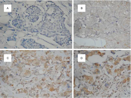
[image:3.612.90.523.71.396.2]gamma synuclein antibody (GeneTex Clone EP 1539Y 2 hours incubation at a dilution of 1:400),IHC staining was carried out according to standard streptavidin-biotin-peroxidase me- thod. A section of tonsil and hippocampal tis-sue were used as a positive control for BubR1 and SNCG, respectively. Sections incubated without the primary antibody served as nega-Figure 1. Stained by immunohistochemistry using the primary antibody againts BubR1 (×200); A. No staining; B. Weak (1+); C. Moderate (2+); D. Strong (3+) expression.
[image:3.612.90.523.447.584.2]BubR1 and synuclein-gamma expression in breast cancer
tive controls for both antibody. Expression of both proteins was mainly cytoplasmic, although some positive nuclei were also seen. For BubR1, cytoplasmic staining in tumor cells was scored on the following three-point scale: IHC score 0, no staining (Figure 1A); 1+, weak (Figure 1B); 2+, moderate (Figure 1C); 3+, strong expression, comparable to that of cells in the germinal centers of normal tonsil (Figure 1D). Tumors with 0 and 1+ staining intensity were considered BubR1 negative, and tumors with 2+ and 3+ staining were considered BubR1 positive [21]. For SNCG, positive cases were defined by the presence of any intensity of intracellular staining with red/brown color in breast cancer cells, since it is not expressed in
normal or benign breast tissues (Figure 2B). Negative cases were defined by the absence of specific intracellular staining as seen in nega-tive controls (Figure 2A).
Statistical analysis
Each clinicopathological variable was com-pared between the BubR1 and SNCG-positive and -negative expression groups, and evaluat-ed with χ2 test. Overall survival (OS) time was calculated using the Kaplan-Meier method as the duration from the date of diagnosis to the date of death or last control. Diferences in sur-vival among the groups were compared using the log-rank test. P < 0.05 (two-tailed) was
con-sidered statistically significant. Statistical anal -ysis was performed using SPSS, version 15.
Results
The BubR1 and SNCG expression in breast cancer and their relationship to the clinic ef-fectiveness of taxanes
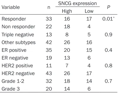
21 tumors (38%) and 34 tumors (62%) showed positive expression for BubR1 and SNCG, respectively. 40% of all patients (22/55) were resistant to taxanes. 76% of BubR1 positive tumors (16/21) had taxane responsive disease, compared with 50% of BubR1 negative tumors (17/34) (P=0.05). Despite statistically border-line correlation, tumors with high BubR1 expression were more sensitive the taxane than those low expression. In contrast, 53% of SNCG positive (18/34) tumors was resistant to taxane, compared with 19% of SNCG negative tomors (4/21) (P=0.01). There was significan differance between the SNCG negative and positive group with respect to clinic effective-ness of taxanes. The association between the response to taxane and the expressions of these proteins is summarized in Tables 1 and 2.
Association of BubR1 and SNCG expression with clinical parameters
[image:4.612.90.290.111.267.2]Three tumors whose diagnosis are available only from metastatic sites were not evaluated for histologic grade. There was no significan association between clinical parameters such as tumor grade, ER positivity, HER2 status and expression levels of BubR1 and SNCG proteins. However, 61% of triple negative tumors (8/13) Table 1. Associations between
clinicopatho-logical variables and the expressions of BubR1
Variable n BubR1 expression P
High Low
Responder 33 16 17 0.05*
Non responder 22 5 17
Triple negative 13 8 5 0.04**
Other subtypes 42 13 29
ER positive 35 12 23 0.3 ER negative 19 9 10
HER2 positive 11 3 8 0.4 HER2 negative 43 18 25
Grade 1-2 32 20 12 0.7
Grade 3 20 12 8
*P=0.05; **P < 0.05.
Table 2. Associations between clinicopatho-logical variables and the expressions of SNCG
Variable n SNCG expression P
High Low
Responder 33 16 17 0.01*
Non responder 22 18 4
Triple negative 13 8 5 0.9 Other subtypes 42 26 16
ER positive 35 20 15 0.4 ER negative 19 13 6
HER2 positive 11 7 4 0.8 HER2 negative 43 26 17
Grade 1-2 32 18 14 0.7
Grade 3 20 14 6
[image:4.612.90.289.333.491.2]were BubR1 positive, compared with 31% of other molecular subtypes (13/42) (P=0.04). There was significant association between tri-ple negative tumors and level of BubR1 expres-sion (Tables 1 and 2).
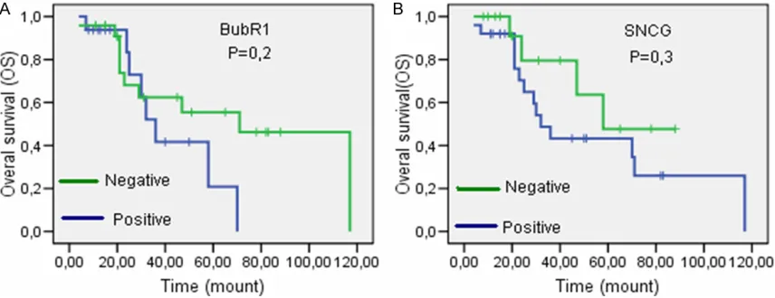
Association of BubR1 expresssion with survival After 15 patients with short term follow up tak-ing neoadjuvant treatment were exluded, the remaining 40 metastatic patients were evalu-ated for OS. 18 of 40 patients died in the dura-tionfrom the date of diagnosis to last control. The longest survival 117 mounts. Median sur-vival time in patient with positive and negative BubR1 expression was 36 and 71 mounts, respectively (Log-rank test, P=0.2, Kaplan Meier curve; Figure 3A). Median survival time in patient with positive and negative SNCG expression was 32 and 58 mounts, respective-ly (Log-rank test, P=0.3, Kaplan Meier curve; Figure 3B). There was no significant associa-tion between the BubR1 and SNCG-positive and -negative groups with respect to OS. Discussion
The association of SAC proteins with response to taxanes and clinical significance of their overexpression have been recently investigat-ed [22-24]. In this study, we focusinvestigat-ed on wheth-er BubR1 and SNCG expressions in breast can-cer tissues were predictive marker of taxane sensitivity. The contradictory results had been reported for BubR1 in various studies per-formed in cancer cell lines. While two study reported that low level expression of BubR1
was associated with increased sensitivity to taxanes [22, 25], conversely, other two study reported that low BubR1 expression was asso-ciated with decreased sensitivity to taxanes [24, 26]. However, so far there have been no clinical study performed to confirm these pre-clinical observations except for our study in prostate cancer. Previously, we reported that high BubR1 expression was not associated with respons to docataxel, but significantly associated with OS in patients with prostate cancer [19]. Here we showed that tumors with high BubR1 expression were more sensitive to taxane than those low expression (borderline significance P=0.05).
BubR1 was reported to be upregulated in vari-ous cancers suck as lung, breast, colon, esoph-agus, stomach, kidney, bladder, ovary, thyroid and liver [27-34]. In a number of these studies, its overexpression has been related to chromo-somal instability, DNA aneuploidy, more advanced pathologic stage and a higher histo-logic grade. In our study, 38% of patients with breast cancer had BubR1 positive tumors. This proportion was slighty greater compared with those of previous two reports in breast cancer (25% and 32%) [16, 21]. There were no correla-tion with tumor grade, ER positivity and HER2 status. Hovever, triple negative tumors had sig-nificantly more high BubR1 expression than those other molecular subtypes.
[image:5.612.92.520.72.237.2]BubR1 and synuclein-gamma expression in breast cancer
selectively inhibits proliferation of basal breast cancer cell lines [35]. BubR1 kinase activity is essential the mitotic checkpoint and is directly stimulated by CENP-E binding to it [36]. Considering that the interaction between CENP-E and BubR1 is curicial for correct mitotic checkpoint fonction, our findings have support-ed that like CENP-E, BubR1 may be also likeley to a important target for triple negative breast cancer. Maciejczyk et al. analyzed the expres-sion of BubR1 in 98 stage II breast cancer patients with a median follow-up of 15 years. They reported that Elevated BubR1 expression was associated with precense of metastasis to lympf node and poor survival in early stage breast cancer patients but without any assso-ciation of BubR1 with the histological tumor grade, estrogen ve progesterone receptor posi-tivity, HER2 status and tumor stage [18]. Du et al reported that high BubR1 expression was associated with high Ki67 labeling index and high histological grade [37].
There are several contradictory results between high BubR1 expression and survival in litera-ture. Ovarian and breast cancer studies showed that patients with high BubR1 expression had significantly shorter recurrence-free survival (RFS) rates [17, 18]. Conversely, patients with low BubR1 expression showed shorter survival in colorectal cancer [20], and oral squamose cell cancer study showed no correlation between BubR1 and survival [21]. We found that despit no statisticaly significant corelation, patients with low BubR1 expression showed longer overall survival period, compared with those of high BubR1 expression. Our results related to triple negative tumors and survival may reflect some limitation due to the small size of the semple and presence of censored data.
SNCG was first named Breast cancer-specific
gene 1 (BCSG1) as it is highly expressed in advanced breast cancer. Then, its overexpres-sion was showed in many other solid tumors including, gastric, lung, pancreatic, colon and ovarian cancer [38-40]. At the cellular level, SNCG increases metastasis and promotes genetic instability. At the molecular level, SNCG functions like a heat-shock protein (Hsp)-based multiprotein chaperone complex for stimulation of ER signalling. It increases the ligand-binding capacity of ER [41]. Moreover, SNCG protects
the function of HER2 by preventing disruption of Hsp90 [42]. Wu et al confirmed the preclini-cal observation mentioned above by showing that patients whose tumors expressed SNCG had a significantly shorter DFS and a high prob-ability of death when compared with those whose tumors did not express SNCG [15]. Moreover, they reported that there was no sig-nificant correlation between the BubR1 expres-sion and clinicopathologic parameters includ-ing ER, PR and HER2 status except for lenf nodu involvement and stage. In contrast, Martin et al reported that high SNCG expres-sion was associated with tumour grade but not with survival of patients with breast cancer [11].
At the cellular level, a number of study showed that overexpressed SNCG resulted in resis-tance to microtubilising drugs [12, 43-45]. However, there is no clinic study performed to confirm these preclinic observations. In our study we confirmed the findings at the cellular level by showing the strong association between the high SNCG expression and the resistanse to taxane (P=0.01). But we did not find any association with clinicopathologic paremeters includin tumor grade, ER, HER2, molecular sub-types and survival. Our results related to clinic parameters and survival may reflect some limi-tations related to the small size of the sample, heterogeneity in the distribution of cases for ER, PR, HER2 and molecular subtypes, and to the presence of censored data.
Disclosure of conflict of interest
None.
Address correspondance to: Dr. Yalcin Cirak,De- partment of Medical Onkology, Goztepe Medical Park Hospital, Bahcesehir University School of
Medicine, Kadıköy, Istanbul 34746, Turkey. Tel: +90
532-1597650; Fax: +90 216-4684567; E-mail: [email protected]
References
[1] Autier P, Boniol M, La Vecchia C, Vatten L, Gavin A, Héry C and Heanue M. Disparities in breast cancer mortality trends between 30 European countries: retrospective trend analy-sis of WHO mortality database. BMJ 2010; 341: 3620.
[2] Paridaens R, Biganzoli R, Bruning P, Klijn JG, Gamucci T, Houston S, Coleman R, Schachter J, Van Vreckem A, Sylvester R, Awada A, Wildiers J and Piccart M. Paclitaxel versus
doxorubicin as first-line single-agent chemo -therapy for metastatic breast cancer: a European Organization for Research and Treatment of Cancer Randomized Study with cross-over. J Clin Oncol 2000; 18: 724-33. [3] McGrogan BT, Gilmartin B, Carney DN and
McCann A. Taxanes, microtubules and chemo resistant breast cancer. Biochim Biophys Acta 2008; 1785: 96-132.
[4] Rieder C, Cole R, Khodjakov A and Sluder G. The checkpoint delaying anaphase in response to chromosome monoorientation is mediated by an inhibitory signal produced by unattached kinetochores. J Cell Biol 1995; 130: 941-948. [5] Ha GH, Baek KH, Kim HS, Jeong SJ, Kim CM,
McKeon F and Lee CW. p53 Activation in Response to Mitotic Spindle Damage Requires Signaling via BubR1-Mediated Phosphorylation. Cancer Res 2007; 67: 7155-64.
[6] Spillantini MG, Schmidt ML, Lee VM, Trojanowski JQ, Jakes R and Goedert M. Alpha-synuclein in Lewy bodies. Nature 1997; 388: 839-40.
[7] Guo J, Shou C, Meng L, Jiang B, Dong B, Yao L, Xie Y, Zhang J, Chen Y, Budman DR and Shi YE. Neuronal protein synuclein gamma predicts poor clinical outcome in breast cancer. Int J Cancer 2007; 121: 1296-305.
[8] Liu H, Liu W, Wu Y, Zhou Y, Xue R, Luo C, Wang L, Zhao W, Jiang JD and Liu J. Loss of epigene-tic control of synuclein-gamma gene as a mo-lecular indicator of metastasis in a wide range of human cancers. Cancer Res 2005; 65: 7635-43.
[9] Hibi T, Mori T, Fukuma M, Yamazaki K, Hashiguchi A, Yamada T, Tanabe M, Aiura K, Kawakami T, Ogiwara A, Kosuge T, Kitajima M, Kitagawa Y and Sakamoto M. Synuclein-gamma is closely involved in perineural inva-sion and distant metastasis in mouse models and is a novel prognostic factor in pancreatic cancer. Clin Cancer Res 2009; 15: 2864-71. [10] Fung KM, Rorke LB, Giasson B, Lee VM and
Trojanowski JQ. Expression of alpha-, beta-, and gamma-synuclein in glial tumors and me-dulloblastomas. Acta Neuropathol 2003; 106: 167-75.
[11] Martin TA, Gomez K, Watkins G, Douglas-Jones A, Mansel RE and Jiang WG. Expression of
breast cancer specific gene-1 (BCSG-1/gam -ma-synuclein) is associated with tumour grade but not with clinical outcome of patients with breast cancer. Oncol Rep 2006; 16: 207-12. [12] Miao S, Wu K and Zhang B. Synuclein γ com
-promises spindle assembly checkpoint and renders resistance to antimicrotubule drugs. Mol Cancer Ther 2014; 13: 699-713.
[13] Munro A, Cameron D, Thomas J, Twelves C and Bartlett J. BUBR1 and MAD2: novel markers
for predicting benefit from adjuvant anthracy -clines? abstracts: thirty-second annual ctrc-aacr san antonio breast cancer symposium 2009; San Antonio. J Cancer Res 2009; 69: 2124.
[14] Wan F, Dong L, Zhang F, Wang Y, Chen F, Ni S, Chen Y and Long J.Clinical study of the
rela-tionship between γ-synuclein and the response
of neoadjuvant chemotherapy in breast can-cer. J Int Med Res 2013; 41: 743-53.
[15] Wu K, Quan Z, Weng Z, Li F, Zhang Y, Yao X, Chen Y, Budman D, Goldberg ID and Shi YE. Expression of neuronal protein synuclein gam-ma gene as a novel gam-marker for breast cancer prognosis. Breast Cancer Res Treat 2007; 101: 259-67.
[16] Guo J, Shou C, Meng L Jiang B, Dong B, Yao L, Xie Y, Zhang J, Chen Y, Budman DR and Shi YE. Neuronal protein synucleinc γ predicts poor clinical outcome in breast cancer. Int J Cancer 2007; 121: 1296-1305.
[17] Lee YK, Choi E, Kim MA, Park PG, Park NH and Lee H. BubR1 as a prognostic marker for recur-rence-free survival rates in epithelial ovarian cancers. Br J Cancer 2009; 101: 504-10. [18] Maciejczyk A, Szelachowska J and Czapiga B.
Elevated bubr1 expression is associated with poor survival in early breast cancer patients: 15-year follow-up analysis. J Histochem Cytochem 2013; 61: 330-9.
re-BubR1 and synuclein-gamma expression in breast cancer
lationship to the gleason score. Med Oncol 2013; 30: 526-7.
[20] Shichiri M, Yoshinaga K, Hisatomi H, Sugihara K and Hirata Y.Genetic and epigenetic inactiva-tion of mitotic checkpoint genes hBUB1 and hBUBR1 and their relationship to survival. Cancer Res 2002; 62: 13-7.
[21] Rizzardi C, Torelli L, Barresi E, Schneider M, Canzonieri V, Biasotto M, Di Lenarda R and Melato M. Bubr1 expression in oral squamous cell carcinoma and its relationship to tumor stage and survival. Head Neck 2011; 33: 727-33.
[22] Sudo T, Nitta M, Saya H and Ueno NT. Dependence of paclitaxel sensitivity on a func-tional spindle assembly checkpoint. Cancer Res 2004; 64: 2502-8.
[23] Fang Y, Liu T, Wang X, Yang YM, Deng H, Kunicki J, Traganos F, Darzynkiewicz Z, Lu L and Dai W. BubR1 is involved in regulation of DNA dam-age responses. Oncogene 2006; 25: 3598-605.
[24] Lee EA, Keutmann MK, Dowling ML, Harris E, Chan G and Kao GD. Inactivation of the mitotic
checkpoint as a determinant of the efficacy of
microtubule-targeted drugs in killing human cancer cells. Mol Cancer Ther 2004; 3: 661-9. [25] Fu Y, Ye D, Chen H, Lu W, Ye F and Xie X.
Weakened spindle checkpoint with reduced BubR1 expression in paclitaxel-resistant ovari-an carcinoma cell line SKOV3-TR30.Gynecol Oncol 2007; 105: 66-73.
[26] Tanaka K, Mohri Y, Ohi M and Yokoe T. Mitotic checkpoint genes, hsMAD2 and BubR1, in oe-sophageal squamous cancer cells and their
associationwith 5-fluorouracil and
cisplatin-based radiochemotherapy. Clin Oncol (R Coll Radiol) 2008; 20: 639-46.
[27] Seike M, Gemma A, Hosoya Y, Hosomi Y, Okano T, Kurimoto F, Uematsu K, Takenaka K, Yoshimura A, Shibuya M, Ui-Tei K and Kudoh S.
The promoter region of the human BUBR1 gene and its expression analysis in lung can-cer. Lung Cancer 2002; 38: 229-234.
[28] Yuan B, Xu Y, Woo JH, Wang Y, Bae YK, Yoon DS, Wersto RP, Tully E, Wilsbach K and Gabrielson E. Increased expression of mitotic checkpoint genes in breast cancer cells with chromosomal instability. Clin Cancer Res 2006; 12: 405-410.
[29] Burum-Auensen E, De Angelis PM, Schjølberg AR, Røislien J, Mjaland O and Clausen OP. Reduced level of the spindle checkpoint pro-tein BUB1B associated with aneuploidy in colorectal cancers. Cell Prolif 2008; 41: 645-659.
[30] Ando K, Kakeji Y, Kitao H, Iimori M, Zhao Y, Yoshida R, Oki E, Yoshinaga K, Matumoto T, Morita M, Sakaguchi Y and Maehara Y. High
expression of BUBR1 is one of the factors for inducing DNA aneuploidy and progression in gastric cancer. Cancer Sci 2010; 101: 639-645.
[31] Pinto M, Vieira J, Ribeiro FR, Soares MJ, Henrique R, Oliveira J, Jerónimo C and Teixeira MR. Overexpression of the mitotic checkpoint genes BUB1 and BUBR1 is associated with ge-nomic complexity in clear cell kidney carcino-mas. Cell Oncol 2008; 30: 389-395.
[32] Yamamoto Y, Matsuyama H, Chochi Y, Okuda M, Kawauchi S, Inoue R, Furuya T, Oga A, Naito K and Sasaki K. Overexpression of BUBR1 is associated with chromosomal instability in bladder cancer. Cancer Genet Cytogenet 2007; 174: 42-47.
[33] Wada N, Yoshida A, Miyagi Y, Yamamoto T, Nakayama H, Suganuma N, Matsuzu K, Masudo K, Hirakawa S, Rino Y, Masuda M and Imada T. Overexpression of the mitotic spindle assembly checkpoint genes hBUB1, hBUBR1 andhMAD2 in thyroid carcinomas with aggres-sive nature. Anticancer Res 2008; 28: 139-144.
[34] Liu AW, Cai J, Zhao XL, Xu AM, Fu HQ, Nian H and Zhang SH. The clinicopathological signifi-cance of BUBR1 overexpression in hepatocel-lular carcinoma. J Clin Pathol 2009; 62: 1003-1008.
[35] Kung PP, Martinez R and Zhu Z. Chemogenetic evaluation of the mitotic kinesin CENP-E re-veals a critical role in triple-negative breast cancer. Mol Cancer Ther 2014; 13: 2104-15. [36] Guo Y, Kim C, Ahmad S, Zhang J and Mao Y.
CENP-E–dependent BubR1 autophosphoryla-tion enhances chromosome alignment and the mitotic checkpoint J Cell Biol 2012; 198: 205-17.
[37] Du J, Du Q, Zhang Y, Sajdik C, Ruan Y, Tian XX and Fang WG. Expression of cell-cycle regula-tory proteins BUBR1, MAD2, Aurora A, cyclin A and cyclin E in invasive ductal breast carcino-mas. Histol Histopathol 2011; 26: 761-8.
[38] Bruening W, Giasson BI, Klein-Szanto AJ, Lee VM, Trojanowski JQ and Godwin AK. Synucleins are expressed in the majority of breast and ovarian carcinomas and in preneoplastic le-sions of the ovary. Cancer 2000; 88: 2154-63. [39] Liu H, Liu W, Wu Y, Zhou Y, Xue R, Luo C, Wang
L, Zhao W, Jiang JD and Liu J. Loss of epigene-tic control of synuclein-gamma gene as a mo-lecular indicator of metastasis in a wide range of human cancers. Cancer Res 2005; 65: 7635-43.
[41] Jiang Y, Liu YE, Goldberg ID and Shi YE. Gamma synuclein, a novel heat-shock protein-associat-ed chaperone, stimulates liganddependent estrogen receptor alpha signaling and mam-mary tumorigenesis. Cancer Res 2004; 64: 4539-4546.
[42] Shao Y, Wang B, Shi D, Miao S, Manivel P, Krishna R, Chen Y, Eric Shi Y. Synuclein gamma protects HER2 and renders resistance to Hsp90 disruption Mol Oncol 2014; 8: 1521-31.
[43] Cheng SX, Zhang S, Zhang H, Song DQ, Wang YP, Li YH, You XF, Wang YM, Jiang JD. Overexpression of synuclein-gamma confers resistance to antimicrotubule drugs against human hepatoma cells. Yao Xue Xue Bao 2010; 45: 724-9.
[44] Pan ZZ, Bruening W, Giasson BI, Lee VM, Godwin AK. Gamma-synuclein promotes can-cer cell survival and inhibits stress- and che-motherapy drug-induced apoptosis by modu-lating MAPK pathways. J Biol Chem 2002; 277: 35050-60.