Original Article
siRNA-mediated Loc554202 knockdown regulates the
proliferation, invasion and apoptosis of
human bladder cancer cells
Yan Liu1*, Tingting Song2*, Yuexia Song3, Shuling Yin1, Haitao Gao1, Kaolu Zhao1, Dawei Cui1, Hui Shi1, Jing Shi1
1Institute of Tangshan Gongren Hospital Affiliated to Hebei Medical University, Tangshan, Hebei, China; 2Shanghai
Putuo District Changfeng Lane Baiyu Community Health Service Center, China; 3Institute of Tangshan Renmin
Hospital Affiliated to North China University of Science and Technology, Tangshan, Hebei, China. *Co-first authors. Received October 26, 2015; Accepted December 24, 2015; Epub February 1, 2016; Published February 15, 2016
Abstract: This study is aimed to investigate the effect of small interfering RNAs (siRNAs)-mediated Loc554202 knockdown on the proliferation, invasion and apoptosis of human bladder cancer cells. The expression of long non-coding RNA (lncRNA) Loc554202 in bladder cancer and adjacent noncancerous tissue, and bladder cancer cell lines (T24, J82 and Biu87) and normal uroepithelium SV-HUC-155 cells was compared by qRT-PCR. The siRNAs tar-geting Loc554202 and negative control si-Scramble were transfected into these cells. Loc554202 expression was detected by qRT-PCR. Cell proliferation was examined by MTT assay. Cell apoptosis was detected by flow cytometry. The expression of key apoptotic proteins including caspase-3, caspase-9 and Bcl-2 was measured by Western blot analysis. Cell invasion was assessed by Matrixgel invasion assay. Loc554202 expression in tumor and bladder can-cer cells was significantly higher than that in normal tissue and SV-HUC-155cells, respectively. The highest inhibitory effect on Loc554202 expression was observed in T24 and J82 cells transfected with siRNA-1, which were used for subsequent experiments. Cell viability of T24 and J82 cells in siRNA-1 group was significantly lower compared with si-Scramble group at 72, 96 and 120 h after transfection (P<0.05). The percentage of necrotic T24 and J82 cells in siRNA group was significantly higher compared with si-Scramble group (P<0.05). The percentage of transmembrane T24 and J82 cells in siRNA-1 group was significantly decreased compared with si-Scramble group (P<0.05). Cas-pase-3 and caspase-9 expression in T24 and J82 cells transfected with siRNA-1 was significantly higher compared with si-Scramble group (P<0.05), whereas the expression of the anti-apoptotic proteins Bcl-2 was significantly lower (P<0.05). The siRNA-mediated Loc554202 knockdown significantly inhibits cell proliferation and invasion of T24 and J82 cells, and promotes their apoptosis.
Keywords: Human bladder cancer cells, lncRNA, small interference RNA, Loc554202, apoptosis
Introduction
Human bladder cancer is one of the most com-mon malignancies worldwide [1]. In the United States, it is the second most common cancer of urinary tract causing over 13,000 deaths annually [2]. In China, bladder cancer is the eighth most common malignancy with gradual-ly increasing incidence and mortality rate in recent years [3]. Bladder cancer is character-ized by a high recurrence rate, and strong cell invasion and metastasis. There are two major types of bladder cancer: low grade papillary tumors and high grade invasive tumors. The typical treatment for papillary bladder tumors is surgical removal of the tumor followed by
post-may even develop invasive and metastatic blad-der cancer [5, 6]. Invasive bladblad-der cancer is much more likely to spread, and thereby have a much worse prognosis with approximately a 5-year survival rate of 50% [7]. Therefore, inves-tigation of biological characteristics of bladder cancer cells and identification of novel molecu-lar targets are important for effective treatment and prognosis of bladder cancer.
several types of cancer including bladder can-cer [12, 13]. Recent studies have demonstrat-ed that lncRNAs play important roles in carcino-genesis and cancer metastasis [14, 15] probably through interaction with microRNAs [16]. In recent years, it has been suggested that lncRNAs may serve as a molecular thera-peutic target during cancer treatment [17, 18]. Loc554202, the gene located on human chro-mosome 9 for a lncRNA, is also the host gene of miR-31 [19], a microRNA with oncogenic properties [20]. ACpG is land upstream of the miR-31 locus which also spans the first exon of Loc554202 has been identified. Studies have confirmed an epigenetic transcriptional down-regulation of lncRNA Loc554202 and miR-31 by hypermethylation of the CpG island in breast cancer [21], which inhibits cancer metastasis. Therefore, Loc554202 might be a promising molecular target for effective treatment of malignant cancers such as breast cancer and bladder cancer with exceptionally high rates of recurrence and metastasis.
In the present study, we determined the expres-sion of lncRNA Loc554202 in both bladder can-cer tissues and cells. We further explored the effect of small interfering RNAs (siRNAs)-medi-ated lncRNA Loc554202 knockdown on the proliferation, invasion and apoptosis of bladder cancer cells in order to investigate whether Loc554202 represents a novel therapeutic tar-get in bladder cancer. The study provides a theoretical basis for molecular targeting thera-py for bladder cancer.
Materials and methods
Cells and reagents
The cell lines used in this study including one normal uroepithelium cell line, SV-HUC-155, two muscles invasive (T24 and J82) bladder cancer cell lines, one superficial transitional bladder cancer cell lines (Biu87) were pur-chased from RPMI culture media, fetal bovine serum (FBS) and penicillin-streptomycin dou-ble-resistance were purchased from Sigma (Gbico, St. Louis, MO, USA). RNA extraction kit, reverse transcription kit, DreamTaq Green PCR Master Mix (2x) were purchased from Takara (Japan). qRT-PCRβ-actin was purchased from Beyotime Institute of Biotechnology (Shanghai, China). The siRNAs targeting Loc554202 and negative control (si-Scramble) were synthe-sized by GenePharma Biotech. (Shanghai, China). The sequences were as follows: of
siRNA-1 sequence, siRNA 2-sequence, siRNA-3 sequence, and si-Scramble. Lipofectamine 2000 transfection reagent was purchased from Shanghai Bioleaf Technology Co (Shanghai, China). Tetrazolium reagent (MTT) was pur-chased from Sigma (St. Louis, MO, USA). AnnexinV-FITC/PI apoptosis detection kit was purchased from BD Biosciences (San Jose, CA, USA). Protein extraction kit and BCA kit were purchased from Beyotime Institute of Bio- technology. Rabbit anti-caspase-3, caspase-9, Bcl-2 and β-actin antibody were purchased from Abcam (Cambridge, MA, USA). Transwell chambers were purchased from Corning (Corning, NY, USA). Matrigel and crystal violet were purchased from BD Biosciences. All other reagents were purchased from Sigma unless otherwise specified. The collection of human tissues was approved by the Research Ethics Committee at Tangshan Gongren Hospital Affiliated to Hebei Medical University and per-formed in strict accordance with international standards. All patients were required to sign the informed consent.
Culture of cells
Cells were cultured in RPMI 1640 medium con-taining 10% FBS at 37°C in an incubator with 5% CO2. After 48 h of incubation, cells at expo-nential phase were subjected to subsequent experiments.
RNA extraction and real-time reverse transcrip-tion-PCR (qRT-PCR) analysis
non-cancerous tissues were also collected and subjected to qRT-PCR analysis.
Transfection of T24 and J82 cells
Aliquots of 2 ml of T24 and J82 cells were inoc-ulated into each well on 6-well plates and cul-tured at 37°C in a 5% CO2 incubator. Cells at 70% confluent were transfected respectively with 0.1 nmol siRNA-1, -2, -3 and 0.1 nmol si-Scramble using Lipofectamine 2000 according to the manufacture’s instruction. The sequenc-es of siRNAs were as follows:
si-1: 5’-GCAGGTAGAGATGGATTCCTGGAAA-3’; si- 2: 5’-CAGGTAGAGATGGATTCCTGGAAAT-3’; si-3: 5’-TGGATTCCTGGAAATACCTCCTCAA-3’; si-Scra mble: 5’-GCAAGATGTAGTTAGGTCCGGGAAA-3’. After 48 h of incubation, cells were collected and Loc554202 expression was quantified as described in 2.3.
MTT assay
Aliquots of 100 μl of T24 and J82 cells were inoculated into each well of 96-well plates. Cells at 70% confluence were transfected with siRNAs and si-Scramble as described earlier. A total of 20 μl of 5 mg/ml MTT was added to the appropriate well after 24, 48, 72, 96 and 120 h, respectively. Medium was discarded after 4 h, and 150 μl of DMSO was added to each well. The optical density of dissolved MTT crystals was measured by a plate reader (Bio-Rad Laboratories) at 490 nm.
Detection of cell apoptosis by Annexin V-FITC/ PI double staining
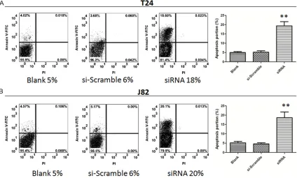
T24 and J82 cells in exponential phase in all groups were digested with trypsin, collected, washed twice with cold PBS, and resuspended in 100 μl of Annexin V binding buffer. A total of 5 μl of Annexin V-FITC solution were added and the mixture was incubated at room tempera-ture in the dark for 15 min. Next, 5 μl of PI stain-ing solution were added and the mixture was incubated for another 5 min. A total of 400 μl of Annexin V binding buffer was added and cell apoptosis was detected by flow cytometry with-in 1 h. The experiment was repeated three times and results were analyzed using Cellquest Pro software. The left lower quadrant (Q3) rep-resents normal viable cells with low staining intensity of both Annexinv and PI, whereas the upper left quadrant (Q1) represents necrotic cells. Q2 and Q3 represent late and early apop-totic cells, respectively.
Western blot analysis
The expression of caspase-3, caspase-9, Bcl-2 in T24 and J82 cells and normal uroepithelium SV-HUC-155 cells was compared by Western blot analysis. Briefly, after transfection, cells at 70% confluence were collected. Total protein was extracted using protein extraction kits and quantified using a BCA kit according to the manufacture’s instruction. Equal amounts of total protein (20 μg) were separated by SDS-PAGE electrophoresis and transferred to polyvi-nylidene difluoride membranes. The membrane was blocked in TBS buffer containing 5% skim milk and 0.1% Tween 20 at room temperature for 1 h, and incubated with the appropriate pri-mary antibody (1:200) overnight at pH 7.6 at 4°C with gentle shaking. The peroxidase-labeled secondary antibodies (1:200) were added and the membranes were incubated at 37°C for 1 h. The membranes were washed 3 times with TBST for 5 min each and subjected to ECL detection. The intensity of bands was detected by a Molecular Imager® ChemiDoc- TMXRS System (Bio-Rad Laboratories). The gray value of bands was analyzed by Image Lab 2.0 software (Bio-Rad Laboratories).
Cell invasion assay
Matrigel (BD Biosciences) was spread evenly on the micro-film of a transwell chamber. Cells were transfected with siRNA with the highest inhibitory effect and si-Scramble as described in 2.4. After 72 h, cells were digested and added into the upper chamber of transwell. DMEM medium containing 5% FBS was added into the lower chamber. After 24 h of culture, transmembrane cells were washed, fixed and stained with crystal violet. The cells were count-ed under an invertcount-ed microscopy. Mean values were obtained from five randomly selected fields for each well.
Statistical analysis
All data were expressed as mean ± standard deviation. Statistical analyses were performed using SPSS 11.0. Difference between groups was analyzed by t tests. P<0.05 is considered statistically significant.
Results
Increased Loc554202 expression in tumor tis-sue and bladder cancer cell lines
Figure 1. A. qRT-PCR analysis comparing lncRNA Loc554202 expression in bladder cancer tissues and adjacent non-cancerous tissues (control). *, P<0.05 compared with the control. B. qRT-PCR analysis of lncRNA Loc554202 expression in bladder cancer cells and normal SV-HUV-1 cells, *, P<0.05 compared with the control.
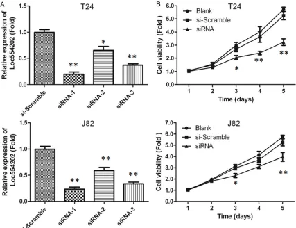
Figure 2. A. The relative Loc554202 expression in T24 and J82 cells. B. MTT assay of T24 and J82 cells transfected with siRNA targeting Loc554202. *, P<0.05 compared with si-Scramble group.
by qRT-PCR analysis. It was shown that relative
[image:4.612.93.520.316.644.2]of Loc554202 in bladder cancer cell lines T24, J82 and BIU-87, and normal uroepithelium SV-HUC-155 cells was also compared. As shown in Figure 1B, relative Loc554202 expression in T24, J82 and BIU-87 cells was significantly higher compared with SV-HUC-155 cells (P<0.05). Moreover, relative Loc554202 expression in T24, J82 cells (4.5±0.1 and 3.0±0.1, respectively) was higher than that in BIU-87 cells, which were used for subsequent experiments.
Inhibitory effect of siRNA targeting Loc554202 on cell viablity of T24 and J82 cells
The expression of Loc554202 in T24 and J82 cells transfected with siRNA-1, -2 and -3 was quantified by qRT-PCR analysis. As shown in
Figure 2A, the relative Loc554202 expression in these cells was significantly reduced com-pared with the si-Scramble group (P<0.05). The highest inhibitory effect was observed in siRNA-1 group, which was used for the transfection in subsequent experiments. Further, the prolifera-tion of T24 and J82 cells transfected with siRNA-1 was detected by MTT assay. As shown in Figure 2B, cell viability of T24 and J82 cells
in siRNA-1 group was significantly lower com-pared with si-Scramble group at 72, 96 and 120 h after transfection (P<0.05), suggest- ing that the siRNA-mediated inhibition of Loc554202 expression markedly reduced the proliferation of T24 and J82 cells.
Effects on of siRNA targeting Loc554202 on cell apoptosis
As shown in Figure 3, the percentage of necrot-ic T24 cells in siRNA group was 18%, whnecrot-ich was significantly higher compared with both the blank control (5%) and si-Scramble group (6%, P<0.05). The percentage of necrotic J82 cells in siRNA group (20%) was also significantly higher compared with si-Scramble group (6%, P<0.05), indicating that siRNA-mediated inhibi-tion of Loc554202 expression significantly pro-moted apoptosis of T24 and J82 cells.
Effects of siRNA on the expression of proteins of the mitochondrial apoptosis pathway
[image:5.612.95.519.76.331.2]The expression of proteins of the mitochondrial apoptosis pathway including caspase-3, cas-pase-9, Bcl-2 in T24 and J82 cells in each group was compared by Western blot analysis. As
shown in Figure 3, caspase-3 and caspase-9 expression in T24 and J82 cells transfected with siRNA-1 was significantly higher compared with si-Scramble group (P<0.05), whereas the expression of the anti-apoptotic proteins Bcl-2 was significantly lower (P<0.05), suggesting that siRNA-mediated inhibition of Loc554202 expression induced cell apoptosis by activating the mitochondrial apoptosis pathway.
Effects of siRNA on the invasion of T24 and J82 cells
As shown in Figure 4, cell invasion assay dem-onstrated that the percentage of transmem-brane T24 and J82 cells in siRNA-1 group was significantly decreased (approximately 30%) compared with the blank control and si-Scram-ble group (P<0.05), which suggested that the invasion ability of T24 and J82 cells was signifi-cantly reduced after the downregulation of Loc554202 expression by siRNA-1.
Discussion
In recent years, many studies have suggested that lncRNAs may serve as a novel target for
cancer treatment. As a host gene of miR-31, Loc554202 controls its transcription. It has been know that hypermethylation of the CpG island in the promoter region of Loc554202 is the primary mechanism for miR-31 expression, which plays an important regulatory role in the development and metastasis of breast cancer [22]. Studies have shown that Loc554202 expression is markedly increased in breast can-cer, and its expression level is positively corre-lated with the size and clinical stage of tumors [22]. Moreover, Loc554202 expression in tri-ple-negative breast cancer (TNBC) cells was significantly higher compared with normal breast cells. Consistent with these findings, our results showed that Loc554202 expression in bladder cancer tissues and cells was signifi-cantly higher than that in normal tissues and SV-HUC-155 cells, respectively.
siRNA silencing has drawn increasingly more attention in the field of functional genomics research, and has been widely used in cancer research [23, 24]. Shi et al. have found that siRNA-mediated Loc554202 knockdown pro-hibited the proliferation, colony formation,
[image:6.612.93.528.79.364.2]sion and migration of TNBC cells as well as the breast tumor in vivo [25]. In this study, Loc554202 in bladder cancer cells was silenced by siRNAs to explore its effects on cell proliferation and apoptosis. Results demon-strated that lncRNA Loc554202 expression in all three siRNA groups was significantly reduced compared with control group, suggesting that siRNA could be used to establish an in vitro model for function analysis of Loc554202. Infinite division and proliferation of tumor cells is an important feature of malignancy, and inva-sive ability of tumor cells is a key factor affect-ing the prognosis and survival rate of cancer. Studies have shown that lncRNA Loc554202 regulates the proliferation, invasion and metas-tasis of breast cancer cells mainly through the regulation of miR-31 [21]. Consistent with previ-ous studies, our results showed that the cell viability of T24 and J82 cells transfected with siRNA-1 was significantly lower compared with control group at 72 h after transfection as indi-cated by MTT assay, suggesting the inhibitory effect of Loc554202 knockdown on the prolif-eration of these cancer cells. Results in the transwell matrigel invasion assay showed that the number of invasive cells in siRNA group was substantially decreased compared with control group, indicating a reduced invasion capacity of bladder cancer cells with siRNA-mediated Loc554202 knockdown. Furthermore, flow cytometry analysis demonstrated that the pro-portion of apoptotic cells in siRNA group was significantly higher than that in control group, suggesting an induction effect of Loc554202 silencing on apoptosis of T24 and J82 cells. Caspase-3 and caspase-9 are important pro-teins of mitochondrial apoptotic pathway, increased expression of which has been shown to inhibit tumor cell proliferation, and induce cell cycle arrest and apoptosis [26]. Bcl-2, a key anti-apoptosis protein, is an important indica-tor for the prognosis of cancer. It has been shown that increased expression of Bcl-2 sig-nificantly inhibits apoptosis of tumor cells [27]. This study also investigated the regulatory mechanism of the induced apoptosis by Loc554202 silencing. The expression of cas-pase-3, caspase-9 in siRNA group was signifi-cantly increased compared with control group; whereas bcl-2 expression was significantly reduced, suggesting that siRNA-mediated Loc554202 knockdown might induce
apopto-sis through the activation of mitochondrial apoptotic pathway.
In summary, Loc554202 is closely associated with cell viability and apoptosis of bladder can-cer T24 and J82 cells. siRNA-mediated Loc554202 knockdown inhibits the prolifera-tion and invasion of these cancer cells, and pro-mote their apoptosis.
This study provides insights into the regulatory mechanisms Loc554202 knockdown on inva-sion and metastasis of bladder cancer cells. Loc554202 might be an effective therapeutic target for the treatment of bladder cancers, especially invasive bladder cancers.
Disclosure of conflict of interest
None.
Address correspondence to: Dr. Yan Liu, Institute of Tangshan Gongren Hospital Affiliated to Hebei Medical University, 27 Wenhua Road, Lubei District, Tangshan 063000, Hebei, China. Tel: +86-3152- 821821; Fax: +86-3152821821; E-mail: liuyan- [email protected]
References
[1] Ferlay J, Shin HR, Bray F, Forman D, Mathers C and Parkin DM. Estimates of worldwide burden of cancer in 2008: GLOBOCAN 2008. Int J Can-cer 2010; 127: 2893-2917.
[2] Jemal A, Siegel R, Ward E, Murray T, Xu J, Smi-gal C and Thun MJ. Cancer statistics, 2006. CA Cancer J Clin 2006; 56: 106-130.
[3] Yu J, Zhu T, Wang Z, Zhang H, Qian Z, Xu H, Gao B, Wang W, Gu L, Meng J, Wang J, Feng X, Li Y, Yao X and Zhu J. A novel set of DNA methyla-tion markers in urine sediments for sensitive/ specific detection of bladder cancer. Clin Can-cer Res 2007; 13: 7296-7304.
[4] James ND, Hussain SA, Hall E, Jenkins P, Trem-lett J, Rawlings C, Crundwell M, Sizer B, Sreenivasan T, Hendron C, Lewis R, Waters R and Huddart RA. Radiotherapy with or without chemotherapy in muscle-invasive bladder can-cer. N Engl J Med 2012; 366: 1477-1488. [5] Dalbagni G. The management of superficial
bladder cancer. Nat Clin Pract Urol 2007; 4: 254-260.
[6] Hadaschik BA, Paterson RF, Fazli L, Clinkscales KW, Shalaby SW and Chew BH. Investigation of a novel degradable ureteral stent in a porcine model. J Urol 2008; 180: 1161-1166.
pro-file of cancer mortality in China: 1991-2005. Br J Cancer 2004; 90: 2157-2166.
[8] Ponting CP, Oliver PL and Reik W. Evolution and functions of long noncoding RNAs. Cell 2009; 136: 629-641.
[9] Spizzo R, Almeida MI, Colombatti A and Calin GA. Long non-coding RNAs and cancer: a new frontier of translational research? Oncogene 2012; 31: 4577-4587.
[10] Wapinski O and Chang HY. Long noncoding RNAs and human disease. Trends Cell Biol 2011; 21: 354-361.
[11] Mercer TR, Dinger ME and Mattick JS. Long non-coding RNAs: insights into functions. Nat Rev Genet 2009; 10: 155-159.
[12] Jin G, Sun J, Isaacs SD, Wiley KE, Kim ST, Chu LW, Zhang Z, Zhao H, Zheng SL, Isaacs WB and Xu J. Human polymorphisms at long non-cod-ing RNAs (lncRNAs) and association with pros-tate cancer risk. Carcinogenesis 2011; 32: 1655-1659.
[13] Calin GA, Liu CG, Ferracin M, Hyslop T, Spizzo R, Sevignani C, Fabbri M, Cimmino A, Lee EJ, Wojcik SE, Shimizu M, Tili E, Rossi S, Taccioli C, Pichiorri F, Liu X, Zupo S, Herlea V, Gramantieri L, Lanza G, Alder H, Rassenti L, Volinia S, Schmittgen TD, Kipps TJ, Negrini M and Croce CM. Ultraconserved regions encoding ncRNAs are altered in human leukemias and carcino-mas. Cancer Cell 2007; 12: 215-229. [14] Prensner JR and Chinnaiyan AM. The
emer-gence of lncRNAs in cancer biology. Cancer Discov 2011; 1: 391-407.
[15] Gutschner T and Diederichs S. The hallmarks of cancer: a long non-coding RNA point of view. RNA Biol 2012; 9: 703-719.
[16] Bouyssou JM, Manier S, Huynh D, Issa S, Roc-caro AM and Ghobrial IM. Regulation of mi-croRNAs in cancer metastasis. Biochim Bio-phys Acta 2014; 1845: 255-265.
[17] Hu W, Yuan B, Flygare J and Lodish HF. Long noncoding RNA-mediated anti-apoptotic activi-ty in murine erythroid terminal differentiation. Genes Dev 2011; 25: 2573-2578.
[18] Yu W, Gius D, Onyango P, Muldoon-Jacobs K, Karp J, Feinberg AP and Cui H. Epigenetic si-lencing of tumour suppressor gene p15 by its antisense RNA. Nature 2008; 451: 202-206.
[19] Corcoran DL, Pandit KV, Gordon B, Bhattacha-rjee A, Kaminski N and Benos PV. Features of mammalian microRNA promoters emerge from polymerase II chromatin immunoprecipitation data. PLoS One 2009; 4: e5279.
[20] Valastyan S and Weinberg RA. miR-31: a cru-cial overseer of tumor metastasis and other emerging roles. Cell Cycle 2010; 9: 2124-2129.
[21] Augoff K, McCue B, Plow EF and Sossey-Alaoui K. miR-31 and its host gene lncRNA LOC554202 are regulated by promoter hyper-methylation in triple-negative breast cancer. Mol Cancer 2012; 11: 5.
[22] George J and Patel T. Noncoding RNA as thera-peutic targets for hepatocellular carcinoma. Semin. Liver Dis 2015; 35: 63-74.
[23] Bo J, Yang G, Huo K, Jiang H, Zhang L, Liu D and Huang Y. microRNA-203 suppresses blad-der cancer development by repressing bcl-w expression. FEBS J 2011; 278: 786-792. [24] Guo Y, Ying L, Tian Y, Yang P, Zhu Y, Wang Z, Qiu
F and Lin J. miR-144 downregulation increases bladder cancer cell proliferation by targeting EZH2 and regulating Wnt signaling. FEBS J 2013; 280: 4531-4538.
[25] Shi Y, Lu J, Zhou J, Tan X, He Y, Ding J, Tian Y, Wang L and Wang K. Long non-coding RNA Loc554202 regulates proliferation and migra-tion in breast cancer cells. Biochem. Biophys Res Commun 2014; 446: 448-453.
[26] Mantena SK, Sharma SD and Katiyar SK. Ber-berine, a natural product, induces G1-phase cell cycle arrest and caspase-3-dependent apoptosis in human prostate carcinoma cells. Mol Cancer Ther 2006; 5: 296-308.