Original Article
TREM2 siRNA inhibits cell proliferation of
human liver cancer cell lines
Sui-Liang Zhang1*, Ting-Song Chen2*, Ling Xiao3, Ying Ye3, Wei Xia4, Hong Zhang3
Departments of 1Invasive Therapy, 4Nuclear Medicine, Shanghai Seventh People’s Hospital, Shanghai, P. R. China; 2T-S Chen Department of Traditional Chinese and Western Medicine, Eastern Hepatobiliary Surgery Hospital,
Shanghai, P. R. China; 3Central Laboratory, Shanghai Seventh People’s Hospital, Shanghai, P. R. China. *Equal
contributors.
Received December 24, 2015; Accepted March 8, 2016; Epub April 1, 2016; Published April 15, 2016
Abstract: Liver cancer is one of the most common malignant tumors in digestive system with poor prognosis and high mortality. Recent researches suggested the important role of TREM2 in various cancers. This study aimed at investigating the effect of TREM2 siRNA on cell proliferation, apoptosis and cell circle distribution of two liver cancer cell lines, 97H and HepG2 cells with significant expression level of TREM2. 25 liver cancer samples contain-ing tumor tissues and normal tissues were detected for the TREM2 expression level uscontain-ing real-time PCR. 97H and HepG2 cells were taken out for the following study. CCK8 assay was preformed to evaluate the cell proliferation. Cell apoptosis and cell cycle of 97H and HepG2 cells were identified using flow cytometry. In addition, apoptosis and cell cycle related proteins caspase3, Bax, Bcl2, PCNA, CDK1 and CDC25C were measured for the expression level by using Western blot. Furthermore, in vivo study using nude mice was evaluated for the inhibition ability of TREM2 siRNA on tumor formation of 97H cells. As a result, expression of TREM2 exhibited a higher level in tumor tissues than that in normal tissues. The cell proliferation of 97H and HepG2 cells was inhibited by TREM2 siRNA time-dependently. Besides, TREM2 siRNA induced cell apoptosis and arrested cell cycle at S phase effectively. The regulation of TREM2 siRNA on the proteins revealed the possible mechanism involved in the cell apoptosis and cell cycle. The tumor size obtained from in vivo study indicated the inhibition capability of TREM2 siRNA on the forma-tion of tumor. In conclusion, TREM2 siRNA inhibited cell proliferaforma-tion of human liver cancer cells 97H and HepG2 by targeting caspase/Bcl and CDK1 signaling pathway, suggesting the potential therapy using TREM2 siRNA for liver cancer treatment.
Keywords: Liver cancer, TREM2 siRNA, cell proliferation, in vivo study
Introduction
Liver cancer is one of the most common malig-nant tumors in digestive system with poor prog-nosis and high mortality. As the third of cancer death rate globally, liver cancer threaten the health and life of human beings [1]. At the cur-rent stage, treatment of liver cancer is mainly surgery, which is effective against early solid tumor [2]. However, it is difficult for patients with recurrence, advanced stage or minimal disease to be cured. Therefore, a new molecu-lar marker for early diagnosis and treatment in clinic is needed currently. Triggering receptors expressed on myeloid cells 2 (TREM2) is com-posed of a variant-type extracellular domain, a charged transmembrane domain and a short cytoplasmic tail. It belongs to a family of
In this study, human liver cancer cell lines 97H and HepG2 with high expression level of TREM2 were selected. Cells transfected with TREM2 siRNA were detected for the cell prolif-eration using CCK8 assay. Flow cytometry was employed to determine the cell apoptosis and cell circle distribution of liver cancer cells. The expression levels of caspase3, Bax, Bcl2, PCNA, CDK1 and CDC25C were measured using Western blot for the further investigation of the mechanism involved in cell apoptosis and cell cycle. In addition, the effect of TREM2 siRNA on the tumor progression was evaluated by nude mice inoculated with 97H cells subcutaneously.
Materials and methods
Patients and tissue samples
The gene expression of TREM2 in 25 tissue samples were measured by real-time PCR, among which four tissue samples were detect-ed using Western blot. Each sample including tumor tissue and normal tissue was collected from the same patient with complete clinical and pathological follow-up data. The study pro-tocol was approved by the local independent ethical committee.
Cell culture and transfection
97H and HepG2 cells were cultured in DMEM containing 10% FCS and 1% 100× mycillin and incubated in 5% CO2 at 37°C. Cell viability was detected at 95% through tryphan blue stain. Then cells were digested and seeded into 6-well plate (5×10⁵ cells/well) before transfec-tion. LipofectamineTM 2000 (Invitrogen, Shang- hai, China) was used to transfect 5 μL TREM2 siRNA or negative control siRNA into 97H and HepG2 cells, respectively. After the incubation for 48 h, transfected cells were collected and processed for cell viability, apoptosis, cycle and Western blot assay.
Western blot assay
Every 20 mg tissue samples were cut into piec-es and mixed with 250 μl histiocyte lysis buffer
(RIPA, solarbio, Shanghai) containing 0.01% protease inhibitor cocktail (Sigma, Shanghai, China). While cultured or transfected cells were washed by 1×PBS for twice, followed by the lysis at 4°C. After fully lysed, samples were cen-trifuged at 12,000 RCF for 15 min at 4°C and the supernatant was collected. BCA protein quantification kit (BCA, thermo, Shanghai) was used to quantify the protein contents. Then tis-sue and cell samples were run on 12% (85 μg/ well) or 10% (25 μg/well) SDS-PAGE gel, respec-tively, and transferred to a nitrocellulose filter membrane (Millipore, Shanghai, China) electro-phoretically. Blots were blocked with 5% skim milk at room temperature for 1 h, followed by the incubation with anti-TREM2 (Abcam), PCAN (CST), CDC25C (Abcam), CDK1 (Abcam), cas-pase3 (Abcam), Bax (Santa), Bcl2 (Santa) and GAPDH (CST) antibodies, and then incubated with goat anti-mouse or anti-rabbit secondary antibody (Beyotime, Shanghai, China). En- hanced chemiluminescence (ECL, Thermo Scientific, Shanghai, China) was used to detect the blots visually.
Real-time PCR assay
Total RNA was extracted from tissue samples or cultured cells and quantified by using Trizol Reagent (Invitrogen). Reverse transcription kit (Fermentas, Shanghai) was used to synthesis cDNA through a total volume of 25 μl (12 μl RNA-primer Mix, 5 μl 5×RT reaction buffer, 1 μl 25 mM dNTPs, 1 μl 25 U/μl RNase inhibitor, 1 μl 200 U/μl M-MLV Rtase, 1 μl Oligo(dt)18 and 4 μl DNase-free ddH2O). Then cDNA was amplified through a total PCR system of 25 μl (12.5 μl SYBR Green Mix, 0.5 μl forward primer, 0.5 μl reverse primer, 9.5 μl ddH2O and 2 μl cDNA template) with the PCR conditions consisted of 10 min at 95°C, and then 15 sec at 95°C and 45 sec at 60°C for 40 cycles. Amplification kinetic curves were obtained through 15 sec at 95°C, 1 min at 60°C, 15 sec at 95°C and 15 sec at 60°C. Primers used for the amplification of TREM2 and GAPDH were listed in Table 1.
Cell proliferation assay using CCK8
[image:2.629.99.295.93.161.2]Cultured 97H and HepG2 cells were trypsinized and diluted to 1~5×10⁴ cells/mL. Then cells were plated into 96-well plates (1~5×10³ cells/ well) and incubated at 37°C for 12 h, followed by the transfection with TREM2 siRNA or nega-tive control siRNA, respecnega-tively. Transfected cells in each well were mixed with 100 μl DMEM Table 1. Primers used in RT-PCR analysis
Gene Primers sequence
containing 10% CCK-8 (Dojindo Biochem) at 0, 24, 48 and 72 h, and then incubated in 5% CO2 at 37°C for 1 h. The OD450nm of cell suspension was measured using a spectrophotometer in order to evaluate the cell viability of 97H and HepG2 cells.
Cell apoptosis assay
Transfected cells were formed single cell by 0.25% trypsin and washed by 10% PBS, fol-lowed by the centrifugation at 1000 RCF for 5 min. Supernatant was discarded and then the cells were incubated with Annexin-V fluorescein isothiocyanate (FITC) apoptosis detection Kit
(BD Biosciences) for 10 min at room tempera-ture without light. Cell apoptosis rate was mea-sured and the data was obtained using flow cytom etry (FACSCalibur, BD Biosciences).
Cell cycle distribution
[image:3.629.100.531.81.451.2]After transfection, 97H and HepG2 cells were digested and washed. Samples were fixed by 70% pre-cooled ethanol for 12 h and RNA was removed by using 1 mg/ml RNase A. Then cells were stained with propidium iodide (PI) for 10 min and the DNA content was measured using flow cytometry so as to determine the propor-tion of cells in each stage of cell cycle.
Tumor formation assay using nude mice
Cultured 97H cells tranfected with negative control siRNA or TREM2 siRNA respectively were digested and diluted to 2 × 10⁷ cells/ml. 12 nude mice were divided into 2 groups aver-agely and randomly, followed by the injection of 100 μl cell suspension. Mice were kept in respective cages with standard food and water for 1-2 weeks. After tumor formed, the tumor size was measured using a vernier caliper every 3 days. Then the mice were sacrificed at day 33 and the tumors were weighted on a digital balance.
Statistical analysis
Data were expressed as mean ± standard devi-ation and analyzed using t-test. GraphPad Prism 5.0 software was used to perform and analyze the data. Real-time PCR data was ana-lyzed using ABI Prism 7300 SDS Software. Results
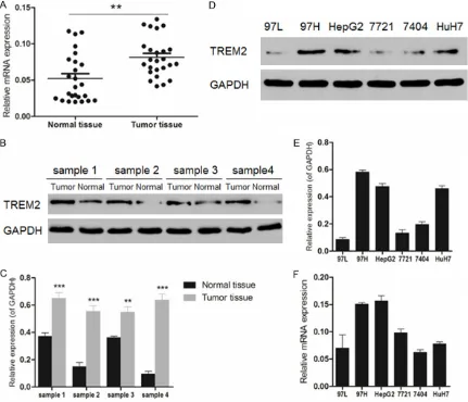
Screen of liver cancer cell line with high ex-pression level of TREM2
According to the gene expression level of TREM2 measured by real-time PCR in 25 liver
cancer patients (Figure 1A), there was signifi-cant difference presented between normal tis-sues and tumor tistis-sues (P < 0.01). Besides, Western blot was used to detect the TREM2 expression levels in 4 tissue samples from liver cancer patients (Figure 1B and 1C). Similarly, an obvious difference was observed in cancer tissues and normal tissues (P < 0.01 or P < 0.001). Then the expression level of TREM2 in various human liver cancer cell lines including 97L, 97H, HepG2, 7721, 7404 and HuH7 was measured using both Western blot and real-time PCR (Figure 1D-F). As a result, 97H, HepG2 and HuH7 cells exhibited high expres-sion levels of TREM2. However, the gene expression of TREM2 in HuH7 cells was not as high as that in 97H or HepG2 cells. Therefore, human liver cancer cell lines 97H and HepG2 were selected for further study of the possible mechanism involved in the siRNA interference against liver cancer.
TREM2 siRNA inhibits cell proliferation
[image:4.629.99.529.81.279.2]The interference effect of TREM2 siRNA on 97H and HepG2 cells was determined using both Western blot and real-time PCR. As shown in Figure 2A and 2B, the expression level of TREM2 declined significantly in 97H and HepG2
cells (P < 0.05 or P < 0.001). The gene expres-sion of TREM2 also presented decrease with significant difference compared with negative control group (Figure 2C, P < 0.001), indicating the efficient interference ability of TREM2 siRNA. Then CCK8 assay was employed to detect the cell viability of 97H and HepG2 cells so as to evaluate the inhibition effect of TREM2 siRNA. Compared with the cell viability in the negative control groups, that of TREM2 siRAN transfected 97H and HepG2 cells declined with a time-dependent manner (Figure 2D and 2E, P
< 0.001), indicating the inhibited cell prolifera-tion due to TREM2 siRNA.
TREM2 siRNA induces cell apoptosis
[image:5.629.97.531.72.568.2]Flow cytometry was used to evaluate the cell apoptosis of 97H and HepG2 cells after the transfection with TREM2 siRNA for 48 h. Cell apoptosis induced by TREM2 siRNA was detect-ed using Annexin-V FITC/PI double staining visually (Figure 3A and 3B). The apoptosis rate shown in Figure 3C was calculated from the
percentages of early apoptotic cells presented in the lower right quadrant of the histograms. As a result, the apoptosis rate of TREM2 siRNA transfected 97H cells increased to 21.57±3.14% in comparison with that of the negative control group which was 2.45±0.81% (n=3, P < 0.001). While the apoptosis rate of HepG2 cells also rose from 3.67±0.42% to 23.63±1.58% (n=3, P
< 0.001) after the transfection, revealing the correlation of cell apoptosis associated with TREM2 siRNA.
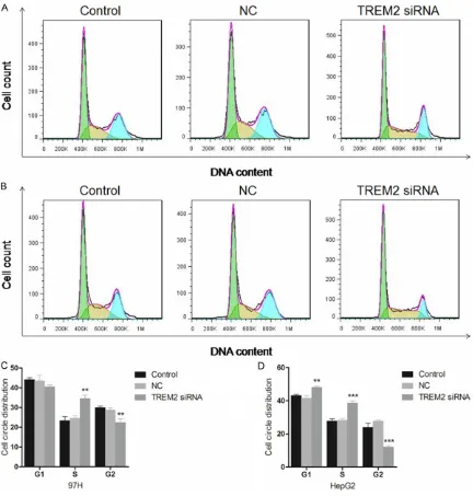
TREM2 siRNA arrests cell cycle
[image:6.629.99.531.78.531.2]After the transfection for 48 h, flow cytometer was also used to analysis the cell circle distri-bution of 97H and HepG2 cells. As shown in Figure 4A and 4B, the cell count in each stage was presented visually and the cell circle dis- tribution was calculated and exhibited in Figure 4C and 4D. According to the results, the percentage of G2 phase in the TREM2 siRNA treated 97H cells dropped significantly from
28.74±1.77% to 22.38±2.81% (n=3, P < 0.01) compared with the negative control group, while that of cells in S phase showed an increase from 24. 62.59±2.65% to 34.48± 2.45% (n=3, P < 0.01). Similarly, there was also a decrease observed in the G2 phase of HepG2 cells from 27.74±0.89% to 12.00±0.92% (n=3,
P < 0.001), and the percentage of S phase rose from 28.16±0.95% to 38.67±1.28% dramati-cally (n=3, P < 0.001), indicating the blocked cell circle distribution in S phase. What inter-ested us is the increased percentage of G1
[image:7.629.100.382.77.466.2]pared with the negative control (n=3, P < 0.001), corresponding to the results obtained from cell circle distribution assay. In addition, Western blot was also used to evaluate the change of cell apoptosis related protein cas-pase3, Bax and Bcl2. As a result, caspase3 and Bax showed an increase in the expression level in both 97H and HepG2 cells significantly in comparison with the negative control (n=3, P < 0.05, P < 0.01 or P < 0.001), while Bcl2 was down-regulated dramatically (n=3, P < 0.001). The up-regulation of the expression levels of
Figure 5. Effect of TREM2 siRNA on the expression of cell apoptosis and cell cycle related proteins. A. The expression of PCNA, CDC25C, CDK1, caspase3, Bax and Bcl2 in 97H and HepG2 cells was exhibited by SDS-PAGE. B. Western blot was used to measure the expression levels of biological pathway related proteins in 97H cells. **P < 0.01, ***P < 0.001, compared with the negative control group (n=3). C. The protein expression levels were also detected by Western blot in HepG2 cells. **P < 0.01, ***P < 0.001, compared with the negative control group (n=3).
phase in transfected HepG2 cells from 41.59±2.13 to 48.02±0.98 (n=3, P < 0.01). While that of 97H cells showed a tendency to de- crease without significant dif-ference compared with the negative control group. The difference between two cell lines might be caused by the different regulation effect of TREM2 siRNA on two cell lines. However, further study might be necessary to investi-gate the potential relation-ship between TREM2 and cell cycle in liver cancer cells.
TREM2 siRNA regulates the biological pathways related proteins
com-apoptotic-promoting proteins caspase3 and Bax, and down-regulated of anti-apoptotic pro-tein Bcl2 eventually resulted in the cell apopto-sis of 97H and HepG2 cells.
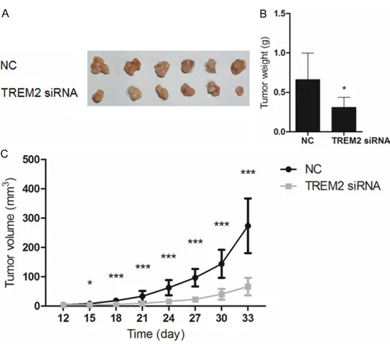
TREM2 siRNA suppressed growth of liver can-cer in vivo
The tumor growth of liver cancer cells transfect-ed with TREM2 siRNA was determintransfect-ed using nude mice in vivo. Mice subcutaneously inject-ed with TREM2 siRNA or negative control siRNA transfected 97H cells were sacrificed on day 33. Tumor volume was recorded every 3 days from day 12 and tumors were weighted on day 33 after the sacrifice. As the result shown in Figure 6B, tumors in interference group were lighter than those in the negative control group (n=6, P < 0.05). In addition, the slower growth of tumor volume indicated the inhibition ability of TREM2 siRNA against liver tumor with a time-dependent manner (n=6, P < 0.05 or P < 0.001).
Discussion
Except for the key role of mutated TREM2 in NHD and Alzheimer’s disease, its
immunoregu-evaluated and the possible mechanism was investigated. Data obtained from our study demonstrated the inhibition capability of TREM2 siRNA on the cell proliferation of 97H and HepG2 cells through induced cell apopto-sis and arrested cell cycle. Besides, delayed tumor formation and progression observed in the in vivo study using nude mice verified the inhibition of TREM2 siRNA against 97H cells, indicating the crucial role of TREM2 in liver cancer.
[image:8.629.100.382.76.325.2]In order to gain further sight into the potential mechanism involved in the function of TREM2 in liver cancer cells, cell apoptosis related pro-teins caspase3, Bax and Bcl2 were investigat-ed. Caspases belonging to cysteine protease family could be activated by apoptotic signal. Activated caspases act on peptide chain and split substrate, resulting in the apoptosis of cells [16]. Caspase3, as one of the effector cas-pases, plays a direct role in cell apoptosis. Activation of caspase-activated deoxyribonu-clease (CAD) induced by caspase3 was report-ed in the degradation of DNA. CAD without activity combined with its inhibitor ICAD in nor-mal cells. When apoptosis occurs, ICAD activated
Figure 6. Effect of TREM2 siRNA on the growth of liver tumor in athymic nude mice. A, B. Mice were sacrificed on day 33 and tumors were weighted. *P < 0.05, compared with the negative control group (n=6). C. TREM2 siRNA in-hibited tumor growth time-dependently. *P < 0.05, ***P < 0.001, compared with the negative control group (n=6).
by caspase3 release CAD with nuclease activity [17]. In addition, nuclear lamina, which is responsible for the stability of chromatin, could be cut by caspase3 at a single site, leading to the degradation of chromatin [18]. Bcl2 gene located on 18q21 contains three exons and two open reading frames. Bcl2 with multiple highly conservative fragments including Bcl2 homology-1 (BH1), BH2, BH3 and BH4 is mainly distributed in the nuclear membrane, endo-plasmic reticulum and mitochondria membrane [19]. Antagonism exists between Bax and Bcl2 both of which belong to B-cell lymphoma/leu-kemia-2 family. Bcl2 harbors anti-apoptosis function inhibiting the release of apoptosis-pro-moting substance from mitochondria and the activation of pro-apoptotic proteins caspase3, Bax and Bak [20, 21]. In correspondence with the cellular function, the up-regulated cas-pase3 expression level in TREM2 siRNA treated 97H and HepG2 cells indicated the activation of cell apoptosis, suggesting the vital role of TREM2 in the survival of liver cancer cells. Similarly, the increased Bcl2 and declined Bax also demonstrated the cell apoptosis induced by TREM2 siRNA, revealing the potential mech-anism involved in the inhibition of cell viability in 97H and HepG2 cells by TREM2 siRNA. In order to gain further sight into the mecha-nism involved in cell cycle, the cell circle distri-bution related proteins PCNA, CDK1 and CDC25C were also measured for the expres-sion levels by Western blot. Proliferating cell nuclear antigen (PCNA) synthesized within the nucleus is a coenzyme of DNA polymerase delta [22]. There exist soluble and insoluble PCNA in nucleus. The former with stable con-tent is expressed in each phase of cell cycle, while the expression of the insoluble one fol-lows the synthesis of DNA [23]. It was detected significant high expression level in S phase and decreased in G2 phase [24], suggesting PCNA as a criterion to evaluate the cell proliferation in tumor cells with strong proliferation activity. Therefore, PCNA was widely studied in tumori-genesis, classification, staging, radiation sensi-tivity, prognosis, recurrence, metastasis causes of death and tumor markers. Cyclin-dependent kinase 1 (CDK1) is a highly conserved protein regulated by cyclins and phosphorylation [25]. CDK1 combined with cyclins leads to the pro-gression of cell cycle. Whi5 phosphorylated by Cln3-CDK1 complex induces the transcription
of G1/S cyclins Cln1 and Cln2, allowing for the entry of S phase. On the other hand, Clb5, 6-CDK1 complexes trigger the origin of replica-tion initially. While in the M phase, phosphory-lased CDK1 by M-cyclins results in the spindle assembly and sister chromatid alignment, fol-lowed by the chromatid segregation. Finally, the dephosphorylation of CDK1 directed by cell division cycle 25C (CDC25C) leads to the final events of mitosis [25]. CDC25C gene encodes S-phase inducer phosphatase 3 regulating cell division as a critical role. It was thought to sup-press p53-induced growth arrest and trigger entry into mitosis [26]. According to the data obtained from cell circle distribution assay (Figure 4); the increased cell count in S phase suggested the blocked entry of G2 phase, which corresponds to the declined expression of PCNA measured by Western blot. At the same time, down-regulated CDK1 and CDC25C also indicated the arrested cell cycle due to TREM2 siRNA, revealing the mechanism involved in the effect of TREM2 siRNA on the cell circle distribution.
In conclusion, our study demonstrated the high expression level of TREM2 in liver cancer tis-sues with significant difference. Interference of TREM2 resulted in the inhibited cell prolifera-tion of liver cancer cells. Besides, the potential mechanisms involved in induced cell apoptosis and arrested cell cycle were revealed using Western blot. In addition, in vivo study using nude mice exhibited inhibition ability of TREM2 siRNA on tumor formation of liver cancer cells, presenting important data for clinical research. Therefore, TREM2 might perform as an onco-gene in the tumor progression and serve as a therapeutic target in the treatment of human liver cancer.
Acknowledgements
2014-12), Star Talent Training Program of the Hospital (QMX2015-01), and the Open Re- search Fund of State Key Laboratory Breeding Base of Systematic Research, Development and Utilization of Chinese Medicine Resources (ME2016012).
Disclosure of conflict of interest None.
Address correspondence to: Hong Zhang, Central Laboratory, Shanghai Seventh People’s Hospital, Shanghai 200137, P. R. China. E-mail: hqzhang51@ 126.com; Wei Xia, Department of Nuclear Medicine, Shanghai Seventh People’s Hospital, Shanghai 200137, P. R. China. E-mail: awingxia@163.com
References
[1] On the rise globally, cancer mortality declines in U.S. Cancer Discov 2014; 4: OF7.
[2] Ahmed I and Lobo DN. Malignant tumours of the liver. Surgery-Oxford International Edition 2009; 27: 30-37.
[3] Turnbull IR, Gilfillan S, Cella M, Aoshi T, Miller M, Piccio L, Hernandez M, Colonna M. Cutting edge: TREM-2 attenuates macrophage activa-tion. J Immunol 2006; 177: 3520-3524. [4] Guerreiro R, Wojtas A, Bras J, Carrasquillo M,
Rogaeva E, Majounie E, Cruchaga C, Sassi C, Kauwe JS, Younkin S, Hazrati L, Collinge J, Pocock J, Lashley T, Williams J, Lambert JC, Amouyel P, Goate A, Rademakers R, Morgan K, Powell J, St George-Hyslop P, Singleton A, Hardy J; Alzheimer Genetic Analysis Group. TREM2 variants in Alzheimer’s disease. N Engl J Med 2013; 368: 117-127.
[5] Hickman SE and El Khoury J. TREM2 and the neuroimmunology of Alzheimer’s disease. Biochem Pharmacol 2014; 88: 495-498. [6] Paloneva J, Manninen T, Christman G, Hovanes
K, Mandelin J, Adolfsson R, Bianchin M, Bird T, Miranda R, Salmaggi A, Tranebjaerg L, Konttinen Y, Peltonen L. Mutations in two genes encoding different subunits of a recep-tor signaling complex result in an identical dis-ease phenotype. Am J Hum Genet 2002; 71: 656-662.
[7] Takahashi K, Rochford CD, Neumann H. Clearance of apoptotic neurons without inflam-mation by microglial triggering receptor ex-pressed on myeloid cells-2. J Exp Med 2005; 201: 647-657.
[8] Ho CC, Liao WY, Wang CY, Lu YH, Huang HY, Chen HY, Chan WK, Chen HW, Yang PC. TREM-1 expression in tumor-associated macro-phages and clinical outcome in lung cancer.
Am J Respir Crit Care Med 2008; 177: 763-770.
[9] Sigalov AB. A novel ligand-independent pep-tide inhibitor of TREM-1 suppresses tumor growth in human lung cancer xenografts and prolongs survival of mice with lipopolysaccha-ride-induced septic shock. Int Immunophar- macol 2014; 21: 208-219.
[10] Zhou J, Chai F, Lu G, Hang G, Chen C, Chen X, Shi J. TREM-1 inhibition attenuates inflamma-tion and tumor within the colon. Int Im- munopharmacol 2013; 17: 155-161.
[11] Liao R, Sun TW, Yi Y, Wu H, Li YW, Wang JX, Zhou J, Shi YH, Cheng YF, Qiu SJ, Fan J. Expression of TREM-1 in hepatic stellate cells and prognostic value in hepatitis B-related he-patocellular carcinoma. Cancer Sci 2012; 103: 984-992.
[12] Li J, Salcedo R, Mivechi NF, Trinchieri G, Horuzsko A. The proinflammatory myeloid cell receptor TREM-1 controls Kupffer cell activa-tion and development of hepatocellular carci-noma. Cancer Res 2012; 72: 3977-3986. [13] Neumann H and Takahashi K. Essential role of
the microglial triggering receptor expressed on myeloid cells-2 (TREM2) for central nervous tissue immune homeostasis. J Neuroimmunol 2007; 184: 92-99.
[14] N’Diaye EN, Branda CS, Branda SS, Nevarez L, Colonna M, Lowell C, Hamerman JA, Seaman WE. TREM-2 (triggering receptor expressed on myeloid cells 2) is a phagocytic receptor for bacteria. J Cell Biol 2009; 184: 215-223. [15] Ito H and Hamerman JA. TREM-2, triggering
re-ceptor expressed on myeloid cell-2, negatively regulates TLR responses in dendritic cells. Eur J Immunol 2012; 42: 176-185.
[16] Lavrik IN, Golks A, Krammer PH. Caspases: pharmacological manipulation of cell death. J Clin Invest 2005; 115: 2665-2672.
[17] Stennicke HR and Salvesen GS. Biochemical characteristics of caspases-3, -6, -7, and -8. J Biol Chem 1997; 272: 25719-25723.
[18] Weng C, Li Y, Xu D, Shi Y, Tang H. Specific cleavage of Mcl-1 by caspase-3 in tumor ne-crosis factor-related apoptosis-inducing ligand (TRAIL)-induced apoptosis in Jurkat leukemia T cells. J Biol Chem 2005; 280: 10491-10500. [19] Shi Y, Chen J, Weng C, Chen R, Zheng Y, Chen
Q, Tang H. Identification of the protein-protein contact site and interaction mode of human VDAC1 with Bcl-2 family proteins. Biochem Biophys Res Commun 2003; 305: 989-996. [20] Hsu YT, Wolter KG, Youle RJ.
[21] Nechushtan A, Smith CL, Hsu YT, Youle RJ. Conformation of the Bax C-terminus regulates subcellular location and cell death. EMBO J 1999; 18: 2330-2341.
[22] Moldovan GL, Pfander B, Jentsch S. PCNA, the maestro of the replication fork. Cell 2007; 129: 665-679.
[23] Shivji KK, Kenny MK, Wood RD. Proliferating cell nuclear antigen is required for DNA exci-sion repair. Cell 1992; 69: 367-374.
[24] Essers J, Theil AF, Baldeyron C, van Cappellen WA, Houtsmuller AB, Kanaar R, Vermeulen W. Nuclear dynamics of PCNA in DNA replication and repair. Mol Cell Biol 2005; 25: 9350-9359.
[25] Enserink JM and Kolodner RD. An overview of Cdk1-controlled targets and processes. Cell Div 2010; 5: 1-41.