Original Article
LPS induces dopamine depletion and iron accumulation
in substantia nigra in rat models of Parkinson’s disease
Jie Huo1, Qu Cui1,2,3, Wei Yang3, Wei Guo1
Departments of 1Emergency, 2Hematology, Beijing Tiantan Hospital, Capital Medical University, Beijing, China; 3Department of Immunology, School of Basic Medicine, Norman Bethune Health Science Center, Jilin University,
Jilin, China
Received May 14, 2018; Accepted September 7, 2018; Epub October 1, 2018; Published October 15, 2018
Abstract: Objective: Intrapallidal inflammation may lead to the pathogenesis of Parkinson’s disease. Pathological changes caused by lipopolysaccharide (LPS)-induced inflammation in Parkinson’s disease rat models were largely unknown. Methods: Male Sprague-Dawley rat models were intra-globuspallidus injected with saline and lipopolysac -charide and divided into two groups, the control group and the LPS-stimulation group. The locomotor activity of the rat models was recorded for 4 consecutive weeks by trajectory analysis software for animal behavior. For the evalu -ation of pathological profiles, the expression levels of tyrosine hydroxylase and OX-42 in the substantia nigra tissues were detected by immunohistochemical staining. Also, the concentrations of dopamine at specific sites were de -tected through high-performance liquid chromatography. Perl’s iron staining was used to evaluate iron accumulation in substantia nigra tissues. Results: LPS-stimulation reduced the locomotor capacity of the rat models compared with the control group. The density of tyrosine hydroxylase-positive cells was reduced and the secretion of striatal dopamine in the substantia nigra pars compacts was lower in the LPS group than it was in the control group. OX-42 positive microglia and ferritin levels were enhanced in the LPS group. Conclusion: Intrapallidal inflammation by LPS induced dopamine depletion and iron accumulation in the substantia nigra of Parkinson’s disease rat models. The management of cerebral inflammation might be pivotal for PD pathogenesis and prognosis.
Keywords: Parkinson disease, lipopolysaccharide, inflammation, iron
Introduction
Parkinson’s disease (PD) is a neurological dis-order characterized by a progressive degenera-tion of dopamine neurons in the substantia nigra pars compacta (SNc) and the loss of pre-dominant dopamine in the striatum [1, 2]. Based on the fact that dopamine modulates neuronal function in the striatum [3, 4], in clini-cal practice, therapies including dopamine replacement and updated neurosurgical tech-niques by subthalamic nucleus (STN) high-fre-quency stimulation in PD have been developed [5-9]. In the past over 2 decades, data from clinical trials have shown the involvement of dopamine in the functional regulation of the basal ganglia [10, 11]. However, dopamine-associated pathophysiological profiles in PD still remain a great challenge.
In PD patients, abnormal activations of sub-stantia nigra and increased levels of
inflamma-tory cytokines have been observed. Increased superoxide dismutase (SOD) and decreased reduced-glutathione (GSH) and catalase are characteristic evidence of local inflammatory oxidative stress. Free radicals generated by oxi-dative stress play a major role in the dampen-ing of adjacent tissues. On the other hand, the enhancement of cerebral iron storage in PD patients is widely recognized [12-14]. Iron pro-motes the generation of oxidative free radicals and the process of neuronal degenerative dis-eases [15-17]. A high concentration of iron can accelerate neuron death by enhancing the pro-cesses of lipid peroxidation and free radical generation. Thus, inflammation associated iron accumulation might induce oxidative stress and contribute to the pathogenesis of PD.
sig-naling inputs via D1 and D2 receptors [20-24]. Meanwhile, GP neurons transmit inhibitory GABAergic projections to the STN and the sub-stantia nigra pars reticulata (SNr) [25, 26]. Recently, Abedi et al. reported that intrapallidal administration of 6-hydroxydopamine would mimic in large part the electrophysiological and behavioral consequences of major dopamine depletion in the classical rat model of Park- inson’s disease [27], supporting the notion that dopamine mediates actions in the striatum and regulates biological processes in basal ganglia circuitry. However, inflammation-induced iron-mediated pathological changes in substantia nigra have not been well investigated in these PD models.
The present study investigated the effects of dopamine neurons on free iron and ferritin expression in substantia nigra by a classical rat model, aiming to decipher the impacts of LPS-induced inflammation on the pathogenesis of PD.
Materials and methods
Animals
Adult male Sprague-Dawley rats that weighed 190-210 g were used in this study. The animals were housed three per cage, kept under artifi-cial light (12 h light/dark cycle, lights on at 7:00 A.M.), with a temperature of 24°C and the humidity at 45%, with food and water available
ad libitum. All efforts were made to minimize the number of animals used and their suffering. The Experiments were carried out in accor-dance with the European Communities Council Directive (EU Directive 2010/63/EU) and the National Institutes of Health’s Guide for the Care and Use of Laboratory Animals.
LPS injection into the GP
The rats were placed in a stereotaxic frame (Kopf, Unimecanique, Beijing, China) under xyl-azine (xylxyl-azine hydrochloride, 10 mg/kg, i.p, Sigma, China) and ketamine anesthesia (ket-amine hydrochloride 75 mg/kg, i.p., Sigma, China). Each animal received a lateral injection of 4-μl LPS (2.5 g/L in sterile NaCl, 0.9%; Sigma, China) into the GP at coordinates 0.9 mm pos-terior to bregma, 3.0 mm lateral to the midline and 6.5 mm below the dura, according to the brain atlas of Paxinos and Watson (1998). The LPS injection was made over a 5-min period
using a cannula connected by polyethylene tub-ing to a 10-μl Hamilton microsyrtub-inge. At the end of each injection, the cannula was left in place for an addition 5 min to prevent a reflux of the solution and to allow for a toxin diffusion, and then was withdrawn slowly. For the sham group, the vehicle (saline plus 0.01% ascorbic acid) was infused into the GP under the same condi-tions as the LPS. Twenty rats were used and distributed into two groups, the LPS-treated group (n=10) and the sham group (n=10). All the rats were tested for locomotor activity before surgery and at 7 days, 14 days, 21 days, and 28 days after surgery. At the end of the locomotor data recording period, all the ani-mals were sacrificed, and their brains were used for histological and biochemical studies.
Locomotor activity
Spontaneous horizontal motor activity on lid-less cages (50 cm×50 cm) was recorded using digital photography (Sony, Japan). Images from 30 min of the active trajectories of the rats were collected. We tested the 30-minute move-ment distance (cm) and the movemove-ment speed (cm/s) using trajectory analysis software for animal behavior (EthoVision, version 2.3, Noldus, Holland). One frame was recorded every six seconds. All testing was done in an isolated room at 10:00 A.M.
Preparation of immunohistochemical sections
sub-stantia nigra, every 6th slice was treated with TH, OX-42, iron staining.
Immunohistochemical analysis
Immunohistochemistry detection was applied for the expression of TH, OX-42, and ferritin in the tissues. Briefly, antigens were unmasked by microwaving sections in a 10 mmol/L citrate buffer for 15 minutes, and immunostaining was undertaken using the avidin-biotinylated en- zyme complex method with antibodies against TH at 1 mg/mL, OX-42 at 1 mg/mL, and
equiva-The effectiveness of the LPS stimulation was confirmed by measuring the DA concentrations at specific brain sites. Sham (n=5) and LPS-treated (n=5) rats were sacrificed, the brain removed and stored at -80°C until analysis. The fresh frozen brains were cryostat-cut at -20°C and the bilateral regions of the striatum were punched. Punches were then stored at -80°C until biochemical assessments were done. The samples were homogenized in perchloric acid. Ho- mogenates were centrifuged, and the superna-tant was assessed for dopamine content by means of reverse phase HPLC with
[image:3.612.95.518.72.216.2]electro-Figure 1. The locomotor capacity of rat models was dampened by LPS stimulation over time. A. The mean move -ment velocity was reduced in the LPS-treated group. B. The mean move-ment distance was also reduced in the LPS-treated group.
Figure 2. LPS-stimulation reduced tyrosine hydroxylase positive cell amounts in the substantia nigra pars compacts. A. TH positive cells in the control group; B. TH positive cells in the LPS group (×50). C and D. The density of TH positive cells and the concentration of striatal dopamine in the control and LPS groups.
lent concentrations of poly-clonal nonimmune IgG con-trols. After incubation with a biotin-conjugated secondary antibody and subsequently with a streptavidin solution, color development was per-formed with 3,3-diaminoben-zidinetetrahydrochloride (Vector Laboratories) as a chromogen. The sections were counterstained using Gill-2 he- matoxylin (Thermo-Shandon, Pittsburgh, PA). After staining, the sections were dehydrated through increasing concentra-tions of ethanol and xylene. The Leica Qwin image analysis system was used to observe and count the morphology.
[image:3.612.91.371.274.498.2]chemical detection, as previously described [28, 29]. The mobile phase consisted of NaH2PO4, disodium EDTA, octane sulfonic acid and methanol, adjusted to PH 3.9 with ortho-phosphoric acid. The potential of the electrodes was set at +350 and -270 mV, and the results were expressed as ng/g of tissue. Each value corresponds to the mean ± SEM. Values were compared using the Mann-Whitney U test (Prism, GraohPad software, San Diego, CA, USA).
Perl’s iron staining
Iron staining was performed as previously described [30]. Briefly, the slides were trans-ferred to 100% ethanol. Individual lobe biopsy materials were placed in processing cassettes for 30-60 seconds, and then hydrated through a serial alcohol gradient at 100%, 100%, 95%, 80%, 70%. The tissue sections were immersed in xylene for 2 min. Then for 30 min they were incubated with 20 g/L hydrochloric acid and ferric ferrocyanide in a solution of 1:1. After rinsing with water and neutral red complex dye-ing, the sections were dehydrated through seri-al ethanol solutions (70%, 80%, 95%, 100%, 100%) each for 30 seconds, respectively. Finally, the slides were input for xylene trans-parentation and neutral balata fixation.
Results
LPS-stimulation reduced locomotor capacity of rat models
After the intra-globus pallidus injection of saline, the movement velocity of the rat models
control group (P=0.01, Figure 1A). Accordingly, the 30-minute movement distance was signifi-cantly shortened in the LPS group, from 1700 cm at 1 week to 1500 cm at 2 weeks, 1000 cm at 3 weeks and 800 cm at 4 weeks in the LPS group, while the distance was maintained at 3000 cm in the control group at those time-points (P=0.01, Figure 1B). These data demon-strated that LPS-stimulation dampened the movement capacity of the rat models, and such an change was progressively devastating over time.
Tyrosine hydroxylase positive cell amounts in substantia nigra pars compacts and the stria-tal dopamine concentration deceased after LPS-stimulation
Immunohistochemistry of the substantia nigra pars compacts at 4 weeks showed that the density of the TH positive cells in the LPS group was 300.2±3 6.2 per slide, significantly less than the level of 607.9±51.2 per slide in the control group (P<0.01, Figure 2). The concen-tration of striatal dopamine was 22.79±1.56 nmol/g in the LPS group, significantly lower than the level of 16.49±1.79 nmol/g in the con-trol group (P<0.05). Tyrosine hydroxylase posi-tive cell amounts in the substantia nigra pars compacts and the striatal dopamine concentra-tion deceased after LPS-stimulaconcentra-tion.
LPS stimulation promoted the proliferation of microglia in substantia nigra pars compacts
After 4 weeks of LPS-stimulation, the OX-42 positive microglia cells were morphologically
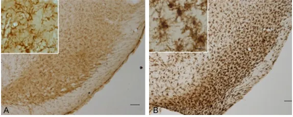
Figure 3. OX-42 positive microglia cells in the substantia nigra pars com -pacts. The left panels (A) showed a representative result of the OX-42 stain -ing of the substantia nigra pars compacts from the control group, while the right panels (B) showed a representative result of the OX-42 staining of the substantia nigra pars compacts from the LPS group (×50 for the base im -ages, and ×400 for the zoomed images).
enlarged, with shortened axons and hyperchro-matic nuclei on the slides. The OX-42 positive microglia cells showed longer axons with pale nuclei. The density of the OX-42 positive cell was more enhanced in the LPS group than it was in the control group (Figure 3).
LPS stimulation caused iron accumulation in the substantia nigra pars compacts
After 4 weeks of LPS stimulation, free ions increased in the substantia nigra pars com-pacts in the LPS group (Figure 4A), compared with almost no iron stains in the control group (Figure 4B). Meanwhile, the expression of ferri-tin in the substantia nigra pars compacts in the LPS group was also enhanced (Figure 4C), com-pared with the control group (Figure 4D).
Discussion
In this study, we established a rat model through the intrapallidal administration of LPS to explore the effects of inflammation-induced pathological changes in the substantia nigra. Our results showed that LPS-induced
inflam-ate motor symptoms. Studies have proposed that inflammatory dysfunctions are associated with psychiatric disorders and neurodegenera-tion in both animal models and human patients [31-33]. In this study, we applied LPS intrapalli-dal administration to triggering neuroinflamma-tion in rat models. The behavioral alternaneuroinflamma-tions implied that inflammation caused by LPS was efficacious in mimicking PD symptoms.
[image:5.612.90.377.71.303.2]Neuroinflammation always triggers downstr- eam signaling pathways and causes subse-quent pathological changes. We further con-firmed that LPS-induced inflammation re- duced the neuron cell amount in substantia nigra pars compacts and suppressed the secre-tion of striatal dopamine. Inflammatory molecu- les can induce the recruitment of peripheral leukocytes into CNS. This neuroinflammatory process could be partially beneficial for neuro-nal tissues since it promotes the clearance of cell debris. Conversely, inflammatory mediators could also act on neurons to cause neurode-generation. The neuronal death further acti-vates inflammatory mechanisms, resulting in a vicious cycle composed of local inflammation
Figure 4. Iron metabolism altered by LPS stimulation. A. Free iron staining showed a pale color in the substantia nigra pars compacts of the control group. B. Free iron staining was obviously increased in the LPS group. C. The immunohistochemical staining of ferritin showed sporadic dots in the substantia nigra pars compacts from the control group. D. Immunohisto -chemical staining of ferritin showed thick dots in the substantia nigra pars compacts from the LPS group. The data were obtained from representative samples (×50 for the base images, and ×400 for the zoomed images).
mation caused dopamine depletion and iron accumula-tion in the substantia nigra and dampened the move-ment capacity of rat PD models.
allevi-and neuronal death. A systemic inflammation due to infection or peripheral injury could exac-erbate symptoms and promote neuronal dam-age in PD. Pro-inflammatory cytokines secreted by leukocytes act on brain tissues through sev-eral pathways, such as the endothelial cells and leakage through damaged BBB. In addi-tion, microglial activation has also been linked with cerebral disorders [34]. Microglia are CNS-resident immune cells activated in response to cerebral injury [35]. As reported, microglial acti-vation induces the release of pro-inflammatory cytokines including interleukin (IL)-1β, IL-6, and tumor necrosis factor (TNF)-α, leading to neuro-nal damage and cellular loss [36, 37]. These cytokines further stimulate microglia to secre- te chronic inflammation mediators, maintain- ing neuroinflammation status. The acute and chronic inflammations might synergistically reduce the neuron cell amount and the sup-pressed secretion of striatal dopamine in the substantia nigra pars compacts, contributing to the pathogenesis of PD.
Our data showed that LPS-induced inflamma-tion altered iron metabolism in the substantia nigra pars compacts. Enhanced iron and ferritin levels in the substantia nigra pars compacts reflects an inflammatory status quo caused by LPS. Iron accumulation in the substantia nigra pars compacts might directly result from LPS injection, but it also contributes to the patho-genesis of PD through its toxic effects on dopa-mine neutrons. As a consequence of LPS-stimulation, iron accumulation leads to slow and progressive neuronal death, which is sup-ported by a gradual reduction in movement capacities over time in our PD models. Despite being essential for tissue homeostasis, our data confirm that inflammatory responses tribute to neuronal injury when they are not con-trolled and/or chronic, and dopaminergic neu-rons from substantia nigra are particularly vulnerable to microglial mediated neurotoxicity, which is in line with previous reports [38, 39].
Collectively, in this study, we investigated the effects of intrapallidal administration of LPS on dopamine depletion and iron accumulation in substantia nigra in a PD rat model. We found that inflammation-induced iron accumulation promotes PD pathogenesis through subse-quent cell damage. Thus, taking measures to prevent the onset of inflammation and to con-trol the magnitude of cerebral inflammation would be pivotal for PD patients.
Acknowledgements
This study was jointly supported by the National Natural Science Foundation of China (No 81500157) and Beijing Municipal Admini- stration of Hospitals’ Youth Programme (QML 20150506).
Disclosure of conflict of interest
None.
Address correspondence to: Dr. Wei Guo, De-partment of Emergency, Beijing Tiantan Hospital, Capital Medical University, 6 Tiantian Xili, Dongcheng District, Beijing 100050, China. Tel: +86-10-67096829; Fax: +86-10-+86-10-67096829; E-mail: guo -wei1010@126.com; Dr. Wei Yang, Department of Immunology, School of Basic Medicine, Norman Bethune Health Science Center, Jilin University, 126 Xinmin Avenue, Chaoyang District, Changchun 130021, Jilin, China. Tel: +86-431-85619476; Fax: +86-431-85619495; E-mail: ywei@jlu.edu.cn
References
[1] Schapira AHV, Chaudhuri KR and Jenner P. Non-motor features of Parkinson disease. Nat Rev Neurosci 2017; 18: 435-450.
[2] Swan M, Doan N, Ortega RA, Barrett M, Nichols W, Ozelius L, Soto-Valencia J, Boschung S, Deik A, Sarva H, Cabassa J, Johannes B, Raymond D, Marder K, Giladi N, Miravite J, Severt W, Sa -chdev R, Shanker V, Bressman S and Saun -ders-Pullman R. Neuropsychiatric characteris-tics of GBA-associated Parkinson disease. J Neurol Sci 2016; 370: 63-69.
[3] Yoshida H, Date I, Shingo T, Fujiwara K, Ko -bayashi K, Miyoshi Y and Ohmoto T. Stereotac-tic transplantation of a dopamine-producing capsule into the striatum for treatment of Par -kinson disease: a preclinical primate study. J Neurosurg 2003; 98: 874-881.
[4] Oiwa Y, Sanchez-Pernaute R, Harvey-White J and Bankiewicz KS. Progressive and extensive dopaminergic degeneration induced by con-vection-enhanced delivery of 6-hydroxydopa -mine into the rat striatum: a novel rodent mod-el of Parkinson disease. J Neurosurg 2003; 98: 136-144.
[5] Pollak P, Benabid AL, Gross C, Gao DM, Lau -rent A, Benazzouz A, Hoffmann D, Gentil M and Perret J. [Effects of the stimulation of the sub -thalamic nucleus in Parkinson disease]. Rev Neurol (Paris) 1993; 149: 175-176.
[7] Poewe W, Seppi K, Tanner CM, Halliday GM, Brundin P, Volkmann J, Schrag AE and Lang AE. Parkinson disease. Nat Rev Dis Primers 2017; 3: 17013.
[8] Sauerbier A, Cova I, Rosa-Grilo M, Taddei RN, Mischley LK and Chaudhuri KR. Treatment of nonmotor symptoms in Parkinson’s disease. Int Rev Neurobiol 2017; 132: 361-379. [9] Taddei RN, Spinnato F and Jenner P. New
symptomatic treatments for the management of motor and nonmotor symptoms of Parkin -son’s disease. Int Rev Neurobiol 2017; 132: 407-452.
[10] Smith Y and Villalba R. Striatal and extrastria-tal dopamine in the basal ganglia: an overview of its anatomical organization in normal and Parkinsonian brains. Mov Disord 2008; 23 Suppl 3: S534-547.
[11] Villalba RM, Lee H and Smith Y. Dopaminergic denervation and spine loss in the striatum of MPTP-treated monkeys. Exp Neurol 2009; 215: 220-227.
[12] Youdim MB. Iron in the brain: implications for Parkinson’s and Alzheimer’s diseases. Mt Si -nai J Med 1988; 55: 97-101.
[13] Youdim MB, Ben-Shachar D, Yehuda S and Rie -derer P. The role of iron in the basal ganglion. Adv Neurol 1990; 53: 155-162.
[14] Jenner P. Oxidative stress as a cause of Parkin -son’s disease. Acta Neurol Scand Suppl 1991; 136: 6-15.
[15] Snyder AM and Connor JR. Iron, the substantia nigra and related neurological disorders. Bio-chim Biophys Acta 2009; 1790: 606-614. [16] Wypijewska A, Galazka-Friedman J, Bauminger
ER, Wszolek ZK, Schweitzer KJ, Dickson DW, Jaklewicz A, Elbaum D and Friedman A. Iron and reactive oxygen species activity in parkin -sonian substantia nigra. Parkinsonism Relat Disord 2010; 16: 329-333.
[17] Nandar W and Connor JR. HFE gene variants affect iron in the brain. J Nutr 2011; 141: 729S-739S.
[18] Herrera AJ, Castano A, Venero JL, Cano J and Machado A. The single intranigral injection of LPS as a new model for studying the selective effects of inflammatory reactions on dopami -nergic system. Neurobiol Dis 2000; 7: 429-447.
[19] Carvey PM, Chang Q, Lipton JW and Ling Z. Pre -natal exposure to the bacteriotoxin lipopoly-saccharide leads to long-term losses of dopa -mine neurons in offspring: a potential, new model of Parkinson’s disease. Front Biosci 2003; 8: s826-837.
[20] Floran B, Aceves J, Sierra A and Martinez-Fong D. Activation of D1 dopamine receptors stimu -lates the release of GABA in the basal ganglia of the rat. Neurosci Lett 1990; 116: 136-140.
[21] Floran B, Floran L, Sierra A and Aceves J. D2 receptor-mediated inhibition of GABA release by endogenous dopamine in the rat globus pal-lidus. Neurosci Lett 1997; 237: 1-4.
[22] Cooper AJ and Stanford IM. Dopamine D2 re -ceptor mediated presynaptic inhibition of stria -topallidal GABA(A) IPSCs in vitro. Neurophar-macology 2001; 41: 62-71.
[23] Shin SS, Bales JW, Yan HQ, Kline AE, Wagner AK, Lyons-Weiler J and Dixon CE. The effect of environmental enrichment on substantia nigra gene expression after traumatic brain injury in rats. J Neurotrauma 2013; 30: 259-270. [24] Mehdi SJ, Rosas-Hernandez H, Cuevas E, Lantz
SM, Barger SW, Sarkar S, Paule MG, Ali SF and Imam SZ. Protein Kinases and Parkinson’s Dis -ease. Int J Mol Sci 2016; 17.
[25] Rommelfanger KS and Wichmann T. Extrastria -tal dopaminergic circuits of the Basal Ganglia. Front Neuroanat 2010; 4: 139.
[26] Hormigo S, Vega-Flores G and Castro-Alaman -cos MA. Basal ganglia output controls active avoidance behavior. J Neurosci 2016; 36: 10274-10284.
[27] Abedi PM, Delaville C, De Deurwaerdere P, Benjelloun W and Benazzouz A. Intrapallidal administration of 6-hydroxydopamine mimics in large part the electrophysiological and be-havioral consequences of major dopamine depletion in the rat. Neuroscience 2013; 236: 289-297.
[28] De Deurwaerdere P, Stinus L and Spampinato U. Opposite change of in vivo dopamine re -lease in the rat nucleus accumbens and stria-tum that follows electrical stimulation of dorsal raphe nucleus: role of 5-HT3 receptors. J Neu -rosci 1998; 18: 6528-6538.
[29] Delaville C, Navailles S and Benazzouz A. Ef -fects of noradrenaline and serotonin deple -tions on the neuronal activity of globus palli -dus and substantia nigra pars reticulata in experimental parkinsonism. Neuroscience 2012; 202: 424-433.
[30] Gebril OH, Simpson JE, Kirby J, Brayne C and Ince PG. Brain iron dysregulation and the risk of ageing white matter lesions. Neuromolecu -lar Med 2011; 13: 289-299.
[31] Onore C, Careaga M and Ashwood P. The role of immune dysfunction in the pathophysiology of autism. Brain Behav Immun 2012; 26: 383-392.
[32] Theoharides TC, Asadi S and Patel AB. Focal brain inflammation and autism. J Neuroinflam -mation 2013; 10: 46.
[33] Theoharides TC and Zhang B. Neuro-inflamma -tion, blood-brain barrier, seizures and autism. J Neuroinflammation 2011; 8: 168.
[35] Stertz L, Magalhaes PV and Kapczinski F. Is bipolar disorder an inflammatory condition? The relevance of microglial activation. Curr Opin Psychiatry 2013; 26: 19-26.
[36] Reus GZ, Fries GR, Stertz L, Badawy M, Passos IC, Barichello T, Kapczinski F and Quevedo J. The role of inflammation and microglial activa -tion in the pathophysiology of psychiatric disor -ders. Neuroscience 2015; 300: 141-154. [37] Zhao Q, Peng C, Wu X, Chen Y, Wang C and You
Z. Maternal sleep deprivation inhibits hippo -campal neurogenesis associated with inflam -matory response in young offspring rats. Neu -robiol Dis 2014; 68: 57-65.
[38] Gao HM and Hong JS. Why neurodegenerative diseases are progressive: uncontrolled inflam -mation drives disease progression. Trends Im-munol 2008; 29: 357-365.