Case Report
Fibrillary glomerulonephritis with anti-glomerular
basement membrane antibody: a
combined glomerular insult
Suhail Al-Salam1, Mohamed Elsharif2, Ayman Al Madani3, Imran Zahid4, Balsam Ahmed5
1Department of Pathology, College of Medicine and Health Sciences, UAE University, ALAIN PO Box 17666, UAE; 2Nephrology Division, Specialist Physician, Mafraq Hospital, Abu Dhabi, UAE; 3Chief of Nephrology Division,
Consultant Nephrologist, Mafraq hospital, Abu Dhabi, UAE; 4Nephrology Division, Consultant Nephrologist, Mafraq
Hospital, Abu Dhabi, UAE; 5Nephrology Division, Specialist Physician, Mafraq Hospital, Abu Dhabi, UAE
Received January 4, 2016; Accepted March 18, 2016; Epub August 1, 2016; Published August 15, 2016
Abstract: Rapidly progressive glomerulonephritis (RPGN) is a clinical syndrome characterized by quick and propagative deterioration of renal function with loosing 50% or more of renal function within three months. RGPN is associated with cellular crescents in more than 50% of glomeruli; hence it is called crescentic glomerulonephritis. We report a 47-year-old patient who presents with RPGN associated with fibrillary glomerulonephritis (FGN) and positive antiglomerular basement membrane (anti-GBM) antibody. In conclusion, FGN should be suspected in any case of RPGN presents with a nephrotic range of proteinuria. Although light microscopic and immunofluorescent studies in a renal biopsy are essential, electron microscopic study is the gold standard in establishing the diagnosis of FGN. FGN and Anti-GBM disease can co-exist as causes of RPGN.
Keywords: Crescentic glomerulonephritis, nephrotic syndrome, fibrillary glomerulonephritis, anti-glomerular base -ment membrane disease
Introduction
Rapidly progressive glomerulonephritis (RPGN) is a clinical syndrome characterized by quick and propagative deterioration of renal function with loosing 50% or more of renal function with-in three months. RPGN is associated with cel-lular crescents in more than 50% of glomeruli; hence it is called Crescentic glomerulone-
phritis (CGN) [1]. CGN is subclassified morpho -logically into three subgroups; pauci-immune, anti-glomerular basement membrane (anti-GBM) antibody-associated and immune-com-plex associated [1]. Most of patients with pau-city- immune CGN have positive serum level of antineutrophil cytoplasmic antibody (ANCA) [1]. While patients with anti-GBM antibody-associ-ated CGN have positive serum level of anti-GBM antibody and linear membranous
immu-noreactivity to IgG and C3 on immunofluores -cent study [1]. On the other hand, patients with immune complex-associated CGN show
char-acteristic immunofluorescent and electron
microscopic patterns of immune complex
depo-sition within affected glomeruli. Fibrillary glo
-merulonephritis (FGN) is a glomerular disease
characterized by the deposition of irregularly
oriented, elongated, nonbranching fibrils of 10-
30 nm thickness in the mesangium and along the capillary walls [2]. Clinically, patients with
FGN present with nephrotic-range proteinuria,
microscopic hematuria, hypertension, and
pro-gressive renal insufficiency, which are similar to our case. These findings occur in the setting of
negative serological investigations, ruling out such entities as systemic lupus erythematosus, light-chain disease, and cryoglobulinemia [3].
CGN is seen in almost 5% of FGN [1, 4-7]. In this
study we report CGN in a 47-yeay-old patient
with FGN and positive serum GBM
anti-body.
Case report
Fibrillary glomerulonephritis with anti-GBM antibody
Room (ER) with nausea, vomiting and bilateral
[image:2.612.89.522.68.565.2]ence generalized body weakness, poor oral intake, nausea, vomiting and hematuria in the past 3 weeks prior to his ER visit. He denied any decrease in urine output, shortness of breath, hemoptysis, and orchest pain. He did not have similar symptoms before. His hyper-tension was well controlled by medication before this presentation.
[image:3.612.89.522.69.563.2]Fibrillary glomerulonephritis with anti-GBM antibody
mole/L (<133), total protein 42 g/L (64-83),
albumin 19 g/L(35-52), Glomerular filtration
rate 5 ml/minute/1.73 m2 (>90), random blood
sugar 5.8 mmol/L (2.8-8.8). Liver function tests were unremarkable. Serologic tests for antinuclear antibodies, anti-double stranded
DNA antibodies, anti-neutrophil cytoplasmic
antibodies, hepatitis B virus and hepatitis C virus were unremarkable. Compliment 3 and 4 levels were within normal range. Anti-glomerular basement membrane antibody was positive. The patient had a nephrotic range of protein-uria with numerous red blood cells in the urine. The complete blood count was remarkable for Hemoglobin of 75 gm/L (131-172), RBC 2.8×10¹²/L (4.2-5.6), MCV 78.6 fL (81-101),
MCH 26.8 pg (27-35), RDW 17.7% (11.6-14.8),
platelets count 108×109/L (140-400), mean
platelets volume 12.2 fL (7.3-9.7), WBC 7.8×10⁹/L, lymphocytes 0.16×10⁹/L (1-3.5),
neutrophils 7.6×10⁹/L (2-8).
The renal function deteriorates rapidly which necessitates starting hemodialysis and arrang-ing for kidney biopsy.
Pathologic findings
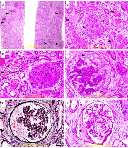
Renal biopsy was performed and we received 3 cores of 1.5 cm in length and 0.1 cm in diam-eter each. Light microscopic examination reveals renal tissue consists of cortex, medulla and contains 50 glomeruli; 3 of which are glob-ally sclerosed. There are 45 cellular crescents, which compress the affected glomeruli (Figure 1). 5 glomeruli are free of crescents and show thickening of glomerular basement membrane with meningeal expansion (Figure 1F). Foci of
acute tubular injury are seen (Figure 1B, 1C).
Foci of mild tubular atrophy are also noticed.
Some of the tubules show intratubular casts. In addition, some tubules show intratubular free
red blood cells (RBC) filling the lumen (Figure 1C) and sometimes forming RBC casts. There
are foci of mild mixed interstitial inflammatory cells infiltration consists mainly of lymphocytes. Periglomerular fibrosis is seen in 6 glomeruli. Few foci of mild interstitial fibrosis are appreci -ated. Sampled blood vessels are
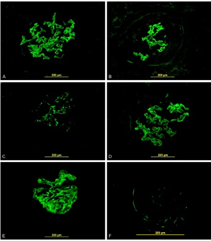
unremark-able. Immunofluorescent examination reveals
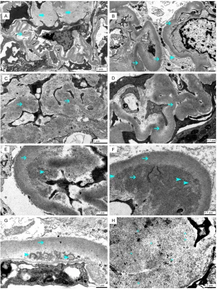
linear membranous staining for IgG (+2), C3 (+2), Kappa light chain (+2) and Lambda light chain (+2) (Figure 2). Electron microscopic
study reveals thickening of glomerular base-ment membrane 0.55 µm±0.15 (0.23-0.48) with a range of (0.41-0.96 µm) as well as
depo-sition of fibrillary material in the basement
membrane and mesangium (Figure 3). The
intramembranous, randomly oriented fibrils
have a mean diameter of 13.6 nm±2.13 and a
range of (10-18) that infiltrate and thicken the glomerular basement membrane. The fibrils
lack hollow centers and parallel stacking.
Mesangial deposition of these fibrils is also
seen (Figure 3). There is severe effacement of foot processes of a podocytes. Congo red stain for amyloid material was negative.
Discussion
Crescentic glomerulonephritis is a serious dis-order that must be diagnosed quickly and accu-rately so that appropriate management can be initiated as quickly as possible, since delay in diagnosis and treatment will have a major neg-ative impact on the outcome due to the rapidly progressing loss of renal function. Clinicians should have a high index of suspicion especial-ly, on facing patients with rapid deterioration of renal function, and quickly referring them to nephrologists. The diagnosis of CGN depends
entirely on the identification of cellular cres -cents in 50% or more of sampled glomeruli. Interestingly, the starting event in crescent for-mation is injury to the glomerular capillary endothelium and GBM. The damaged glomeru-lar endothelium and GBM will permit passage
of fibrin and other plasma proteins to
Bowmann’s space, which stimulate the prolif-eration of visceral and parietal epithelium, as
well as recruitment of inflammatory cells espe -cially T lymphocytes and macrophages [8]. This cellular process is accompanied by a release of
inflammatory cytokines and activation of coag -ulation cascade [8]. In this study we show a severe CGN with more than 90% of glomeruli showing cellular crescents that compress affected glomeruli leading to severe
deteriora-tion of GFR and renal funcdeteriora-tion. We also show a linear glomerular membranous immunofluores -cent staining with IgG and C3 as well as
posi-tive serum level for anti-GBM antibody; findings
support the diagnosis of anti-GBM-associated CGN.
Anti-GBM antibodies bind primarily to the
Fibrillary glomerulonephritis with anti-GBM antibody
continue with immunosuppression therapy or to prescribe more conservative treatment [15]. We also show thickening of GBM with intra-membranous deposition of randomly oriented,
Congo-red negative fibrils of 10-18 nm in diam
-eter that infiltrate and thicken the glomerular basement membrane; features confirm the diagnosis of FGN.
However; the unusual and important features
of this case of FGN are the presence of global
linear IgG staining, largely limited to capillary walls with active cellular crescents in more than 90% of the glomeruli and positive anti-GBM antibody. Cellular crescents can be seen
in FGN but this degree of severity of the dis -ease with more than 90% cellular crescents is
rarely reported in FGN-associated CGN [1]. Although it has been suggested that FGN is
immune mediated [16], the exact mechanism
of the disease remains unclear. Whereas fibril
deposition is almost always limited to the kid-ney, there have been observations suggesting
that FGN is a systemic, rather than a renal-lim
-ited disease as it has been shown that FGN can
recur in renal transplant recipients [3], suggest-ing a systemic factor in the recipient.
An important question can be raised here;
which event starts first; Anti-GBM antibody or FGN? The past history of the patient did not show any feature of acute renal insufficiency,
which usually seen in anti-GBM antibody, besides the patient denied any previous pre-sentation with features similar to the current illness. This might exclude anti-GBM disease as being the primary initiator and support its recent development with the initiation of cur-rent illness. Besides, it is very rare to have nephrotic range of proteinuria in CGN due to anti-GBN disease [17], hence the presence of heavy proteinuria in our case might be mainly
due to FGN.
Guerin et al suggests the possibility of repeat-ed insults to the GBM [18], may cause altera-tions to its structure, possibly through local
inflammation, with infiltrating
leukocytes-rele-asing granular enzymes and free radicals. This chain of type IV collagen and this binding is
associated with the development of progres-sive renal disease [9].
Mutations in the COL4A3 gene which codes the
α3 chain of collagen IV NC1 [10] have not been
found in any experimental or clinical studies, which suggests no role of this gene in the pathogenesis of the disease. There are two dis-tinct epitopes; EA and EB, for the anti-GBM-Ab [11]. Reports have suggested that EA and EB are hidden epitopes for B lymphocytes, covered by the quaternary structure of the hexamers created by bridges among monomers adjacent to NC1 chains [12]. The disconnection of these NC1 chains exposes the pathogenic epitopes
of the α3 and α5 chains, leading to production
of anti-GBM antibody. The crucial decisive fac-tor of exposure of these epitopes remains unknown. However, it is assumed to be the result of a combination of factors such as
genetic susceptibility, post-transcription modifi -cations, epitope extension and environmental factors [13].
Moreover, cell-mediated mechanisms have been incriminated in the initiation of anti-GBM antibody production. T cells play a key role in response to exposure to the cryptic EA and EB epitopes, generating signals that enable B-cell proliferation and production of GBM anti-body [14].
High serum level of anti-GBM antibody in our patient can damage glomerular endothelial cells and GBM leading to cellular crescents [1, 8]. The hematuria and RBC casts within renal tubules also supports anti-GBM antibody dam-aging effect on glomerular endothelial cells and basement membrane leading to cellular cres-cents formation. Our patient has clear lungs and denied any history of hemoptysis, which excludes Goodpasture syndrome and supports restricted renal anti-GBM disease.
Since the establishment of laboratory tests to detect anti-GBM antibodies the number of diagnostic biopsies have been enormously
decreased. Now, biopsies are more significant
for determining a prognosis, computing
epithe-lial and fibrotic crescents to decide whether to
could favor the exposure of the globular domain
NC1 of α3 chain of collagen IV, which contains
the cryptic EA and EB epitopes, triggering an autoimmune response. This can be triggered with different kinds of insults including extra-corporeal short wave therapy [18], ureteric obstruction [19], and monoclonal gammopathy [20].
We propose glomerular basement membrane insult might have been occurred due to
deposi-tion of fibrils that might uncover cryptic epit -opes and initiates anti-GBM antibody. However, these are just speculations and necessitate further studies.
We think that the high percentage of cellular crescents in our case is due to the combined insults to glomerular endothelium and GBM by
deposited fibrils and anti-GBM antibody.
Our literature search identified a patient with FGN who was diagnosed as having anti-GBM
-glomerulonephritis based on immunofluores
-cence microscopy findings [21] and another case of FGN which was diagnosed of having
anti-GBM antibody by immunoassay test [22].
In conclusion, FGN should be suspected in any
case of RPGN presents with a nephrotic range of proteinuria. Although light microscopic and
immunofluorescent studies in a renal biopsy
are essential, electron microscopic study is the gold standard in establishing the diagnosis of
FGN. FGN and Anti-GBM disease can co-exist
as causes of RPGN.
Disclosure of conflict of interest
None.
Address correspondence to: Dr. Suhail Al-Salam, Department of Pathology, College of Medicine and Health Sciences, UAE University, ALAIN PO Box 17666, UAE. Tel: +97137137464; Fax: +9713767-1966; E-mail: suhaila@uaeu.ac.ae
References
[1] Jennette JC. Rapidly progressive crescentic glomerulonephritis. Kidney Int 2003; 63: 1164-77.
[2] Nasr SH, Valeri AM, Cornell LD, Fidler ME, Sethi S, Leung N, Fervenza FC. Fibrillary glomerulo -nephritis: a report of 66 cases from a single
institution. Clin J A Soc Nephrol 2011; 6: 775-784.
[3] Pronovost PH, Brady HR, Gunning ME, Espin- oza O, Rennke HG. Clinical features, predictors of disease progression and results of renal transplantation in fibrillary/immunotactoid glomerulopathy. Nephrol Dial Transplant 1996; 11: 837-842.
[4] Sharma P, Kuperman M, Racusen L, Geetha D. Fibrillary glomerulonephritis presenting as rap -idly progressive glomerulonephritis. Am J Kidney Dis 2012; 60: 157-9.
[5] Mahajan S, Kalra V, Dinda AK, Tiwari SC, Agarwal SK, Bhowmik D, Dash SC. Fibrillary glomerulonephritis presenting as rapidly pro-gressive renal failure in a young female: a case report. Int Urol Nephrol 2005; 37: 561-4. [6] Soma J, Sato K, Nakaya I, Yahata M, Sakuma T,
Sato H. Systemic non-amyloidotic fibril deposi -tion disease: a probable variant form of fibril -lary glomerulonephritis. Clin Nephrol 2011; 75: 74-9.
[7] Ovuworie C, Volmar K, Charney D, Kravet S, Racusen L. Rapidly progressive renal failure with nephrotic syndrome in a patient with type 2 diabetes mellitus: the differential of fibrillary deposits. Am J Kidney Dis 2000; 35: 173-7. [8] Thorner PS, Ho M, Eremina V, Sado Y, Quaggin
S. Podocytes contribute to the formation of glo-merular crescents. J Am Soc Nephrol 2008; 19: 495-502.
[9] Kambham N. Crescentic Glomerulonephritis: an update on pauci-immune and anti-GBM dis-eases. Adv Anat Pathol 2012; 19: 111-24. [10] Persson U, Hertz JM, Carlsson M, Hellmark T,
Juncker I, Wieslander J, Segelmark M. Patients with Goodpasture’s disease have two normal COL4A3 alleles encoding the NC1 domain of the type IV collagen alpha 3 chain. Nephrol Dial Transplant 2004; 19: 2030-5.
[11] Borza DB, Netzer KO, Leinonen A, Todd P, Cervera J, Saus J, Hudson BG. The Goodpasture autoantigen: Identification of multiple cryptic epitopes on the NC1 domain of the alpha3(IV) collagen chain. J Biol Chem 2000; 275: 6030-7.
[12] Vanacore RM, Friedman DB, Ham AJ, Sun-daramoorthy M, Hudson BG. Identification of S-hydroxylysyl-methionine as the covalent cross-link of thenoncollagenous (NC1) hexam-er of the alpha1alpha1alpha2 collagen IV net-work: a role for the post-translational modifica -tion of lysine 211 to hydroxylysine 211in hex-amer assembly. J Biol Chem 2005; 280: 29300-10.
Fibrillary glomerulonephritis with anti-GBM antibody
[14] Kluth D and Rees A. Anti-glomerular basement membrane disease. J Am Soc Nephrol 1999; 10: 2446-2453.
[15] Kambham N. Crescentic Glomerulonephritis: an update on pauci-immune and anti-GBM dis-eases. Adv Anat Pathol 2012; 19: 111-24. (Lee RW, D’Cruz DP. Pulmonary renal vasculitis syn -dromes. Autoimmun Rev 2010; 9: 657-60. [16] Rostagno A, Vidal R, Kumar A, Chuba J,
Niederman G, Gold L, Frangione B, Ghiso J, Gallo G. Fibrillary glomerulonephritis related to serum fibrillar immunoglobulinfibrinectin com -plexes. Am J Kidney Dis 1996; 28: 674-684. [17] Quiroga B, Vega A, Rivera F, López-Gómez
JM; Spanish Registry of Glomerulonephritis. Crescentic glomerulonephritis: data from the Spanish Glomerulonephritis Registry. Intern Med J 2015; 45: 557-62.
[18] Guerin V, Rabian C, Noel LH, Droz D, Baron C, Lallemand F, Jungers P. Anti-glomerular-basement-membrane disease after lithotripsy. Lancet 1990; 335: 856-7.
[19] Takeuchi Y, Takeuchi E, Kamata K. A possible clue for the production of anti-glomerularbase-mentmembrane antibody associated with ure-teral obstruction and hydronephrosis. Case Rep Nephrol Dial 2015; 5: 87-95.
[20] Maes B, Vanwalleghem J, Kuypers D, Van Damme B, Waer M, Vanrenterghem Y. IgA anti -glomerular basement membrane disease as-sociated with bronchial carcinoma and mono-clonal gammopathy. Am J Kidney Dis 1999; 33: E3.
[21] Nilajgi S, Killen JP, Baer R, Renaut P, Mantha M. Fibrillary glomerulonephritis. NDT Plus 2011; 4: 413-415.