Original Article
Up-regulated expression of lncRNA
NEAT1 promotes progression of osteosarcoma
by regulating the activity of Wnt/β-catenin pathway
Hongxing Zhao, Yilei Zhao, Jingang Tao, Chao Ma, Jun Zhang, Haibin Xu, Yuzhen Dong
Department of Orthopedics, The First Affiliated Hospital of Xinxiang Medical University, Weihui, Henan, China Received August 12, 2016; Accepted August 24, 2016; Epub November 1, 2016; Published November 15, 2016
Abstract: Background: Accumulating evidence showed that lncRNA NEAT1 play an important role in various tumors. However, the expression and function of NEAT1 on osteosarcoma (OS) have not been studied extensively. Methods: The relative expression level of NEAT1 was determined by qRT-PCR in a total of 37 patients with OS. We inhibited NEAT1 expression by transfecting NEAT1 specific siRNA (si-NEAT1). Cell proliferation was determined by using both CCK-8 assay and EdU assay. Cell migration was determined by using Scratch assay. Cell invasion was determined by Transwell invasion assay. Western blot was used to investigate the activity of the Wnt/β-catenin pathway in si-NEAT1 transfected OS cells. Results: We first reported that si-NEAT1 was consistently up-regulated in OS tissues, and the expression levels of NEAT1 were associated with clinical stage and distant metastasis. Kaplan-Meier analysis suggested that patients with high NEAT1 expression had poorer overall survival in OS patients. Silencing NEAT1 significantly inhibited OS cell proliferation, migration and invasion. Moreover, we found that decreased expression of NEAT1 inhibited the Wnt/β-catenin signaling pathway activity. Conclusion: Our study suggested that NEAT1 could act as an oncogene in OS progression.
Keywords: Long non-coding RNA, NEAT1, osteosarcoma, Wnt/β-catenin pathway
Introduction
Osteosarcoma (OS), occurs most frequently in the second decade of life or after the age of 60, is the most common form of primary malignant bone tumor [1]. With a high tendency of local invasion and early lung metastasis, the 5-year overall survival rate of those diagnosed with metastatic OS is about 20-30% [2, 3]. In the past decades, multidisciplinary therapeutic strategy significantly improved the prognosis, the rate rises to 60-70% for patients with local-ized disease [4]. However, many patients show no improvement in their condition due to resis-tance to the treatment and some presenting severe side effects in other organs of the body [5]. Therefore, there is an urgent need to inves-tigate a novel therapeutic target for OS patients. Long non-coding RNAs (lncRNAs) are a class of RNA transcripts, with over 200 nucleotides in length and no protein-coding potential [6]. Various studies found vast dysregulated
lncRNAs in many human diseases, especially in human cancers [7]. For example, Zhang et al showed that lncRNA MALAT1 was increased in clear cell renal cell carcinoma and correlated with tumor progression and poor prognosis [8]. Zhang et al suggested that lncRNA CASC11 interacted with hnRNP-K and activated the Wnt/β-catenin pathway to promote growth and metastasis in colorectal cancer [9]. Wang et al showed that up-regulated lncRNA UCA1 contrib-uted to progression of hepatocellular carcino-ma through inhibition of miR-216b and activa-tion of FGFR1/ERK signaling pathway [10]. However, the potential functions of lncRNAs are still poorly understood in OS.
NEAT1 regulate OS progression by wnt/β-catenin
human malignancies. For example, Ma et al showed that NEAT1 was significantly incre- ased in gastric cancer and associated with the tumor progression [12]. Sun et al suggested that NEAT1 promoted non-small cell lung can-cer progression through regulation of miR-377-3p-E2F3 pathway [13]. Gao et al found that overexpression of NEAT1 mitigated multidrug resistance by inhibiting ABCG2 in leukemia [14]. However, little is known about whether NEAT1 affects carcinogenesis and biological behavior of OS.
In this study, we first reported that lncRNA NEAT1 was up-regulated in OS tissues com-pared to matched adjacent non-tumor tissues. NEAT1 expression level in OS was correlated clinicopathologic features and prognosis. De- creased expression of NEAT1 inhibited OS cell proliferation, migration and invasion. Fur- thermore, suppressing NEAT1 expression aff- ected the Wnt/β-catenin signaling pathway. Those data suggested that NEAT1 acted as a potential oncogene in OS, and provided a novel approach for OS treatment.
Materials and methods
Patient and specimens
Matched OS tissues and adjacent non-tumor tissues were acquired from 37 patients at The First Affiliated Hospital of Xinxiang Medical University between January 2006 and Dece- mber 2010. None of the patients received chemotherapy or radiotherapy before surgery. All specimens were frozen in liquid nitrogen immediately after collection and stored at -80°C until use. All the human tissues were obtained with informed consent and this study was approved by the Clinical Research Ethics Committee of Xinxiang Medical University. Cell lines and culture conditions
Human OS cell line Saos2 was purchased from American Type Culture Collection (ATCC). Cells were cultured at 37°C in a CO2 incubator in Dulbecco’s modified Eagle’s media (DMEM, Invitrogen) supplemented with 10% fetal bovine serum (FBS) and 1% penicillin-streptomycin. siRNA transfection
Cells were transfected with siRNA using Lipo- fectamine 2000 (Invitrogen) according to the
manufacture’s protocol. The siRNAs se- quences targeting NEAT1 were as foll- ows: 5’-GUGAGAAGUUGCUUAGAAACUUUCC-3’ (si-NEAT1-1) and 5’-GATCCCTAAGCTGTAGAAC- AT-3’ (si-NEAT1-2). si-NEAT1 and negative control siRNA (si-NC) were purchased from GenePharma (Shanghai, China).
Quantitative real-time PCR
Total RNA was extracted by using Trizol rea- gent (Invitrogen). 2 μg of total RNA were reve- rsely transcribed to cDNA by using High Capacity cDNA Reverse Transcription Kits (Applied Biosystems). Quantitative PCR was performed using Power SYBR Green RT-PCR Reagents (Applied Biosystems). Primers were as follows: NEAT1 forward, 5’-CTTCCTCCC- TTTAACTTATCCATTCAC-3’; reverse, 5’-CTCTT- CCTCCACCATTACCAACAATAC-3’; All reactions were carried out on the Applied Biosystems 7000 Sequence Detection System (Applied Biosystems). Relative expression levels were normalized against GAPDH, and calculated using the 2-ΔΔCt method.
CCK-8 assay
To detect cell growth ability, 5×10³ cells were seeded in a 96 well plate after transfection with siRNA for 48 h. A 10 μl CCK-8 (Beyotime Institute of Biotechnology) was added to each well and incubated at 37°C for 3 h. The proliferation of OS cells was assessed at 24, 48 and 72 h. Absorbance values at 490 nm were detected by the microplate reader.
EdU assay
5-ethynyl-20-deoxyuridine (EdU) assay kit (Ribobio) was detected for cell proliferation according to the manufacturer’s instructions. Pictures were taken from each well quickly using a digital camera system.
Scratch assay
inside the upper chamber were obliterated with cottons swabs, while cells on the lower mem-brane surface were fixed and then stained with 0.5% crystal violet solution. Five fields were counted randomly in each well.
Western blot
The transfected cells were washed with PBS twice and lysed in RIPA lysis buffer (Thermo Scientific) containing a protease inhibitor cock-tail (Roche) for 30 min on ice. Then, an equal quantity of protein (50 μg) was separated by 10% SDS-PAGE and transferred onto a PVDF membrane (Roche) by electroblotting. Mem- branes were blocked for 1 h in TBST buffer contaminated 5% nonfat milk in room tempera-ture, incubated with primary antibodies over-night at 4°C, and subsequently washed and incubated for 2 h with HRP Goat-anti-Rabbit antibody. Protein signals were detected with enhanced chemiluminescence reagent (ECL, Thermo Scientific) according to the manufac-turer’s instructions.
Statistical analysis
Statistical analyses were conducted using SPSS software version 17.0. The data are expressed as the mean ± SD. An independent Student t test was used to determine statistical significance. The Kaplan-Meier method was performed for patients’ overall survival analy-sis. P value less than 0.05 was considered sta-tistically significant.
Transwell invasion assay
[image:3.612.94.524.75.245.2]For cell invasion assay, 24 well Transwell cham-bers with 8 μm pore size polycarbonate mem-brane were used (Corning). Cells were planted on the top side of the membrane pre-coated with Matrigel (BD) and incubated for 24 h. Cells
Figure 1. LncRNA NEAT1 expression in OS. A: Relative expression of NEAT1 in OS tissue (n=37) and adjacent non-tumor tissue (n=37) was examined by qRT-PCR and normalized to GAPDH expression. B: The correlation between NEAT1 expression and prognosis. 5-year overall survival was analyzed by the Kaplan-Meier survival curve. *P<0.05.
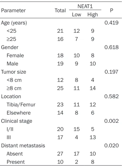
Table 1. Correlation between NEAT1 expres-sion and clinicopathological features of OS patients
Parameter Total NEAT1 P
Low High
Age (years) 0.419
<25 21 12 9
≥25 16 7 9
Gender 0.618
Female 18 10 8
Male 19 9 10
Tumor size 0.197
<8 cm 12 8 4
≥8 cm 25 11 14
Location 0.582
Tibia/Femur 23 11 12
Elsewhere 14 8 6
Clinical stage 0.002
I/II 20 15 5
III 17 4 13
Distant metastasis 0.020
Absent 27 17 10
[image:3.612.91.288.343.610.2]NEAT1 regulate OS progression by wnt/β-catenin
However, there is no correlation between NEAT1 expression level and other parameters, such as age, gender, tumor size and tumor location (P>0.05).
Up-regulated NEAT1 expression predicts poor prognosis of OS
The Kaplan-Meier survival analysis and log-rank test was performed to detect the correla-tion between NEAT1 expression and OS patients’ outcome. As shown in Figure 1B, patients with high levels of NEAT1 expression have a shorter overall survival rate than low level groups (P<0.05). Those data suggested that NEAT1 play an important role in OS progression.
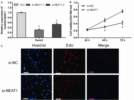
Inhibition of NEAT1 inhibits OS cell prolifera-tion
Because NEAT1 was overexpressed in OS, we investigated the function of NEAT1 using siRNA-mediated disruption of NEAT1
expres-Results
Up-regulation of NEAT1 in OS and its correla-tion with clinicopathologic features
[image:4.612.95.523.74.394.2]To explore the function of lncRNA NEAT1 in OS, we determined the expression level of NEAT1 in 37 OS tissues and adjacent non-tumor tissues by qRT-PCR, and normalized to GAPDH. The results showed that NEAT1 expression was significantly up-regulated com-pared to adjacent non-tumor tissues (Figure 1A, P<0.05). According to the median ratio of relative NEAT1 expression in OS tissues, the 37 OS patients were classified into two groups: the relative low group (n=19, NEAT1 expres- sion ratio≤median ratio) and the relative high group (n=18, NEAT1 expression ratio>median ratio). The association between NEAT1 expres-sion and clinicopathologic features was exam-ined. As shown in Table 1, the NEAT1 expres-sion level was significantly associated with clinical stage and distant metastasis (P<0.05).
cells (Figure 2C, P<0.05). Taken together, these results suggested that NEAT1 is associated with proliferative ability in OS cells.
Inhibition of NEAT1 suppresses OS cell migra-tion and invasion
To investigate the roles of NEAT1 on the migra-tion and invasion of OS cells, Scratch assay and Transwell invasion assay was performed to determine the migratory and invasive abilities of cells after transfection of siRNA. Scratch assay revealed that the migration capacity was significantly suppressed in Saos2 cells when si-NEAT1 was transfected (Figure 3A, P<0.05). Transwell invasion assay indicated that the cell number of invading cells was obviously lower in the si-NEAT1 group than in the si-NC group (Figure 3B, P<0.05). These findings showed that NEAT1 plays an important role in the migra-tion and invasion capacity in OS cells.
Mechanisms of NEAT1 exert its function
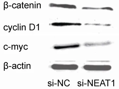
To determine the possible mechanism by which NEAT1 regulated proliferation and me- tastasis of OS cells, we performed western blot analysis to investigate the effects of NEAT1 knockdown on Wnt/β-catenin pathway, which is often aberrantly activated in human sion in Saos2 cells. qRT-PCR assay showed
[image:5.612.95.523.75.339.2]that NEAT1 was significantly decreased in si-NEAT1 transfected Saos2 cells (Figure 2A, P<0.05). In order to examine the proliferative ability of NEAT1, CCK-8 assay and EdU assays were performed. As shown in Figure 2B, CCK-8 assay showed inhibition of NEAT1 significantly inhibited cell proliferation in Saos2 cells com-pared to the control group (si-NC) (Figure 2B, P<0.05). In addition, compared with si-NC group, the relative fold of EdU positive cell was significantly decreased by si-NEAT1 in Saos2
Figure 3. Inhibited NEAT1 expression suppressed OS cells migration and invasion in vitro. A: Scratch assay showed that down-regulated NEAT1 in Saos2 cells significantly reduced their migration capacities. B: Transwell invasion assay showed that down-regulated NEAT1 in Saos2 cells significantly decreased their invasion capacities. *P<0.05.
[image:5.612.91.287.380.526.2]NEAT1 regulate OS progression by wnt/β-catenin
suppress cell proliferation, migration and inva-sion. Thus, NEAT1 plays an oncogene role in OS and represents a potential target for OS treatment.
Wnt/β-catenin signaling pathway is a highly conserved signaling pathway that regulates multiple biological processes, such as cell pro-liferation, invasion and other signaling path-ways, through regulating the ability of the multi-functional β-catenin protein, which is a crucial signaling molecule in the Wnt/β-catenin path-way [20]. Recent studies showed that Wnt/β-catenin pathway plays a critical role in the development and progression of OS [21]. Thus, we analyzed the activity of Wnt/β-catenin sig-naling in si-NEAT1 transfected OS cells. In our study, we found that decreased expression of NEAT1 suppressed the expression levels of β-catenin. Additionally, cyclin D1 and c-myc (classic downstream genes of the Wnt/β-catenin signaling pathway) protein expression were also down-regulated in si-NEAT1 transfected OS cells. Those data indicated that NEAT1 might regulate tumor progression through the Wnt/β-catenin pathway in OS. However, the exact mechanisms for NEAT1’s regulation of the Wnt/β-catenin pathways require further investigation.
In conclusion, the present study suggested for the first time that lncRNA NEAT1 acted as a functional oncogene in OS, and the up-regula-tion of NEAT1 expression is closely associa- ted with OS progression. Thus, lncRNA NEAT1 could potentially serve as a required thera- peutic method targeting for OS.
Acknowledgements
The project was supported by Scientific and Technological Project of Henan Province (No. 201403141).
Disclosure of conflict of interest
None.
Address correspondence to: Yuzhen Dong, Depart- ment of Orthopedics, The First Affiliated Hospital of Xinxiang Medical University, Weihui 453100, Henan, China. E-mail: [email protected]
References
[1] Kansara M, Teng MW, Smyth MJ and Thomas DM. Translational biology of osteosarcoma. Nat Rev Cancer 2014; 14: 722-735.
cancers and contributes to enhance cell prolif-eration and metastasis. Western blot showed that decreased expression of NEAT1 leads to significant decreased β-catenin expression in si-NEAT1 transfected OS cells compared with si-NC group. In addition, our study showed that cyclin D1 and c-myc (classic downstream genes of the Wnt/β-catenin signaling pathway) pro- tein expression were reduced in the si-NEAT1 transfected OS cells (Figure 4). Those results suggested that lncRNA NEAT1 may regulate OS progress through regulation of the Wnt/β-catenin signaling pathway.
Discussion
In recent years, lncRNA has gained increasing attention as a novel class of molecules that demonstrate an important role in carcinogene-sis [15]. Previous studies have proven that the expression of lncRNAs was dysregulated in various human cancers, and these aberrant lncRNAs might play important roles during cancer progression [16]. Recently, numerous lncRNAs have been reported to be abnorm- ally expressed in OS. For example, Sun et al revealed that lncRNA HULC was increased expression in OS and indicated a poor pro- gnosis and promoted cell metastasis in OS [17]. Li et al showed that HOTTIP was overex-pressed in OS and increased chemoresis- tance of OS cell by activating the Wnt/β- catenin pathway [18]. Dong et al showed that lncRNA MALAT1 promoted the prolifera- tion and metastasis of OS cells by activating the PI3K/Akt pathway [19].
LncRNA NEAT-1 has been reported to play an important role in types of tumors, but its role in OS is still unclear. In the present study, we showed that NEAT1 was significantly up-regu-lated in OS tissue compared to adjacent non-tumor tissues by qRT-PCR. The association analysis showed that clinical stage and distant metastasis were associated with NEAT1 expres-sion levels. Furthermore, the overall survival of the high NEAT1 expression group was poor than the low NEAT1 expression group, indicat-ing that lncRNA NEAT1 plays an important role in OS progression.
[2] Koshkina NV and Kleinerman ES. Aerosol gem-citabine inhibits the growth of primary osteo-sarcoma and osteoosteo-sarcoma lung metastases. Int J Cancer 2005; 116: 458-463.
[3] Ottaviani G and Jaffe N. The epidemiology of osteosarcoma. Cancer Treat Res 2009; 152: 3-13.
[4] Ando K, Heymann MF, Stresing V, Mori K, Re-dini F and Heymann D. Current therapeutic strategies and novel approaches in osteosar-coma. Cancers (Basel) 2013; 5: 591-616. [5] Chou AJ and Gorlick R. Chemotherapy
resis-tance in osteosarcoma: current challenges and future directions. Expert Rev Anticancer Ther 2006; 6: 1075-1085.
[6] Mercer TR, Dinger ME and Mattick JS. Long non-coding RNAs: insights into functions. Na-ture Reviews Genetics 2009; 10: 155-159. [7] Esteller M. Non-coding RNAs in human
dis-ease. Nat Rev Genet 2011; 12: 861-874. [8] Zhang HM, Yang FQ, Chen SJ, Che J and Zheng
JH. Upregulation of long non-coding RNA MALAT1 correlates with tumor progression and poor prognosis in clear cell renal cell carcino-ma. Tumour Biol 2015; 36: 2947-2955. [9] Zhang Z, Zhou C, Chang Y, Hu Y, Zhang F, Lu Y,
Zheng L, Zhang W and Li X. Long non-coding RNA CASC11 interacts with hnRNP-K and acti-vates the WNT beta-catenin pathway to pro-mote growth and metastasis in colorectal can-cer. Cancer Lett 2016; 376: 62-73.
[10] Wang F, Ying HQ, He BS, Pan YQ, Deng QW, Sun HL, Chen J, Liu X and Wang SK. Upregulated lncRNA-UCA1 contributes to progression of he-patocellular carcinoma through inhibition of miR-216b and activation of FGFR1/ERK sig-naling pathway. Oncotarget 2015; 6: 7899. [11] Choudhry H, Albukhari A, Morotti M, Haider S,
Moralli D, Smythies J, Schödel J, Green C, Camps C and Buffa F. Tumor hypoxia induces nuclear paraspeckle formation through HIF-2α dependent transcriptional activation of NEAT1 leading to cancer cell survival. Oncogene 2015; 34: 4482-4490.
[12] Ma Y, Liu L, Yan F, Wei W, Deng J and Sun J. Enhanced expression of long non-coding RNA NEAT1 is associated with the progression of gastric adenocarcinomas. World J Surg Oncol 2016; 14: 41.
[13] Sun C, Li S, Zhang F, Xi Y, Wang L, Bi Y and Li D. Long coding RNA NEAT1 promotes non-small cell lung cancer progression through regulation of miR-377-3p-E2F3 pathway. Onco-target 2016; [Epub ahead of print].
[14] Gao C, Zhang J, Wang Q and Ren C. Overex-pression of lncRNA NEAT1 mitigates multidrug resistance by inhibiting ABCG2 in leukemia. Oncol Lett 2016; 12: 1051-1057.
[15] Gibb EA, Brown CJ and Lam WL. The functional role of long non-coding RNA in human carcino-mas. Mol Cancer 2011; 10: 38.
[16] Cheetham SW, Gruhl F, Mattick JS and Dinger ME. Long noncoding RNAs and the genetics of cancer. Br J Cancer 2013; 108: 2419-2425. [17] Sun XH, Yang LB, Geng XL, Wang R and Zhang
ZC. Increased expression of lncRNA HULC indi-cates a poor prognosis and promotes cell me-tastasis in osteosarcoma. Int J Clin Exp Pathol 2015; 8: 2994-3000.
[18] Li Z, Zhao L and Wang Q. Overexpression of long non-coding RNA HOTTIP increases chemo-resistance of osteosarcoma cell by activating the Wnt beta-catenin pathway. Am J Transl Res 2016; 8: 2385-2393.
[19] Dong Y, Liang G, Yuan B, Yang C, Gao R and Zhou X. MALAT1 promotes the proliferation and metastasis of osteosarcoma cells by acti-vating the PI3K/Akt pathway. Tumour Biol 2015; 36: 1477-1486.
[20] Clevers H and Nusse R. Wnt/beta-catenin sig-naling and disease. Cell 2012; 149: 1192-1205.