Systems/Circuits
The Generation of Antiphase Oscillations and Synchrony by
a Rebound-Based Vertebrate Central Pattern Generator
Wen-Chang Li,
1Robert Merrison-Hort,
2Hong-Yan Zhang,
1and Roman Borisyuk
2,31School of Psychology and Neuroscience, University of St. Andrews, St Andrews, Fife, KY16 9TS, United Kingdom,2School of Computing and Mathematics,
Plymouth University, Plymouth, Devon, PL4 8AA, United Kingdom, and3Institute of Mathematical Problems in Biology, Russian Academy of Sciences,
142290, Pushchino, Russia
Many neural circuits are capable of generating multiple stereotyped outputs after different sensory inputs or neuromodulation. We have previously identified the central pattern generator (CPG) forXenopustadpole swimming that involves antiphase oscillations of activity between the left and right sides. Here we analyze the cellular basis for spontaneous left–right motor synchrony characterized by simul-taneous bursting on both sides at twice the swimming frequency. Sponsimul-taneous synchrony bouts are rare in most tadpoles, and they instantly emerge from and switch back to swimming, most frequently within the first second after skin stimulation. Analyses show that only neurons that are active during swimming fire action potentials in synchrony, suggesting both output patterns derive from the same neural circuit. The firing of excitatory descending interneurons (dINs) leads that of other types of neurons in synchrony as it does in swimming. During synchrony, the time window between phasic excitation and inhibition is 7.9⫾1 ms, shorter than that in swimming (41⫾2.3 ms). The occasional, extra midcycle firing of dINs during swimming may initiate synchrony, and mismatches of timing in the left and right activity can switch synchrony back to swimming. Computer modeling supports these findings by showing that the same neural network, in which reciprocal inhibition mediates rebound firing, can generate both swimming and synchrony without circuit reconfiguration. Modeling also shows that lengthening the time window between phasic excitation and inhibition by increasing dIN synaptic/conduction delay can improve the stability of synchrony.
Key words: central pattern generator; locomotion; oscillations; spinal cord; swimming; synchrony
Introduction
Multiple stereotyped behavioral patterns are common in both invertebrates and vertebrates. The underlying neural mecha-nisms have been better understood in some invertebrate prepa-rations (Kupfermann and Weiss, 2001; Jing and Weiss, 2005;
Kristan et al., 2005;Marder et al., 2005;Marder and Bucher, 2007;
Briggman and Kristan, 2008). Studies show some neurons can be active in multiple tasks (Weimann et al., 1991; Weimann and Marder, 1994) and circuits can be dynamically reconfigured to generate different outputs (Dickinson et al., 1990;Meyrand et al.,
1991,1994;Popescu and Frost, 2002;Norris et al., 2006;White and Nusbaum, 2011). Much less is known about circuit architec-ture in vertebrates. It is widely thought that polymorphic neural networks can mediate multiple motor outputs (Getting, 1989;
Soffe, 1993; Marder and Calabrese, 1996; Marder et al., 2005;
Briggman and Kristan, 2008;Doi and Ramirez, 2008;Rauscent et al., 2009;Klein et al., 2010). However, a number of recent non-mammalian vertebrate studies suggest that circuit reconfigura-tion is needed to generate different motor responses (Ritter et al., 2001;Kimura et al., 2006;Berkowitz, 2007,2008;Li et al., 2007;
McLean et al., 2007,2008;Liao and Fetcho, 2008;Frigon, 2009;
Satou et al., 2009;Wyart et al., 2009;Berkowitz et al., 2010). There are three basic categories of neural activity in central pattern generator (CPG) circuits that control motor outputs: tonic firing, antiphase oscillations, and synchronous oscillations (termed “synchrony” here for simplicity). Movements like walk-ing, runnwalk-ing, and axial swimming involve antiphase oscillatory left–right and flexor– extensor muscle activities, known to be co-ordinated by reciprocal inhibition (Kiehn, 2006). In galloping, hopping, flying, frog- or turtle-style limb-based swimming, left– right synchrony is the operational mode. Whereas neural mech-anisms underlying normal left–right synchrony have received little attention, synchrony has been reported in many experimen-tal conditions (Cohen and Harris-Warrick, 1984; Clarke et al., 1991;Cowley and Schmidt, 1995;Kullander et al., 2003). In these studies, genetic mutations or pharmacological blockade led to Received Oct. 1, 2013; revised Feb. 19, 2014; accepted March 21, 2014.
Author contributions: W.-C.L. designed research; W.-C.L., R.M.-H., and R.B. performed research; W.-C.L. and R.M.-H. analyzed data; W.-C.L., R.M.-H., H.-Y.Z., and R.B. wrote the paper.
This work was supported by the Wellcome Trust and a Royal Society Fellowship to W.-C.L. Many analyses on synchrony in this study took advantage of the big dataset accumulated in our past collaborations with Drs. Alan Roberts and Steve Soffe. This is essential since spontaneous synchrony is not common in normal conditions. The computational model was partially developed with support from a grant funded by UK Biotechnology and Biological Sciences Research Council. We thank Drs. Ying Zhang, Avis Cohen, Andrew McClellan, Keith Sillar, Alan Roberts, Steve Soffe, and David Parker for helpful discussions.
The authors declare no competing financial interests.
This article is freely available online through theJ NeurosciAuthor Open Choice option.
Correspondence should be addressed to Wen-Chang Li at the above address. E-mail: [email protected]. H.-Y. Zhang’s present address: Centre for Neuroregeneration, University of Edinburgh, Edinburgh, UK. DOI:10.1523/JNEUROSCI.4198-13.2014
Copyright © 2014 Li et al.
functional reorganization of the circuits, and crossed excitation was proposed to couple left–right motor rhythms in synchrony.
Xenopustadpoles have been used as a simple vertebrate model for studying neural mechanisms controlling locomotion, and the configuration of the tadpole spinal circuit has been sys-tematically characterized recently using paired whole-cell re-cordings (Roberts et al., 2010). During tadpole swimming, neuronal activity alternates between the two sides of the spinal cord and hindbrain by way of reciprocal inhibition (Moult et al., 2013). Occasionally, however, the activity on the two sides can become spontaneously locked in synchrony, similar to that re-ported byKahn and Roberts (1982)in curarized tadpoles. Syn-chrony disrupts propulsive swimming and bears no identified behavioral significance to tadpoles. We want to understand how synchrony rhythms are generated by this well studied neural cir-cuit. In contrast to the prevalent evidence that circuit reconfigu-ration is needed for generating different network outputs, we reveal that the same CPG can generate both swimming and syn-chrony and that both types of rhythmic activity are coupled by fast reciprocal inhibition.
Materials and Methods
Experimental procedures were similar to those described previously (Li and Moult, 2012), approved by the local Animal Welfare Ethics Commit-tee, and complied with UK Home Office regulations. Pairs of adult Xe-nopus of either sex were given injection of Human Chorionic Gonadotropin to induce mating. Tadpoles at stage 37/38 (Nieuwkoop and Faber, 1956), whose sex cannot be determined at this stage, were briefly anesthetized with 0.1% MS222 (3-aminobenzoic acid ester; Sigma-Aldrich) to allow immobilization with␣-bungarotoxin (12.5M; Tocris Cookson) after the dorsal fin was cut open. The animal then was pinned onto a sylgard stage for dissections to expose myotome blocks, spinal cord, and caudal hindbrain. Additional dissections were then made to remove ependymal cells lining the central canal to reveal neuro-nal somata. Saline contained (in mM) 115 NaCl, 3 KCl, 2 CaCl2, 2.4 NaHCO3, 1 MgCl2, and 10 HEPES, and pH was adjusted to 7.4 with 5M NaOH. Current-clamp or voltage-clamp whole-cell recordings were made from exposed somata under a Nikon E600FN microscope. Patch pipettes were filled with 0.1% neurobiotin (Vector Laboratories) in the intracellular solution (in mM: 100 K-gluconate, 2 MgCl2, 10 EGTA, 10 HEPES, 3 Na2ATP, 0.5 NaGTP, pH adjusted to 7.3 with KOH). Pipette DC resistances ranged from 10 to 20 M⍀. Standard current-clamp or voltage-clamp recordings were made with the Axon-2B or Axon Multi-clamp 700B amplifier (Molecular Devices). Junction potentials (14.7 mV) were not corrected in voltage-clamp recordings. Data were digitized using either CED 1401 plus or Power 1401 mkII, sampled with Signal 5 (Cambridge Electronic Design). Fictive swimming (we use “swimming” in the text for an easier description) was initiated by applying single or repetitive 1 ms current pulses to tadpole skin on the tail or head. Motor nerve (m.n.) recordings from muscle clefts were made normally between the 5th and 10th muscle segments caudal to the otic capsule by using glass suction electrodes. Whole-cell recordings were made from neurons in the hindbrain (caudal to the fifth rhombomere segment) and rostral spinal cord (rostral to the fifth myotome). They were in current-clamp mode for looking at neuronal activities and in voltage-clamp mode for the examination of synaptic currents. Most neurons were successfully filled with neurobiotin after recording and stained as described previ-ously (Li and Moult, 2012). Neuronal anatomy was used for final classi-fication of neurons. Microperfusion of strychnine was done by positioning a glass pipette (⬃10m in diameter) in the hindbrain area. The microperfusion pipette was connected to a 50 ml syringe where a gentle pressure (⬃0.01 bar) was kept.
Dynamic clamping was configured in Signal 5 to inject artificial syn-aptic currents into the recorded excitatory descending interneurons (dINs) to evoke extra midcycle firing. The currents were defined by an␣ synapse model and triggered by action potentials in the recorded dIN
(autapse) with a delay of 20 –25 ms. Each dynamic-clamping period lasted about 200 ms to a few seconds.
Data processing and analyses were performed using Dataview (version 6.1, courtesy of Dr. W. J. Heitler, University of St. Andrews) and Excel. Synchrony activity was initially compared with swimming in recordings where a left dIN and a right m.n. were recorded simultaneously. In these recordings, dIN spike timing was adjusted to compensate for rostral-to-caudal delays and also dIN-to-m.n. activity delays (Roberts et al., 2010). dIN spike time was shifted for 6.5–11.2 ms so that dIN spikes appeared, on average, in the middle of swimming cycles defined by m.n. bursts (phase, 0.5). Neuronal firing reliability during swimming or synchrony is defined as the percentage of cycles with action potentials. The measure-ments of EPSCs and IPSCs were made by clamping dIN membrane po-tential at approximately⫺55 or 0 mV, respectively. The size of EPSCs and IPSCs during synchrony was compared with that in swimming im-mediately before or after the synchrony bouts. Statistical analyses were done using PASW statistics 18 (SPSS). Normality tests were performed first, and means were given with SEs (Mean⫾SEM) and compared using
ttests for normally distributed data. The Wilcoxon signed rank test was used for other datasets unless otherwise stated.
[image:2.594.304.540.62.257.2]currents in dINs lack a fast component (W.-C. Li, unpublished observa-tions). All other parameters associated with neurons’ membrane prop-erties were the same as given byRoberts et al. (2014).
The model includes glutamatergic (AMPARs, NMDARs with Mg2⫹ -dependent voltage dependency) and glycinergic synapses established in paired whole-cell recordings (Sautois et al., 2007). We introduced syn-aptic saturation in the present model by increasing synsyn-aptic openingo(t)
and closingc(t)variables using a varying, rather than constant, increment after a spike. The postspike increment⌬o,c(t)is given by the following
equation:
⌬o,c共t兲⫽s
冉
1⫺c共t兲⫺o共t兲
冊
.Here parametersis the original increment size (AMPA/NMDA, 1.25; glycine, 3.0), andis the saturation level (AMPARs and glycine recep-tors, 1.0; NMDA, 5.0). These values were chosen so that saturation only had a major effect on NMDAR activation during high-frequency activity. In most cases, we used the same synaptic parameters as given by Rob-erts et al. (2014), but in some cases, minor changes were made. First, to obtain realistic and stable swimming activity after removal of the fast potassium current in dINs and to enable synaptic saturation, it was nec-essary to increase the strength of dIN– dIN NMDA synapses from 0.15 to 0.29 nS. This new value agrees with the standard NMDA conductance given bySautois et al. (2007). To obtain realistic measures of firing reli-ability of CPG neurons during swimming and synchrony, we made small changes to the conductances of synapses that dINs make onto
commis-sural interneurons (cINs) (from 0.593 to 0.65 nS) and motoneurons (MNs) (from 0.593 to 0.54). For the same reason, we made a larger change to the strength of dIN to ascending in-terneuron (aIN) synapses (from 0.1 to 0.3 nS). Finally, the conductance of the inhibitory syn-apses made by aINs onto all other neuron types was reduced to 0.0435 nS so that these synapses provided only 10% of the inhibitory strength of corresponding cIN synapses.
As in the study byRoberts et al. (2014), all synaptic connections contained a constant lay of 1 ms and a conduction delay that de-pends on the difference in rostrocaudal position of the presynaptic and postsynaptic neurons according to a conduction speed of 3.5 ms/mm.
The simulation was implemented using cus-tom C code. The simulator uses the fourth-order adaptive Runge-Kutta-Felberg ODE solver from the GNU Scientific Library (ver-sion 1.15). The absolute and relative error tol-erances for step-size control and adjustment were 1.0e⫺5, and spikes are detected by mem-brane potential zero crossing after every time step (maximum step size, 0.5 ms). Small vari-ability in neuron and synapse strengths was in-troduced by adding Gaussian noise (SD of 2% of mean values) to cell membrane capacitance and channel conductances and Gaussian noise (SD of 5% of mean values) to the conductance of each synapse.
Results
Basic features of synchrony in tadpoles
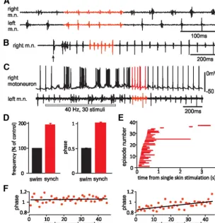
Left–right synchrony in tadpoles involves simultaneous m.n. bursts on both sides, in contrast to the alternation during swim-ming (Fig. 2A). We first analyzed the basic properties of synchrony. We arbitrarily define periods with more than five con-secutive bursts of synchronous activity (four cycles) as a synchrony bout. Initially, we analyzed sponta-neous synchrony bouts in 14 tadpoles where the activity of a left m.n. (normally eighth/ninth myotome cleft) and a right dIN in the caudal hindbrain was recorded simultaneously (see Materials and Methods;n⫽17 synchrony bouts, 419 cycles). Periods of swimming were chosen either immediately before or after the synchrony for comparison (175 cycles). During synchrony, the phase of left-side activity relative to the right side was 1 (1.01⫾ 0.01; one-sample, two-tailedttest,p⫽0.18;Fig. 2A,D), indicat-ing activity on the two sides was synchronous. In 13 long bouts with 30⫾4 cycles (range, 14 – 62), we examined whether one side clearly led the synchronous activity. In four bouts, there were
[image:3.594.55.359.69.384.2]⬎80% of cycles with phase value above or below 1, indicating one side being active consistently earlier than the other side (Fig. 2F, left). If synchrony maintenance relies on the synchronous firing of neurons in the network, the gradual drifting of phase during synchrony may end synchrony itself. Linear regression was ap-plied to the phase measurements of each of the 13 long bouts to see whether there was drifting of phase with the progression of synchrony. Significance was found in 6 of the 13 bouts. The drift-ing rate (regression coefficient) was 0.0029⫾0.0008 per cycle, equivalent to 0.11⫾0.03 ms assuming one synchrony cycle is about 40 ms. In three bouts, the leading side remained the same but with increased phase lags, whereas in the other three, it lost its Figure 2. Left–right motor synchrony in tadpoles and its basic features.A, A synchrony bout with left and right m.n. activity
recorded simultaneously (both fifth/sixth muscle cleft).B, Synchrony after single skin stimulation (arrow).C, Synchrony after repetitive skin stimulation (gray bar) with a simultaneous motoneuron recording.D, Synchrony rhythm frequency and phase (red bars) compared with that of adjacent swimming (black bars).E, Starting time (beginning of red lines) and duration (length of lines) for 37 synchrony bouts after single skin stimulation in 13 tadpoles.F, Cycle-by-cycle phase measurements of two synchrony bouts. Left, Example lacking a phase drift (linear regression coefficient is 0.0004 per cycle;R2⫽0.006;p⫽0.62). Right, Example with the
clearest phase drift with the progression of synchrony (coefficient is 0.0036 per cycle;R2⫽0.41;p⬍0.001). Solid lines are for
lead toward the end of synchrony (Fig. 2F, right). The small phase drifts (0.07⫾0.02; n⫽6, estimated by regression) all ended abruptly with phases jumping to⬃0.5 when synchrony switched to swimming. Synchrony frequency (24⫾0.6 Hz) was reliably twice the frequency of adjacent swimming (12.4 ⫾ 0.4 Hz; 195.5⫾4.5%; one-sample, two-tailedttest,p⫽0.34;Fig. 2D). This feature of abruptly-doubled m.n. frequency fits in with pre-vious descriptions (Clarke et al., 1991). Since swimming fre-quency only changes gradually after initiation, we used this simple unique feature to identify synchrony when activity was not recorded simultaneously from both sides.
In a bigger sample of 37 tadpoles, we analyzed how often and when synchrony occurred. Within 6 s after single skin stimula-tion, 37 synchrony bouts were seen in 13 tadpoles (17.5% of 212 trials; Fig. 2B). Within 2.5 s after repetitive skin stimulation (30 – 40 Hz, 20 –50 pulses), 50 bouts of synchrony were seen in 22 tadpoles (26.6% of 188 trials;Fig. 2C). Overall, we observed syn-chrony in 26 of 37 tadpoles (70.3%) when both types of stimula-tion were considered. The 37 synchrony bouts evoked by single skin stimulation lasted 0.35⫾0.03 s (range, 0.09 –2.04) or 11⫾ 0.8 cycles (range, 4 – 66). Most of them started within 0.5 s and stopped within 1 s after skin stimulation (32 of 37;Fig. 2E). When the hindbrain was sectioned at the fifth/sixth rhombomere bor-der in another sample of 11 tadpoles, synchrony was seen in 1 of 82 trials after single skin stimulation and in 59 of 521 trials after repetitive skin stimulation of nine tadpoles. This suggests that tadpole spinal cord and caudal hindbrain, which contain the highest density of dINs and are critical for swimming (Li et al., 2006;Soffe et al., 2009), can also generate synchrony.
Neuronal activity during synchrony
To understand how synchrony is generated, we next identified neurons that were active during synchrony. We found that only neurons that were rhythmically active during swimming fired action potentials during synchrony (Table 1). They included all swimming CPG neurons (Roberts et al., 2010): dINs, cINs, aINs, and motoneurons (Fig. 3). We define their firing reliability as the percentage of cycles with action potentials. Their firing reliabili-ties during synchrony were lower than those during swimming immediately before or after synchrony (Table 1). To see whether there is a change of firing reliability with the progression of syn-chrony, we compared the reliability in the first five synchrony cycles with that in the last five cycles. In 26 long bouts with an average of 19.4⫾1.3 synchrony cycles (range, 12–35) from 11 neurons (3 motoneurons, 4 dINs, 2 cINs, and 2 aINs), no
differ-ence was found (pairedttest,p⫽0.74). Neurons that were silent during swimming [dorsolateral commissural interneurons (dlcs), dor-solateral ascending interneurons (dlas), excitatory commissural interneurons (ecINs), and repetitive-firing descending interneu-rons (dINrs)] remained silent during synchrony (Table 1). This lack of recruitment or exclusion of network neurons between the two motor patterns strongly suggests that synchrony is a motor output derived from the swimming circuit.
Analyses of dIN activity during synchrony
dINs are the only excitatory interneurons in the swimming CPG, and their activity drives swimming (Soffe et al., 2009;Li, 2011). We made 11 paired whole-cell recordings with one dIN and an-other (non-dIN) CPG neuron at similar locations on the same side. This allowed us to determine directly which neuron fired first in each synchrony or swimming cycle. dIN firing during synchrony preceded the firing of non-dINs (seven cINs, two mo-toneurons, two unidentified). The latency between dIN and non-dIN firing in synchrony (2.6⫾0.68 ms;n⫽11 bouts) was similar to that in swimming (2.47⫾0.46 ms;p⬎0.05,ttest;Fig. 4A–C). Since there was no recruitment of extra types of neurons in syn-chrony (see above), this suggests that the excitatory dINs were also responsible for driving the firing of other neurons during synchrony, as they are for swimming (Soffe et al., 2009). dIN rebound firing had been proposed as the main mechanism in sustaining swimming (Moult et al., 2013). To see whether the same rebound mechanism could sustain synchrony, we mea-sured the delay between the start of IPSPs and the time point when the following dIN spike crossed 0 mV. This time (rebound time) should reflect the interaction between different voltage-dependent ionic channels and receptor channels that give rise to dIN rebound firing. No difference was found between re-bound time during swimming (36.3⫾2.6 ms) and that during synchrony (34.3⫾2.2 ms;n⫽7 dINs; two-tailed pairedttest, p⬎0.05;Fig. 4D,E). These data suggest that the same rebound mechanism that underlies swimming is also responsible for synchrony.
Synaptic currents in dINs during synchrony
[image:4.594.42.286.83.219.2]Knowing that dIN activity is likely to drive synchrony, we next looked at the synaptic currents dINs receive during synchrony to understand how they could support dIN firing. dINs receive ex-citation from other dINs during swimming, with NMDAR com-ponents contributing to tonic inward currents and AMPAR and nicotinic receptor components giving rise to the phasic on-cycle EPSCs (Fig. 5A). Additionally, cINs from the opposite side pro-duce midcycle IPSCs, and ipsilateral aINs propro-duce early-cycle IPSCs, which appear between the on-cycle dIN EPSCs and cIN IPSCs (Li and Moult, 2012;Fig. 5B, arrows). First, membrane potential was clamped around IPSC reversal (approximately⫺55 mV) to compare EPSCs. EPSCs were smaller in synchrony (⫺119⫾13 pA) than those in swimming just before or after synchrony (⫺150⫾19 pA;n⫽11 bouts in 11 neurons; pairedt test, p ⬍ 0.01; Fig. 5A). Second, membrane potentials were clamped around 0 mV to reveal IPSCs. During synchrony, aINs and cINs fire synchronously, and both can contribute to the IPSCs. The mixed IPSCs during synchrony (262⫾42 pA) were smaller than the midcycle cIN-mediated IPSCs in swimming (322⫾51 pA;n⫽8 bouts; pairedttest,p⬍0.05;Fig. 5B). This fits with the observation above that neuronal firing during syn-chrony is less reliable than during swimming (Table 1). Third, the delay between EPSCs and IPSCs in swimming or synchrony cy-cles was measured when membrane potential was clamped at Table 1. Neuronal firing reliability (percentage of cycles with firing) during
synchrony and adjacent swimming
Cell type
Number of cells
Firing reliability (%), mean⫾SEM or mean (median)
Synchrony Swimming
Swimming CPG
dIN 19 84 (100)* 99 (100)
Motoneuron 13 65⫾7.3* 86 (100)
aIN 13 45⫾9** 64⫾9
cIN 10 52⫾8*** 73⫾9
Other types
dlc 8 0 0
dla 10 0 0
dINr 11 0 0
ecIN 6 5 (0) 2 (0)
approximately⫺20 mV. This was measured as the time differ-ence between the EPSC trough and the IPSC peak. The delay was 7.9⫾1 ms during synchrony, which was much shorter than that during swimming (41⫾2.3 ms;n⫽8 bouts; pairedttest,p⬍ 0.01;Fig. 5C). This delay is important because if inhibition arrives during the rising phase of a dIN action potential, then the spike may be suppressed, which reduces spike reliability (Table 1; Figs.
3D, 5D). Instead, inhibition must arrive during the decaying phase of the dIN action potential to trigger another postinhibi-tory spike. The fact that during synchrony IPSCs arrive very shortly after dIN spiking may contribute to the instability of syn-chrony, since a small difference in the timing of inhibition may cause it to arrive before dIN spiking.
Midcycle firing in dINs may initiate synchrony
A critical feature of synchrony is that its frequency is double that of swimming. Abnormal midcycle dIN firing, which doubles the
IPSPs in dINs (median, 0.54; n ⫽308 cycles in five tadpoles analyzed;p⫽0.69, independent sample nonparametric median test). However, a dIN spike can appear a few milliseconds earlier than IPSPs in individual cases (Fig. 6B), allowing it to drive post-synaptic firing. Therefore, this type of abrupt doubling of dIN firing frequency during swimming in the form of midcycle spikes may be key to synchrony initiation.
As we have shown above, most synchrony bouts occur within 1 s of skin stimulation. Synaptic currents during this initial period differ from the following periods in that there are bigger tonic inward currents and more reliable aIN IPSCs in dINs (Li and Moult, 2012). Higher depolarization can promote firing at mid-cycle position. We injected positive DC (60 –200% threshold cur-rent for evoking firing at rest) into 15 dINs to mimic higher background excitation to see whether this could induce midcycle dIN firing during swimming. In three dINs, this led to doublet on-cycle firing in 48 of 49 swimming cycles. In the other 12 dINs, DC injection led to midcycle dIN spikes on many cycles (Fig. 6C; 269 of 796 cycles). In three trials in three different dINs, this DC injection coincided with synchrony during the injection period (Fig. 6F). However, such coincidence was not repeatable. Inject-ing DC into non-dINs (55–200% threshold current for firInject-ing; five motoneurons, five cINs, four aINs, and two unidentified CPGs) mainly induced on-cycle multiple firing (265 of 502 cycles) with occasional midcycle firing (Fig. 6D; 31 cycles). There were higher percentages of cycles with midcycle firing in dINs (p⬍0.01) and multiple firing in non-dINs (p⬍0.001, independent samples me-dian test;Fig. 6E). These results indicate that dINs, most of which
were recorded in the caudal hindbrain and rostral spinal cord, tend to fire midcycle spikes during enhanced excitation. In some cases, the midcycle firing of dINs may directly lead to synchrony.
The midcycle dIN firing evoked by DC current injections dur-ing swimmdur-ing tended to appear in the latter half-cycle (Fig. 6C, filled triangles), potentially reducing its ability to evoke syn-chrony. This asymmetrical firing pattern is presumably caused by different synaptic events in a swimming cycle, first strong reliable on-cycle dIN excitation and then weak unreliable early-cycle aIN inhibition and strong reliable midcycle cIN inhibition (Li and Moult, 2012; Fig. 5B). To have better control over the timing of evoked midcycle dIN firing, we used the dynamic-clamp technique to time the injection of artificial excitatory synaptic currents into the recorded dINs. The synaptic currents were triggered by the dIN ac-tion potentials with an artificial delay of approximately half of the swimming cycle period (20 –25 ms). We failed to induce any syn-chrony bouts in this way (n⫽12 dINs, 259 trials, 3–50 cycles per trial with current injections, data not shown).
Effects of blocking inhibition on synchrony generation
Blocking glycinergic inhibition can induce synchrony in neonatal rats (Cowley and Schmidt, 1995) and lamprey (Cohen and Harris-Warrick, 1984). In tadpoles, bath-applied 2.5M
strych-nine and 20M SR95531 (GABA antagonist) led to an initial
[image:6.594.84.505.59.373.2]tions of strychnine (0.07– 0.25 M) increased swimming
fre-quency, but the incidence of synchrony did not increase (Li and Moult, 2012).
Fortuitously, two batches of tadpoles showed an unusual ten-dency for synchronous activity (Fig. 7A; data were not included for preparingFig. 2E). In these animals, 21.1⫾4.6% of rhythmic activity (including both synchrony and swimming) was syn-chrony (n ⫽63 swimming episodes). Microperfusing 0.5M
strychnine in the hindbrain area decreased synchrony to 1.8⫾ 0.6% (n⫽66 episodes, 7 tadpoles;p⬍0.01, pairedttest). This recovered to 23.3, 17.8, and 5.3% in wash in three tadpoles (⬎20 min;Fig. 7A). Injecting positive DC in two of the seven dINs, which could induce midcycle dIN firing in control (cycles with midcycle firing are 33 of 57 and 23 of 115, respectively) as shown above, evoked little dIN midcycle firing in strychnine (0 of 20 and 1 of 84;Fig. 7B). These data imply that inhibition may play an important role in the generation of synchrony.
Synchrony initiation and stability in a computational model of swimming
The excitation from single midcycle spiking in one dIN in whole-cell recordings above was very unlikely to be sufficient to drive substantial midcycle cIN firing and start synchrony. Therefore,
we injected brief midcycle currents into multiple dINs simulta-neously in a computational tadpole swimming network to see whether synchrony could be initiated.
[image:7.594.84.503.65.423.2]imental data. During synchrony, neuronal firing reliability was lower than periods of swimming (pairedttest,p⬍0.001 in each case; n⫽12 synchrony bouts; 193 synchrony cycles and equivalent swim-ming periods compared;Fig. 8B). AMPA receptor conductance was smaller during synchrony (4.39⫾0.71 nS) than during swimming (5.12⫾0.79 nS;p⬍0.01, pairedttest;n⫽10 dINs). Similarly, combined inhibitory conductance during synchrony (3.87⫾0.86 nS) was smaller than cIN conductance during swimming (5.28⫾ 1.17 nS;p⬍0.01, pairedttest;n⫽10 dINs;Fig. 8C). Reducing inhibition strength by up to 60% failed to convert normal swimming to synchrony (12 trials). Further depression of inhibition made it difficult (three successes in 12 trials at 70%) or impossible (above 70% reduction) to start swimming, similar to what was seen exper-imentally (Moult et al., 2013).
As shown above, the delay between EPSCs and IPSCs is much shorter during synchrony, and from this we hypothesize that the
short delay (Fig. 5C) reduces neuronal firing reliability and is responsible for the unstable nature of synchrony. To test this, we first advanced dIN activity on one side to further shorten the gap. We injected some brief depolarizing currents into all dINs (3 ms, 100 pA) on the left side to advance neuronal activity by 2.27⫾ 0.15 ms during the fifth synchrony cycle (12 trials). This reliably shortened synchrony bouts (related-samples Wilcoxon signed rank test,p⬍0.01;Fig. 9A) with 10 of 12 bouts ending in the following cycle or with one cycle delay. We next artificially lengthened the EPSC–IPSC gap by increasing the synaptic/con-duction delay at dIN to cIN synapses to see whether we could improve synchrony stability. Synaptic delay is about 1 ms in the tadpole swimming circuit, and dIN spike conduction velocity is 0.36 mm/ms (360m will increase conduction delay by 1 ms;
Soffe et al., 2009). Increasing dIN– cIN transmission/conduction delay is comparable with adding relay interneurons between dINs Figure 6. Extra midcycle dIN firing may initiate synchrony.A, Midcycle dIN firing (filled triangles) before and after a synchrony bout.B, Some examples of midcycle dIN firing showing the relative timing of the firing and cIN inhibition (triangles). Note some spikes are narrowed by inhibition (*). Lack of midcycle burst in the overlapped left m.n. recording implies the absence of synchrony in the circuit. The illustrated cycles are normalized based on dIN spiking.C, Depolarizing DC injection (gray bar) into a dIN during swimming results in reliable midcycle firing (filled triangles). Note the midcycle spikes do not divide the swimming cycle equally.D, DC injections into a motoneuron result in multiple firing (*) in many cycles and some midcycle firing (filled triangles).E, Summary of 12 dINs’ (filled bar) and 16 non-dINs’ (unfilled) firing after DC injections during swimming. Significance values as follows: **p⬍0.01; ***p⬍0.001.
and cINs or increasing dIN projection distance (e.g., as is the case when the spinal cord size increases during development). The percentage of simulations with stable synchrony (continuous synchrony from initiation to the end of the 3 s simulation,⬎100 cycles) initiated by midcycle dIN current injections increased with longer delays (Fig. 9B,C). When the delay was 2 ms and above, all evoked synchrony bouts were stable. With longer dIN– cIN synaptic/conduction delays and stable synchrony, all CPG neurons fired more reliably during synchrony (p⬍0.001 in each neuron type, one-way ANOVA; Fig. 9D). We also examined whether there was significant drift of phase during the stable synchrony bouts in simulation with a 2ms dIN– cIN delay. Linear regression revealed some small drift in 6 of the 12 simulations (drifting rate, 0.000074⫾0.000009 per cycle;p⬍0.05). The modeling, therefore, provides theoretical support that tadpole
swimming circuit can generate synchrony and dIN– cIN synap-tic/conduction delay can directly determine synchrony stability.
Discussion
We propose that left–right motor synchrony in tadpoles is medi-ated by the swimming CPG. The extra midcycle firing of dINs during swimming can initiate synchrony, and the mismatches of timing in left and right activity can convert synchrony to swim-ming (Fig. 10A). dIN midcycle firing will drive ipsilateral cINs to fire action potentials and produce extra crossed inhibition on the opposite side. This extra inhibition means dINs on both sides will fire simultaneously on postinhibitory rebound and start synchrony. When the activity on one side comes earlier than the opposite side, the crossed inhibition it produces will arrive earlier, inhibiting the activity on the opposite side. Figure 7. The effects of weakening inhibition on synchrony and dIN firing.A, Synchrony bouts in control, when 0.5Mstrychnine was applied and during wash. The arrow indicated time of skin stimulation.B, Midcycle dIN firing (arrowheads) induced by DC injection (gray bar) during swimming (control) and its suppression by 0.5Mstrychnine.
Then, the synchrony pattern breaks down and swimming resumes.
The production of different rhythms in CPGs has been asso-ciated with circuit reconfiguration after different sensory inputs (Marder et al., 2005;Briggman and Kristan, 2008;Berkowitz et al., 2010) or under neuromodulatory influences (Rauscent et al., 2009;Combes et al., 2012). The synchrony shown here does not seem to involve reconfiguration of the swimming CPG. Both swimming and synchrony can occur after the same brief skin stimulation, and they involve the same types of neuron, without recruitment of extra or inhibition of existing types of neuron. The same rebound mechanism that sustains swimming appears to operate in synchrony, since dIN firing precedes that of other neurons in both patterns and the rebound times during both outputs are not different. The phase of cIN inhibition during swimming is 0.54, indicating dIN rebound firing can take place within less than half a cycle. This leaves scope for halving the activity cycle period and may explain the strict relationship be-tween synchrony and swimming frequencies (Fig. 10B). In com-parison, the generation of struggling rhythms requires sustained sensory inputs and the recruitment of specialized interneurons, and, furthermore, struggling frequency is not locked to swim-ming rhythms in any fixed manner (Li et al., 2007).
We have identified midcycle dIN firing as one potential trigger for swimming to switch to synchrony. This extra firing can result from enhanced excitation, e.g., at the beginning of a swimming episode when sensory inputs are still present. In accordance with this, most synchrony bouts occur within 0.5–1 s of single skin stimulation. Although we have recently shown that pacemaker
firing is not sufficient to sustain swimming rhythms, higher ex-citation may support firing at high frequencies in some dINs (Fig. 6A–C). Once synchrony is initiated, the narrow window for neu-ronal firing (Fig. 5C) determines that the firing of some dINs may be prevented by IPSPs (Table 1; Figs.3D,5D) and account for the unreliable firing of CPG neurons during synchrony. It also means that synchrony will stop when the activity on both sides drift out of synchrony slightly and explains why most spontaneous syn-chrony bouts are short. Indeed, drifts in phase with time have been seen only in some longer synchrony bouts. In modeling, evoking midcycle dIN firing during swimming can initiate syn-chrony, and advancing activity on one side even for only a couple of milliseconds can convert synchrony back to swimming. Syn-chrony evoked in the model resembles recorded synSyn-chrony in frequency and phase features. Artificially broadening the EPSC– IPSC delay by increasing dIN-to-cIN synaptic/conduction delay increased CPG firing reliability during synchrony and made syn-chrony more stable. Therefore, our biological data and modeling both support that the transitions between swimming and syn-chrony are unlikely to involve circuit reconfiguration.
[image:10.594.84.508.62.365.2]tadpole swimming circuit in a large number of paired whole-cell recordings (Roberts et al., 2010;Li, 2011). This has enabled de-tailed analyses of neuronal activity and synaptic events during swimming and synchrony here and also allows us to build a com-putational swimming model of large populations of neurons with biologically realistic properties (Sautois et al., 2007;Borisyuk et al., 2011,2014). Both our biological and modeling data now pro-vide strong support that reciprocal inhibition, mediated by fast glycinergic synapses, can synchronize network activity. The spontaneous synchrony does not involve different sensory in-puts, circuit configuration (Roberts and Tunstall, 1990), or changes in cellular properties like decreased fast potassium cur-rents (Wall and Dale, 1994;Dale, 1995).
The left–right coordination of spinal activity is mediated by crossed inhibition (Kiehn, 2006), but direct commissural excit-atory interneurons have been found in some vertebrates. Direct commissural glutamatergic synapses onto motoneurons were re-ported in mice (Quinlan and Kiehn, 2007). In lamprey, commis-sural excitatory interneurons have been identified in paired recordings (Buchanan, 1982; Parker and Grillner, 2000), but their role in swimming is primarily unknown (Parker, 2006). Although some interneurons with commissural axons were found to be positive for vesicular glutamate transporters in the larval zebrafish spinal cord (Higashijima et al., 2004), no left– right motor synchrony has been reported in zebrafish. In the tadpole swimming circuit, there is little evidence supporting the existence of crossed excitation from paired recordings except from the sensory interneurons, which are not active during swimming (Dale, 1985;Li et al., 2007). Recording from neurons caudal to hemisections only revealed cIN inhibition during swimming (Soffe and Roberts, 1982).
Blocking glycinergic inhibition may transform alternating flexor– extensor and left–right motor rhythms into bilateral and also flexor– extensor synchrony at lowered frequencies in rodents (Cowley and Schmidt, 1995;Cazalets et al., 1998;Beato and Nis-tri, 1999). Similar left–right synchrony after inhibition blockade was seen in lamprey (Cohen and Harris-Warrick, 1984;Alford
and Williams, 1989;Hagevik and McClel-lan, 1994) and mudpuppy (Jovanoviæ et al., 1999). It is thought that in the absence of strong reciprocal inhibition, the weak crossed excitation becomes dominant and couples the activity on the two sides in synchrony (Cohen and Harris-Warrick, 1984; Quinlan and Kiehn, 2007). It is not known whether strychnine could re-cruit neurons in the circuit that are inhib-ited in normal rhythms in these cases. The spontaneous synchrony we have studied here cannot be explained by increased weak crossed excitation since there is no evidence for its presence during swim-ming or synchrony. Instead, inhibition seems to play a role in sustaining sponta-neous synchrony as demonstrated by our experiments on the tadpoles with a high tendency for synchronous activity.
Left–right synchrony coupled by crossed excitation has also been reported in some mutant mice. One example comes from mice lacking V2a interneurons (ChX10 DTA), which form ipsilateral excitatory synapses onto commissural interneurons. These mice show galloping gait at high-locomotion speed instead of normal trotting (Crone et al., 2009). This was thought to result from weakened activation of crossed inhibition pathways (Crone et al., 2008,2009). A second example comes from mutants lack-ing Ephrin type-A receptor 4, in which ipsilateral excitatory in-terneurons grow aberrant commissural axons. It was proposed that this led to reciprocal overexcitation of CPG that overcame the coordination mediated by reciprocal inhibition and gave mu-tants the rabbit-like hopping behavior (Kullander et al., 2001,
2003). Another example involves the deletion of spinal V0 in-terneurons, which constitute pure commissural interneurons with ascending axons (70% inhibitory, 30% excitatory). This ma-nipulation could increase left–right synchrony during fictive lo-comotion (Lanuza et al., 2004). In notochordless Xenopus tadpoles, normal alternation of activity between the left and right side is replaced with synchrony (Clarke et al., 1991). In these tadpoles, many motoneurons have dendrites on both sides of the spinal cord. It is likely that they are excited by dINs on both sides and mediate synchrony.
It is worth pointing out that the aforementioned left–right synchronies were all observed in experimental conditions. Unlike the cases of flying, galloping, hopping, and frog-style swimming, left–right synchrony in axial swimming animals like tadpoles and lamprey is not locomotive. In our modeling, a small increase in dIN-to-cIN synaptic/conduction delay could transform syn-chrony to a stable regime. Such an increase can take place in the much bigger spinal cord in adultXenopus. This may provide the mechanism for frog-style swimming, which requires left–right synchrony.
Whereas biological evidence from the spinal cord studies above supports that synchronous activity may be coupled by tual excitation, some computer modeling has suggested that mu-tual inhibition can also synchronize network activities. Some studies propose that synchrony takes place when the reciprocal inhibition has slow kinetics (Wang and Rinzel, 1992,1993;Van Vreeswijk et al., 1994;White et al., 1998). This has gained some biological support from studies of pairs of stomatogastric neu-Figure 10. Hypothesis on synchrony initiation and termination.A, Illustrative traces show alternating dIN rebound firing in
[image:11.594.62.359.62.245.2]rons artificially connected with reciprocal inhibition using the dynamic-clamp technology (Sharp et al., 1993; Marder, 1998;
Elson et al., 2002). A recent modeling study has suggested that fast reciprocal inhibition could also synchronize bursting neurons (Jalil et al., 2010). Similar inhibitory synchronization has also been reported in larger modeled networks. In reticular thalamic (Destexhe et al., 1994), hippocampal, and neocortex ( Whitting-ton et al., 1995;Jefferys et al., 1996; Wang and Buzsaki, 1996) computational networks, GABAergic inhibition could synchro-nize neuronal activity. Biological support for this type of inhibi-tory synchronization, however, is scarce (Whittington et al., 1995).
Antiphase oscillations and synchrony are arguably the two most common neural rhythms in the CNS. The former is important in locomotion, chewing, and respiration (Kiehn, 2006; Doi and Ramirez, 2008). The latter is common in many cognitive and per-ceptual functions and shows decrease or enhancement in some neu-rological or psychiatric disorders (Schnitzler and Gross, 2005;
Uhlhaas and Singer, 2006;Brown, 2007;Colgin, 2011;Jiruska et al., 2013). We show that both rhythms are intrinsic to a rebound-based neural circuit that relies on reciprocal inhibition. Additional work needs to thoroughly identify how different factors, including sensory and modulatory inputs and synaptic and neuronal intrinsic proper-ties, stabilize one output preferentially over the other.
References
Alford S, Williams TL (1989) Endogenous activation of glycine and NMDA receptors in lamprey spinal cord during fictive locomotion. J Neurosci 9:2792–2800.Medline
Beato M, Nistri A (1999) Interaction between disinhibited bursting and fic-tive locomotor patterns in the rat isolated spinal cord. J Neurophysiol 82:2029 –2038.Medline
Berkowitz A (2007) Spinal interneurons that are selectively activated during fictive flexion reflex. J Neurosci 27:4634 – 4641.CrossRef Medline Berkowitz A (2008) Physiology and morphology of shared and specialized
spinal interneurons for locomotion and scratching. J Neurophysiol 99: 2887–2901.CrossRef Medline
Berkowitz A, Roberts A, Soffe SR (2010) Roles for multifunctional and spe-cialized spinal interneurons during motor pattern generation in tadpoles, zebrafish larvae, and turtles. Front Behav Neurosci 4:36. CrossRef Medline
Borisyuk R, Al Azad AK, Conte D, Roberts A, Soffe SR (2011) Modeling the connectome of a simple spinal cord. Front Neuroinform 5:20.CrossRef Medline
Borisyuk R, Al Azad AK, Conte D, Roberts A, Soffe SR (2014) A develop-mental approach to predicting neuronal connectivity from small biolog-ical datasets: a gradient-based neuron growth model. Plos One 9:e89461. CrossRef Medline
Briggman KL, Kristan WB (2008) Multifunctional pattern-generating cir-cuits. Annu Rev Neurosci 31:271–294.CrossRef Medline
Brown P (2007) Abnormal oscillatory synchronisation in the motor system leads to impaired movement. Curr Opin Neurobiol 17:656 – 664. CrossRef Medline
Buchanan JT (1982) Identification of interneurons with contralateral, cau-dal axons in the lamprey spinal cord: synaptic interactions and morphol-ogy. J Neurophysiol 47:961–975.Medline
Cazalets JR, Bertrand S, Sqalli-Houssaini Y, Clarac F (1998) GABAergic control of spinal locomotor networks in the neonatal rat. Ann N Y Acad Sci 860:168 –180.CrossRef Medline
Clarke JD, Holder N, Soffe SR, Storm-Mathisen J (1991) Neuroanatomical and functional analysis of neural tube formation in notochordless Xeno-pus embryos; laterality of the ventral spinal cord is lost. Development 112:499 –516.Medline
Cohen AH, Harris-Warrick RM (1984) Strychnine eliminates alternating motor output during fictive locomotion in the lamprey. Brain Res 293: 164 –167.CrossRef Medline
Colgin LL (2011) Oscillations and hippocampal-prefrontal synchrony. Curr Opin Neurobiol 21:467– 474.CrossRef Medline
Combes D, Sillar KT, Simmers J (2012) A switch in aminergic modulation
of locomotor CPG output during amphibian metamorphosis. Front Bio-sci (Schol Ed) 4:1364 –1374.Medline
Cowley KC, Schmidt BJ (1995) Effects of inhibitory amino acid antagonists on reciprocal inhibitory interactions during rhythmic motor activity in the in vitro neonatal rat spinal cord. J Neurophysiol 74:1109 –1117. Medline
Crone SA, Quinlan KA, Zagoraiou L, Droho S, Restrepo CE, Lundfald L, Endo T, Setlak J, Jessell TM, Kiehn O, Sharma K (2008) Genetic ablation of V2a ipsilateral interneurons disrupts left-right locomotor coordination in mammalian spinal cord. Neuron 60:70 – 83.CrossRef Medline Crone SA, Zhong G, Harris-Warrick R, Sharma K (2009) In mice lacking
V2a interneurons, gait depends on speed of locomotion. J Neurosci 29: 7098 –7109.CrossRef Medline
Dale N (1985) Reciprocal inhibitory interneurones in the Xenopus embryo spinal cord. J Physiol 363:61–70.Medline
Dale N (1995) Experimentally derived model for the locomotor pattern generator in the Xenopus embryo. J Physiol 489:489 –510.Medline Destexhe A, Contreras D, Sejnowski TJ, Steriade M (1994) A model of
spin-dle rhythmicity in the isolated thalamic reticular nucleus. J Neurophysiol 72:803– 818.Medline
Dickinson PS, Mecsas C, Marder E (1990) Neuropeptide fusion of two motor-pattern generator circuits. Nature 344:155–158.CrossRef Medline Doi A, Ramirez JM (2008) Neuromodulation and the orchestration of the respiratory rhythm. Respir Physiol Neurobiol 164:96 –104. CrossRef Medline
Elson RC, Selverston AI, Abarbanel HD, Rabinovich MI (2002) Inhibitory synchronization of bursting in biological neurons: dependence on synap-tic time constant. J Neurophysiol 88:1166 –1176.Medline
Frigon A (2009) Reconfiguration of the spinal interneuronal network dur-ing locomotion in vertebrates. J Neurophysiol 101:2201–2203.CrossRef Medline
Getting PA (1989) Emerging principles governing the operation of neural networks. Annu Rev Neurosci 12:185–204.CrossRef Medline
Hagevik A, McClellan AD (1994) Coupling of spinal locomotor networks in larval lamprey revealed by receptor blockers for inhibitory amino acids: neurophysiology and computer modeling. J Neurophysiol 72:1810 –1829. Medline
Higashijima S, Schaefer M, Fetcho JR (2004) Neurotransmitter properties of spinal interneurons in embryonic and larval zebrafish. J Comp Neurol 480:19 –37.CrossRef Medline
Jalil S, Belykh I, Shilnikov A (2010) Fast reciprocal inhibition can synchro-nize bursting neurons. Phys Rev E Stat Nonlin Soft Matter Phys 81: 045201.CrossRef Medline
Jefferys JG, Traub RD, Whittington MA (1996) Neuronal networks for in-duced ‘40 Hz’ rhythms. Trends Neurosci 19:202–208.CrossRef Medline Jing J, Weiss KR (2005) Generation of variants of a motor act in a modular
and hierarchical motor network. Curr Biol 15:1712–1721. CrossRef Medline
Jiruska P, de Curtis M, Jefferys JG, Schevon CA, Schiff SJ, Schindler K (2013) Synchronization and desynchronization in epilepsy: controversies and hypotheses. J Physiol 591:787–797.CrossRef Medline
Jovanoviæ K, Petrov T, Stein RB (1999) Effects of inhibitory neurotransmit-ters on the mudpuppy (Necturus maculatus) locomotor pattern in vitro. Exp Brain Res 129:172–184.CrossRef Medline
Kahn JA, Roberts A (1982) Experiments on the central pattern generator for swimming in amphibian embryos. Philos Trans R Soc Lond B Biol Sci 296:229 –243.CrossRef Medline
Kiehn O (2006) Locomotor circuits in the mammalian spinal cord. Annu Rev Neurosci 29:279 –306.CrossRef Medline
Kimura Y, Okamura Y, Higashijima S (2006) alx, a zebrafish homolog of Chx10, marks ipsilateral descending excitatory interneurons that partici-pate in the regulation of spinal locomotor circuits. J Neurosci 26:5684 – 5697.CrossRef Medline
Klein DA, Patino A, Tresch MC (2010) Flexibility of motor pattern genera-tion across stimulagenera-tion condigenera-tions by the neonatal rat spinal cord. J Neu-rophysiol 103:1580 –1590.CrossRef Medline
Kristan WB Jr, Calabrese RL, Friesen WO (2005) Neuronal control of leech behavior. Prog Neurobiol 76:279 –327.CrossRef Medline
recross-ing, allowing for unilateral motor control. Genes Dev 15:877– 888. CrossRef Medline
Kullander K, Butt SJ, Lebret JM, Lundfald L, Restrepo CE, Rydstro¨mA, Klein R, Kiehn O (2003) Role of EphA4 and EphrinB3 in local neuronal cir-cuits that control walking. Science 299:1889 –1892.CrossRef Medline Kupfermann I, Weiss KR (2001) Motor program selection in simple model
systems. Curr Opin Neurobiol 11:673– 677.CrossRef Medline
Lanuza GM, Gosgnach S, Pierani A, Jessell TM, Goulding M (2004) Genetic identification of spinal interneurons that coordinate left-right locomotor activity necessary for walking movements. Neuron 42:375–386.CrossRef Medline
Liao JC, Fetcho JR (2008) Shared versus specialized glycinergic spinal in-terneurons in axial motor circuits of larval zebrafish. J Neurosci 28: 12982–12992.CrossRef Medline
Li WC (2011) Generation of locomotion rhythms without inhibition in ver-tebrates: the search for pacemaker neurons. Integr Comp Biol 51:879 – 889.CrossRef Medline
Li WC, Moult PR (2012) The control of locomotor frequency by excitation and inhibition. J Neurosci 32:6220 – 6230.CrossRef Medline
Li WC, Soffe SR, Wolf E, Roberts A (2006) Persistent responses to brief stimuli: feedback excitation among brainstem neurons. J Neurosci 26: 4026 – 4035.CrossRef Medline
Li WC, Sautois B, Roberts A, Soffe SR (2007) Reconfiguration of a verte-brate motor network: specific neuron recruitment and context-dependent synaptic plasticity. J Neurosci 27:12267–12276. CrossRef Medline
Li WC, Roberts A, Soffe SR (2010) Specific brainstem neurons switch each other into pacemaker mode to drive movement by activating NMDA receptors. J Neurosci 30:16609 –16620.CrossRef Medline
Marder E (1998) From biophysics to models of network function. Annu Rev Neurosci 21:25– 45.CrossRef Medline
Marder E, Bucher D (2007) Understanding circuit dynamics using the sto-matogastric nervous system of lobsters and crabs. Annu Rev Physiol 69: 291–316.CrossRef Medline
Marder E, Calabrese RL (1996) Principles of rhythmic motor pattern gen-eration. Physiol Rev 76:687–717.Medline
Marder E, Bucher D, Schulz DJ, Taylor AL (2005) Invertebrate central pat-tern generation moves along. Curr Biol 15:R685–R699.CrossRef Medline McLean DL, Fan J, Higashijima S, Hale ME, Fetcho JR (2007) A topographic map of recruitment in spinal cord. Nature 446:71–75.CrossRef Medline McLean DL, Masino MA, Koh IY, Lindquist WB, Fetcho JR (2008) Contin-uous shifts in the active set of spinal interneurons during changes in locomotor speed. Nat Neurosci 11:1419 –1429.CrossRef Medline Meyrand P, Simmers J, Moulins M (1991) Construction of a
pattern-generating circuit with neurons of different networks. Nature 351:60 – 63. CrossRef Medline
Meyrand P, Simmers J, Moulins M (1994) Dynamic construction of a neu-ral network from multiple pattern generators in the lobster stomatogas-tric nervous system. J Neurosci 14:630 – 644.Medline
Moult PR, Cottrell GA, Li WC (2013) Fast silencing reveals a lost role for reciprocal inhibition in locomotion. Neuron 77:129 –140. CrossRef Medline
Nieuwkoop PD, Faber J (1956) Normal tables of Xenopus laevis (Daudin). Amsterdam: North Holland.
Norris BJ, Weaver AL, Morris LG, Wenning A, GarcíaPA, Calabrese RL (2006) A central pattern generator producing alternative outputs: tem-poral pattern of premotor activity. J Neurophysiol 96:309 –326.CrossRef Medline
Parker D (2006) Complexities and uncertainties of neuronal network func-tion. Philos Trans R Soc Lond B Biol Sci 361:81–99.CrossRef Medline Parker D, Grillner S (2000) The activity-dependent plasticity of segmental
and intersegmental synaptic connections in the lamprey spinal cord. Eur J Neurosci 12:2135–2146.CrossRef Medline
Popescu IR, Frost WN (2002) Highly dissimilar behaviors mediated by a multifunctional network in the marine molluskTritonia diomedea. J Neu-rosci 22:1985–1993.Medline
Quinlan KA, Kiehn O (2007) Segmental, synaptic actions of commissural interneurons in the mouse spinal cord. J Neurosci 27:6521– 6530. CrossRef Medline
Rauscent A, Einum J, Le Ray D, Simmers J, Combes D (2009) Opposing aminergic modulation of distinct spinal locomotor circuits and their
functional coupling during amphibian metamorphosis. J Neurosci 29: 1163–1174.CrossRef Medline
Ritter DA, Bhatt DH, Fetcho JR (2001) In vivo imaging of zebrafish reveals differences in the spinal networks for escape and swimming movements. J Neurosci 21:8956 – 8965.Medline
Roberts A, Tunstall MJ (1990) Mutual re-excitation with post-inhibitory rebound: a simulation study on the mechanisms for locomotor rhythm generation in the spinal cord of Xenopus embryos. Eur J Neurosci 2:11– 23.CrossRef Medline
Roberts A, Li WC, Soffe SR (2010) How neurons generate behavior in a hatchling amphibian tadpole: an outline. Front Behav Neurosci 4:16. CrossRef Medline
Roberts A, Conte D, Hull M, Merrison-Hort R, al Azad AK, Buhl E, Borisyuk R, Soffe SR (2014) Can simple rules control development of a pioneer vertebrate neuronal network generating behavior? J Neurosci 34:608 – 621.CrossRef Medline
Satou C, Kimura Y, Kohashi T, Horikawa K, Takeda H, Oda Y, Higashijima S (2009) Functional role of a specialized class of spinal commissural inhib-itory neurons during fast escapes in zebrafish. J Neurosci 29:6780 – 6793. CrossRef Medline
Sautois B, Soffe SR, Li WC, Roberts A (2007) Role of type-specific neuron properties in a spinal cord motor network. J Comput Neurosci 23:59 –77. CrossRef Medline
Schnitzler A, Gross J (2005) Normal and pathological oscillatory communi-cation in the brain. Nat Rev Neurosci 6:285–296.CrossRef Medline Sharp AA, O’Neil MB, Abbott LF, Marder E (1993) Dynamic clamp:
computer-generated conductances in real neurons. J Neurophysiol 69: 992–995.Medline
Soffe SR (1993) Two distinct rhythmic motor patterns are driven by com-mon premotor and motor neurons in a simple vertebrate spinal cord. J Neurosci 13:4456 – 4469.Medline
Soffe SR, Roberts A (1982) Tonic and phasic synaptic inputs to spinal cord motoneurones active during fictive locomotion in frog embryos. J Neu-rophysiol 48:1279 –1288.Medline
Soffe SR, Roberts A, Li WC (2009) Defining the excitatory neurons that drive the locomotor rhythm in a simple vertebrate: insights into the origin of reticulospinal control. J Physiol 587:4829 – 4844.CrossRef Medline Uhlhaas PJ, Singer W (2006) Neural synchrony in brain disorders: relevance
for cognitive dysfunctions and pathophysiology. Neuron 52:155–168. CrossRef Medline
Van Vreeswijk C, Abbott LF, Ermentrout GB (1994) When inhibition not excitation synchronizes neural firing. J Comput Neurosci 1:313–321. CrossRef Medline
Wall MJ, Dale N (1994) A role for potassium currents in the generation of the swimming motor pattern of Xenopus embryos. J Neurophysiol 72: 337–348.Medline
Wang XJ, Buzsa´kiG (1996) Gamma oscillation by synaptic inhibition in a hippocampal interneuronal network model. J Neurosci 16:6402– 6413. Medline
Wang X-J, Rinzel J (1992) Alternating and synchronous rhythms in recip-rocally inhibitory model neurons. Neural Comput 4:84 –97.CrossRef Wang XJ, Rinzel J (1993) Spindle rhythmicity in the reticularis thalami
nu-cleus: synchronization among mutually inhibitory neurons. Neurosci-ence 53:899 –904.CrossRef Medline
Weimann JM, Marder E (1994) Switching neurons are integral members of multiple oscillatory networks. Curr Biol 4:896 –902.CrossRef Medline Weimann JM, Meyrand P, Marder E (1991) Neurons that form multiple
pattern generators: identification and multiple activity patterns of gastric/ pyloric neurons in the crab stomatogastric system. J Neurophysiol 65: 111–122.Medline
White JA, Chow CC, Ritt J, Soto-Trevin˜oC, Kopell N (1998) Synchroniza-tion and oscillatory dynamics in heterogeneous, mutually inhibited neu-rons. J Comput Neurosci 5:5–16.CrossRef Medline
White RS, Nusbaum MP (2011) The same core rhythm generator underlies different rhythmic motor patterns. J Neurosci 31:11484 –11494.CrossRef Medline
Whittington MA, Traub RD, Jefferys JG (1995) Synchronized oscillations in interneuron networks driven by metabotropic glutamate receptor activa-tion. Nature 373:612– 615.CrossRef Medline