REPAIR AND COHESION ESTABLISHMENT
Christopher Wasson
A Thesis Submitted for the Degree of PhD
at the
University of St Andrews
2011
Full metadata for this item is available in
St Andrews Research Repository
at:
http://research-repository.st-andrews.ac.uk/
Please use this identifier to cite or link to this item:
http://hdl.handle.net/10023/3134
The role of ChlR1 in DNA replication, DNA damage
repair and cohesion establishment
Christopher Wasson
070013753
This thesis is submitted in partial fulfilment for the degree of
PhD
at the
University of St Andrews
1. Candidate’s declarations:
I, Christopher Wasson, hereby certify that this thesis, which is approximately 43,558 words in length, has been written by me, that it is the record of work carried out by me and that it has not been submitted in any previous application for a higher degree.
I was admitted as a research student in October, 2007 and as a candidate for the degree of Medical Research in October, 2007; the higher study for which this is a record was carried out in the University of St Andrews between 2007 and 2011.
(If you received assistance in writing from anyone other than your supervisor/s):
I, …..., received assistance in the writing of this thesis in respect of [language, grammar, spelling or syntax], which was provided by ……
Date signature of candidate
2. Supervisor’s declaration:
I hereby certify that the candidate has fulfilled the conditions of the Resolution and Regulations appropriate for the degree of Medical Research in the University of St Andrews and that the candidate is qualified to submit this thesis in application for that degree.
Date signature of supervisor
3. Permission for electronic publication: (to be signed by both candidate and supervisor)
In submitting this thesis to the University of St Andrews I understand that I am giving permission for it to be made available for use in accordance with the regulations of the University Library for the time being in force, subject to any copyright vested in the work not being affected thereby. I also understand that the title and the abstract will be
published, and that a copy of the work may be made and supplied to any bona fide library or research worker, that my thesis will be electronically accessible for personal or research use unless exempt by award of an embargo as requested below, and that the library has the right to migrate my thesis into new electronic forms as required to ensure continued access to the thesis. I have obtained any third-party copyright permissions that may be required in order to allow such access and migration, or have requested the appropriate embargo below.
The following is an agreed request by candidate and supervisor regarding the electronic publication of this thesis:
(i) Access to printed copy and electronic publication of thesis through the University of St Andrews.
Date signature of candidate
signature of supervisor
Phd1c.doc - PhD Candidate’ Report Form – 2009/2010 version UNIVERSITY OF ST ANDREWS
REGISTRY – STUDENT OFFICE
EXAMINATION FOR THE DEGREE OF Ph.D, D.Lang, Eng.D or M.D (by research thesis)
Special Circumstances Form
(to be completed by the candidate)
Name of Candidate: Christopher Wasson
ID Number: 070013753
Department / School: Medicine
Are there Special Circumstances? NO
Student’s Signature:
Date:
N.B. The form should state any special circumstances that have affected the progress of the thesis or its final presentation e.g. ill health, bereavement or other temporary incapacity, and which should be brought to the attention of the examining committee and the Faculty Officer before the thesis is examined. Failure to report any special circumstances without good reason may affect any subsequent appeal. This form must be submitted with the thesis. It is expected that the form will normally be blank.
Circumstances:-Acknowledgements
First of all I would like to thank my supervisor Joanna for the opportunity to study for a
PhD and for the invaluable advice and support throughout my studies (including the
nagging about controls).
I would like to thank Katherine for the advice and the time spent training me up in lab.
Sorry for the annoying questions.
I would like to thank the rest of the Parish lab for the advice and support throughout my
studies.
I would like to thank my family for the support provided during my studies. Thank you,
mum, dad and Laura for the encouragement.
Enough with the serious stuff, Katherine sorry for all the innuendoes at work, I’m sure you
loved it, Katherine you have made the time spent in the lab an enjoyable experience.
I would like to offer an apology to Mary, the abuse would justify a sexually harassment
complaint. So thank you for not filing a complaint. Mary you have made the time spent in
the lab an enjoyable experience and I’m sure I won’t meet another person like you, p.s
you, Feeney and Young got me a peach in your office with that prank.
Sorry to the rest of the folk in the Bute Medical School research labs (Keeley, Parks, Eva,
Laura, Christian, Lotte, Vita and Sarah) for my inappropriate behaviour. I’m sure you all
loved it.
To the St Andy’s lads (Adam, Keeks, Neil, Debar, Sam, Feezer) cheers for the nickname
that will stick with me for life (c c c unit). More importantly cheers for the great laughs.
To my EK brothers (Mick, Steve and Alan) love you lads, we’ll always remain bros.
To the Bobby Jones Place lads (Scott, Alan and Andy) cheers boys for the banter and sorry
for the loud calls on the white telephone (Hope I made up for it with the wingman duties).
Finally, big hugh thanks for providing the wine that kick started many a good night in St
Table of contents
1. Introduction 1
1.1 The cell cycle 2
1.2 DNA replication in S phase 3
1.3 Sister chromatid cohesion 15
1.4 Mitosis 23
1.5 Spindle assembly checkpoint 25
1.6 Removal of cohesin in mitosis 27
1.7 The role of ChlR1 in sister chromatid cohesion 29
1.8 The role of cohesin in gene expression 32
1.9 DNA damage responses 36
1.10 The role of cohesin in DNA damage responses 47
1.11 Diseases associated with mutations in cohesin and ChlR1 52
1.12 Aims and hypothesis 55
2. Material and Methods 59
2.1 Transformation of E.coli with plasmid DNA 59
2.2 Purification of plasmid DNA 59
2.3 Protein expression in E.coli 60
2.4 Nickel affinity protein purification 60
2.5 GST affinity protein purification 61
2.6 GST affinity protein purification ‘double lysis’ method 63
2.7 PCR reaction 64
2.8 Restricition digestion 65
2.9 Ligation of DNA 65
2.10 DNA electrophoresis 65
2.11 Reverse transcriptase PCR 66
2.12 Antibody purification 67
2.13 Routine cell culturing 68
2.14 Cell counting 68
2.15 Cryopreservation and thawing 69
2.16 Cell synchronisation 69
2.17 Calicum phosphate transfection 70
2.18 Fugene transfection 70
2.19 siRNA transfection 71
2.20 Immunoprecipitation 71
2.21 Western blotting 72
2.22 Alkaline comet assay 74
2.23 Neutral comet assay 75
2.24 Immunocytochemistry 76
2.25 Chromosome spreads 77
2.26 PI/phosphor-H3 staining for flow cytometry 77
2.27 DNA combing 78
2.28 Chromatin immunoprecipitation 79
Results 83
3. A novel interacting partner of ChlR1 83
3.1 The function of FHL2 83
3.2 Aims and hypothesis 87
3.3In vivoanalysis of ChlR1 interaction with FHL2 87
3.4In vitroanalysis of the interaction between ChlR1 and FHL2 94
3.5 The role of FHL2 in cohesion establishment 101
4. The role of ChlR1 in DNA damage repair 109
4.1 Introduction 109
4.2 Aims and hypothesis 111
4.3 The knockdown of ChlR1 results in inefficient repair of DNA damage 112
4.4 The function of ChlR1 in DNA damage repair 124
5. The role of ChlR1 in DNA replication 142
5.1 Introduction 142
5.2 Aims and hypothesis 146
5.3 ChlR1 depleted cells display S phase DNA double strand break repair defects 146
5.4 ChlR1 is required for efficient DNA replication 149
5.5 ChlR1 is required for efficient DNA replication after Hydroxurea treatment 154 5.6 Analysis of proteins that associate with newly replicated DNA 156
6. Discussion 159
References 178
Table of Figures
1. Introduction
1. The replication fork complex 5
2. The structure of the cohesin complex 18
3. Models of sister chromatid cohesion 22
4. The model for the role of cohesin at silent domains 34
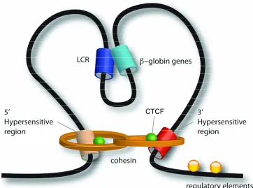
5. The structure of the β-globin locus 36
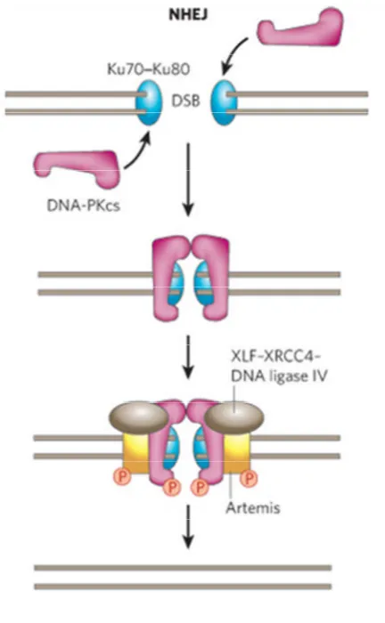
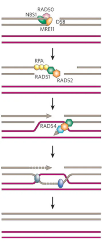
6. Non-homologous end joining pathway of DNA double strand break repair 39 7. Homologous recombination pathway of DNA double strand break repair 41
8. Nucleotide excision repair pathway 44
9. Recruitment of cohesin to DNA double strand breaks 49
3. A novel interacting partner of ChlR1
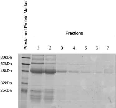
10. Elution fractions following affinity purification of rabbit anti-ChlR1 serum 88 11. Characterisation of affinity purified rabbit anti-ChlR1 antibody 89 12. Calcium phosphate precipitation transfection of FHL2 NF and CF in HEK 293 cells 90 13. Interaction between endogenous ChlR1 and FLAG FHL2 91 14. Co-immunoprecipitation of endogenous ChlR1 with FLAG tagged FHL2 92
15. Expression of HA-FHL2 in HEK-293 cells 94
16. Expression of His-FHL2 protein from the pEHISTEV expression vector 95 17. Nickel affinity purification of Hexa-Histidine-FHL2 96 18. Expression of GST-FHL2 from the pGEX-4T-2 expression vector 97
19. Purification of GST-FHL2 98
20. Purification of GST-FHL2 protein from RosettaE.coli 100 21. Transfection of HEK 293 cells with siRNA specific for endogenous FHL2 102 22. Analysis of cohesion defects in FHL2 knockdown cells 103 23. Interaction between endogenous Smc1 and FLAG-FHL2 106 24. Cell cycle analysis of FHL2 knockdown cells by flow cytometry 107
4. The role of ChlR1 in DNA damage repair
25. Treatment of HeLa cells with siRNA specific for endogenous ChlR1 113 26. Transfection of HeLa cells with siRNAs targeting either the 5’ and 3’ UTR of ChlR1 114 27. Detection of DNA damage in HeLa cells using the alkaline comet assay 116
28. Alkaline comet assay dose response curve 117
29. Alkaline comet assay repair curve 118
30. Alkaline comet assay following ChlR1 depletion 119
31. Neutral comet assay dose response curve 121
32. Neutral comet assay repair curve 122
33. Neutral comet assay following ChlR1 depletion 123
34. HeLa cells damaged and stained with gamma H2AX antibody 126
35. Expression of mCherry-ChlR1 in HeLa cells 128
36. Schematic representation of the integrated Tet Operator with the Sce-I 131 37. Localisation of TetR-GFP foci in U2OS TetO/R I-Sce clones 132
38. PCR Amplification of integrated sequence 133
39. PCR amplification of the Sce-I cut site with and without TA treatment 134 40. The phosphorylation of H2AX to the TETO/R I-SceI GFP integration site 135 41. The recruitment of phosphorylated Smc1 to the TetO/R I-SceI GFP integration site 138 42. The recruitment of ChlR1 to the TetO/R I-Sce GFP integration site 140
5. The role of ChlR1 in DNA replication
43. Neutral comet assay following ChlR1 depletion 147
44. Schematic representation of the DNA combing assay 149 45. Analysis of replicated DNA in hTERT-RPE1 cells 150 46. Analysis of replication fork dynamics in hTERT-RPE1 cells 151 47. Analysis of replication fork dynamics in ChlR1 siRNA and control siRNA transfected cells 152 48. Analysis of replication fork restart dynamics in ChlR1 and control siRNA transfected cells 154
49. BrdU incorporation and Reverse ChIP for PCNA 157
6. Discussion
Abstract
Sister chromatid cohesion is essential for the equal distribution of genetic material in
mitosis. The cohesin complex plays a central role in the establishment of sister chromatid
cohesion. The cohesin complex is a ring shaped structure that encircles sister chromatids
prior to the onset of anaphase ensuring equal distribution of genetic material. The
DEAD/H DNA helicase ChlR1 is important in the establishment of sister chromatid
cohesion. ChlR1 interacts with the cohesin complex and is required for the loading of
cohesin onto DNA. Cohesin is loaded onto the DNA during DNA replication.
Here I identified a novel interacting partner of ChlR1. The multifunctional DNA binding
protein FHL2 was shown to interact with ChlR1 and FHL2 was shown to have a role in
sister chromatid cohesion since depletion of FHL2 resulted in abnormal metaphase spreads
and reduced centromeric cohesion. These sister chromatid cohesion defects also result in a
G2/M delay.
Here I show an additional function of ChlR1 in the repair of DNA damage. ChlR1 was
required for the repair of DNA double strand breaks and ChlR1 was recruited to DNA
double strand breaks. Furthermore the function of ChlR1 in DNA double strand break
repair is S phase specific. This suggests that ChlR1 is important in the homology
I also show that ChlR1 is important in DNA replication. Depletion of ChlR1 results in
inefficient DNA replication. In addition depletion of ChlR1 results in defects in DNA
replication after hydroxyurea treatment.
The results in this thesis shed light on novel functions of the DNA helicase ChlR1 in DNA
replication and DNA damage repair and the multifunctional DNA binding protein FHL2 in
Introduction
Genetic integrity is important for all species. Organisms have evolved to protect the
integrity of their genomes in a number of ways including assault from DNA damage
reagents (UV radiation, reactive oxygen species, replication errors and viruses) and the
co-ordinated segregation of the genome into daughter cells.
The genome is protected from DNA damage reagents by a number of DNA damage repair
pathways including homologous recombination, non-homologous end joining and
nucleotide excision repair. These pathways will be explained in detail later. The majority
of DNA damage repair occurs during DNA replication. Defects in the DNA damage repair
pathways can cause increased cancer susceptibility, progeria (accelerated aging disease),
growth and mental retardation.
Co-ordinated segregation of the genome occurs during mitosis. Sister chromosomes are
paired together during mitosis by the cohesin complex. The cohesin complex is a ring
shaped structure that encircles the sister chromatids and holds them together (termed sister
chromatid cohesion) until segregation occurs. At this stage the cohesin ring is cleaved by
the enzyme separase to releases the sister chromatids (discussed later). The accurate
separation of the chromosomes is controlled by the spindle assembly checkpoint. The
spindle assembly checkpoint and the cohesin complex will be explained in detail later.
Defects in mitosis that result in disruption of chromosome segregation can cause
aneuploidy (where there is an abnormal number of chromosomes in daughter cells) and
translocations (when a portion of one chromosome is transferred to another chromosome).
Work completed in this thesis looks at the role of the DEAD/H DNA helicase ChlR1 in the
protecting the integrity of the genome. ChlR1 is important in the establishment of sister
chromatid cohesion. ChlR1 interacts with the cohesin complex and plays a role in loading
the cohesin complex onto the chromosomes (explained in detail later). ChlR1 has recently
been linked to the Warsaw breakage syndrome. Patients with mutations in ChlR1 have a
phenotype of microcephaly, growth retardation, aneuploidy and abnormal skin
pigmentation. Results in this thesis help explain the phenotype in Warsaw breakage
syndrome. The results suggest that ChlR1 has a role in DNA double strand break repair
and in DNA replication. Therefore the phenotype associated with Warsaw breakage
syndrome may be a result of DNA damage repair defects due to mutations in the gene
encoding ChlR1.
1.1 The Cell Cycle
ChlR1 and the cohesin complex are important in a number of phases of the cell cycle. The
cell cycle is divided into 4 main phases G1, S, G2and mitosis. G1phase (also known as the
gap phase) is marked with an increase in the biosynthetic activity of the cells. Proteins
required for S phase such as the replication machinery proteins are synthesised in G1. S
phase starts when DNA replication (explained in detail below) begins, and ends when all
the chromosomes are replicated. Rates of protein synthesis are very low in S phase. In G2
the level of protein synthesis increases again and proteins important for mitosis are
synthesised. In mitosis the replicated sister chromosomes in the nuclei are separated into
1.2 DNA Replication during S phase
DNA replication occurs in S phase and begins at specific sites in the DNA called origins of
replication complexes
Origin recognition complex
The origin recognition complex (ORC) binds to the origins of replication. The ORC was
identified in S.cerevisiae as a heterohexameric complex of proteins Orc1-6 that binds to
origins of DNA replication [1]. Orc 1-6 were shown by chromatin immunoprecipitation
(ChIP) to localise to 70 of the 96 replication origins inS.cerevisiae [2]. Cell division cycle
6 (Cdc6) is a replication factor in mammalian cells that is involved in the loading of the
MCM complex has strong similarities to Orc1 inS.cerevisiae suggesting it is a member of
the ORC in mammalian cells [3]. Orc 1-6 as well as Cdc6 have conserved AAA+ (ATPase
Associated Activity) folds including walker A and B ATP binding domains that are
characteristic of ATP dependent clamp loading proteins, which allow ring shaped protein
complexes to encircle DNA [4].
ATP binding by Orc1 promotes its association to DNA in S.cerevisiae [5]. The other Orc
proteins are believed to bind to the DNA through AT hooks [6]. Origin of replication
sequences are AT rich and Orc4 protein inS.pombe has been shown to have an AT hook
domain that allows it to bind to the replication origins [6]. In the human embryonic kidney
cell line HEK293 a High Mobility Group A protein (HMGA1) protein that contains an AT
hook, acts as a mediator for the ORC recruitment to origins [7]. HMGA1 also localises to
AT rich sequences and co-immunoprecipitation assays showed an interaction of HMGA1
HMGA1 and the Orc proteins co-localise at AT rich regions [7]. From these data it is
hypothesised that Cdc6 binds ATP and associates with the Orc1-6 proteins causing a
conformational change in the complex that results in increased specificity for origin DNA
[8].
When the ORC and Cdc6 bind to replication origins, Cdc6 interacts with Cdt1 (chromatin
licensing and DNA replication factor 1) of the pre-replication complex to promote
association of the replicative helicase, the MCM (minichromosome maintenance) complex
with the origin DNA in S.cerevisiae [9]. Depletion of Orc6 in late G1 results in
displacement of the MCM complex from the chromatin [9]. ATP hydrolysis by Orc6 is
thought to result in increased binding affinity of MCM complex to the origins and
recruitment of additional MCM complexes to the origins [8]. The ORC complex is
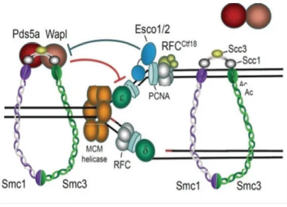
Figure 1:The replication fork complex.The MCM helicase unwinds the DNA. The clamp
loading complex RFC ctf18 loads PCNA onto the DNA. The Polymerases δ and ε are then
loaded to the PCNA-primer-template complex. Esco1 (Eco1) is involved in the establishment of cohesion during DNA replication. Esco1 acetylates the cohesin complex and this is thought to allow for the progression of the replication complex through the cohesin complex [10]. Image taken with permission from the Nature Publishing Group
MCM Replicative Helicase
The canonical MCM complex consists of 6 subunits, Mcm2 to 7 that together form a
heterohexameric helicase. Electron microscopy images of the MCM complex from
methanobacterium thermoautotrophicumshows a ring shaped structure [11]. Mutations of
each subunit inS.cerevisiaeandS.pomberesulted in a variety of replication defects [12].
The Mcm2-7 complex is transported to the origin of replication in an active state by Cdt1
[13]. This occurs during G1. At the replication origins it forms the pre-replication complex
(pre-RC) with the AAA+ proteins Cdc6 and the ORC [14]. The loading of the MCM
complex to the origin of replication requires the hydrolysis of ATP by Orc1-6 and Cdc6
[9]. Once the MCM complex is loaded then Cdc6 and Orc1-6 are removed from the origin
dependent kinases (CDKs) and Cdc7 [13]. Upon activation the MCM complex unwinds
the DNA resulting in the formation of the replication fork [13].
All six Mcm proteins have DNA dependent ATPase motifs in the central domains of the
complex suggesting that the helicase activity of complex is dependent on ATP hydrolysis.
These motifs are conserved from yeast to mammals [15]. The MCM complex was purified
from HeLa cells and this purified complex was able to displace oligonucleotides annealed
to single stranded circular DNA [16]. Furthermore the complex had a preference to
displace oligonucleotides annealed to the single stranded DNA in a 3’-5’ direction [16].
The helicase activity of the complex was only seen in the presence of hydrolysable ATP
[16]. The role of ATP in the helicase activity of MCM was further analysed in Xenopus
egg extracts [17]. Recombinant Mcm2-7 complexes with mutations in the walker A motif
of Mcm6 that abolishes ATP binding and hydrolysis of the complex was added toXenopus
egg extracts with endogenous MCM complexes depleted [17]. This assay showed that ATP
hydrolysis was not required for chromatin loading and pre-RC assembly but was required
for origin unwinding and DNA replication [17].
Cdc45 and GINS (from the Japanese go-ichi-ni-san meaning 5-1-2-3, after the four related
subunits of the complex Sld5, Psf1, Psf2 and Psf3) proteins were shown to activate the
helicase activity of the MCM complex purified fromDrosophila[18]. The helicase activity
of the MCM complex was enhanced when associated with either Cdc45 or GINSin vitro
[19]. The ATP hydrolysis rates of the MCM complex increased two fold upon association
with these proteins and the binding of these proteins to the MCM complex resulted in the
complex having an increased affinity to DNA. These two proteins were shown to interact
with Mcm4 [18].
GINS proteins (Sld5, Psf1, Psf2 and Psf3 inS.pombe) have an important function in DNA
deletion of any of the GINS proteins results in cell cycle arrest and the yeast are unable to
undergo DNA replication [20]. DNA replication is also blocked in Xenopus egg extracts
where GINS protein Sld5 is immunodepleted [20].
In the event of DNA damage during S phase the replication fork stops and stabilises. This
stabilisation involves the strengthening of the association of the MCM complex with the
replication fork [21]. This stabilisation of the fork requires the phosphorylation of the
MCM complex by Cds1 (Checking DNA synthesis 1 (a checkpoint kinase)). In the
absence of this protein the MCM complex continues to unwind the DNA in the presence of
DNA damage which results in collapsed replication forks [21]. Furthermore in metazoans
the MCM subunits are phosphorylated by the ATM (Ataxia telangiectasia mutated) kinases
promoting the interaction of the MCM complex with Rad51 (involved in homologous
recombination) in mammalian cells [21]. These results suggest that DNA damage repair
pathways target the MCM complex to stabilise the replication forks until the DNA damage
has been repaired.
The removal of the MCM complex after DNA replication involves the MCM binding
protein (McmBP) [22]. This protein interacts with Mcm7 and accumulates in the nuclei in
late S phase. Immunodepletion of McmBP in Xenopus egg extracts inhibits MCM
dissociation from DNA. Furthermore the addition of recombinant McmBP toXenopusegg
extracts results in the release of the MCM complex from chromatin isolated in late S phase
[22].
Replication licensing
The pre-replication complex (pre-RC) can only be assembled from late mitosis to G1
phase [23]. CDKs inhibit the assembly of the pre-RC for the remainder of the cell cycle
sensitive mutants of Cdk1 cause pre-RC re-assembly in S phase [24]. CDKs inhibit pre-RC
assembly by inhibiting each component of the complex [25]. In S.cerevisiae,
phosphorylation of Cdc6 by CDKs results in ubiquitin-mediated proteolysis of Cdc6 by
SCFcdc4 (Skp1–Cdc53/cullin–F box) [25]. This proteolysis occurs in late G1 through S
phase [25] and is important as it stops the ORC from assembling therefore preventing
re-replication.
Mcm2-7 enters the nucleus at the end of mitosis and remains in the nucleus during G1
phase [13]. After which it is transported to the cytoplasm. Inactivation of Cdk1 in
S.cerevisiaeresults in the nuclear accumulation of Mcm2-7 [26]. This suggests that CDKs
are involved in the transporting of the MCM complex to the cytoplasm in S phase.
Furthermore expression of a stable mitotic CDK (Clb2) results in the re-distribution of the
MCM complex to the cytoplasm [27].
Regulation of Cdt1 in S.cerevisiae is connected to that of Mcm2-7 [13]. Cdt1 protein
levels remain constant through the cell cycle but are localised in the nucleus in G1 and the
cytoplasm later in the cycle [13]. Cdt1 binds to free Mcm2-7 and in the absence of Cdt1,
Mcm2-7 do not localise in the nucleus during G1 [13].
In mammalian cells CDKs play an important role in the regulation of replication licensing.
Depletion of Cdk1 results in multiple rounds of replication [28]. Inhibition of Cdk1 in G2
causes Mcm2-7 binding to the DNA and re-licensing of replication [29]. Cdk2 also plays
an important role in the regulation of replication licensing. Cyclin A-Cdk2 targets Cdc6 for
proteolysis in mammalian cells [30]. Cdk2 phosphorylates Cdt1 and this targets Cdt1 for
proteolysis by SCFcdc4[31]. Orc1 is phosphorylated by Cdk2 and results in the proteolysis
of the protein by SCFcdc4[32].
In mammalian cells a further protein called Geminin is involved in the regulation of
absent in G1 but accumulates in the nucleus in S phase through to mitosis [33, 34].
Geminin binds to Cdt1, thereby preventing Cdt1 association with Mcm2-7 and thus
preventing the formation of pre-RC during S phase [34]. Geminin is degraded in late
mitosis by the anaphase promoting complex/cyclosome (APC/C) and it has been shown
that inhibition of ubiquitination by APC/C results in stabilization of Geminin during
mitosis [33].
CDKs may also have a positive role in licensing of replication. Cyclin E deficient cells
result in replication defects. These cells are unable to load Mcm2-7 onto DNA [35].
Furthermore cyclin E activates the E2F transcription factor. Cdc6, Cdt1 and Orc1 are
transcribed throughout this pathway [36].
PCNA
Proliferating cell nuclear antigen (PCNA) is a member of the DNA sliding β clamp family.
These sliding clamps form ring shaped complexes (homotrimers in eukaryotes and
heterotrimers in archaea) that encircle the DNA and are able to slide along the DNA in
both directions [37]. PCNA monomers have two similar globular domains linked by a long
flexible loop called the interdomain connecting loop [37]. The head to tail arrangement of
the three monomers forms the ring structure [37].
PCNA is loaded onto the DNA by the replication factor C (RFC) complex which
recognises the template primer 3’ ends of the fork and loads PCNA at these sites [38]. The
binding of RFC to PCNA is dependent on ATP binding, as well as the loading of this
complex onto DNA [39]. The binding of RFC to DNA results in the activation of the
and DNA [39]. The RFC complex binds to the C terminus of PCNA resulting in
positioning of this end of PCNA toward the 3’ end of the elongating DNA [40]. This
ensures that the polymerase is oriented towards the elongating DNA as the polymerase
binds to the C terminus of PCNA. Dissociation of RFC from the DNA upon loading of
PCNA suggests that PCNA alone is required for the loading of the DNA polymerases to
the DNA [41]. Indeed, PCNA was shown to be required for the loading of DNA
polymerasesin vitro, DNA polymerases δ and ε are unable to bind to DNA in the absence
of PCNA [42].
Studies have shown that PCNA stimulates the activity of the DNA polymerases as well as
loading the polymerases onto the DNA. The archaeal Pyrococcus furiosus (Pfu) PCNA
stimulates the activity of DNA polymerases [43]. Pfu PCNA protein has a similar amino
acid sequence to eukaryotic PCNA and the three dimensional structure is highly
conserved. The incorporation of radio-labelled nucleotides by DNA polymerases I and II
increased 3.6 fold whenPfuPCNA was added [43]. In this study the addition of high salt
to create physiological conditions inhibited the activity of polymerase I but the activity
was restored after the addition ofPfuPCNA [43].PfuPCNA was also able to stimulate the
activity of the eukaryotic DNA polymerase δ [43].
PCNA also has a function in lagging strand processing as both flap endonuclease 1 (Fen1)
and DNA ligase 1, which are both important in lagging strand processing are specifically
recruited to PCNA. Both proteins have PCNA interacting protein (PIP) box motifs [44]. It
has been shown that PCNA stimulates the activity of Fen1. In this study in vitro
biochemical assays were used to analyse the flap endonuclease activity of Fen1 and it was
shown that the addition of PCNA resulted in a 7 fold increase in the activity of the protein
[44]. The addition of a mutated PCNA that no longer binds to Fen1 resulted in only a 1.7
Fen1 activity by PCNA is dependent on physical association of these proteins [44]. PCNA
has also been shown to increase the activity of the S.cerevisiae Fen1 protein by 50-fold
[44].
PCNA is also involved in the prevention of re-replication by the destruction of Cdt1 [45].
Cdt1 also contains a PIP box and it is thought that PCNA remains on DNA after the
completion of replication and this may recruit free Cdt1 and subsequently promote its
destruction by DDB1-Cul4 (DNA damage binding- Cullin 4) ubiquitin ligase [45].
Fen1 Endonuclease
The lagging strand of the replication fork is synthesised discontinuously. These series of
short segments of DNA are known as Okazaki fragments. In eukaryotic cells these
Okazaki fragments are usually of 100-150 nucleotides in length [46]. The synthesis of an
Okazaki fragment is initiated by the DNA polymerase α-primase complex (pol α). The
primase domain of the complex produces an 8–12 oligoribonucleotide primer, and then the
polymerase extends the primer by adding another 20 deoxyribonucleotides (dNTPs) [47].
When the synthesis of one fragment encounters the following fragment, the elongating
DNA strand displaces the 5’end of the preceding strand [48]. This creates a 5’ unannealed
flap that has to be removed before ligation of the fragments. These 5’ unannealed flaps are
removed by Fen1 [48]. Fen1 is a structure specific 5’ endo/exonuclease that specifically
recognises double stranded DNA with a 5’-unannealed flap [49]. On encountering this flap
Interestingly, ChlR1 may have a role in lagging strand processing as it is an interacting
partner of Fen1 [50]. ChlR1 was shown to stimulate the endonuclease activity of Fen1
[50]. In the presence of low levels of Fen1 (5 fmol), increasing concentrations of
hChlR1stimulated the cleavage reaction approximately 3-fold.
DNA Polymerases
Eukaryotes contain 5 distinct families of DNA polymerases and in humans there have been
15 DNA polymerases characterised [51]. The large number of polymerases is required to
perform a wide range of actions ranging from DNA replication to DNA repair. All the
DNA polymerase families share a common core catalytic structure with ‘palm’, ‘finger’
and ‘thumb’ domains [51].
DNA polymerases require a single stranded DNA template to begin the synthesis of the
complementary strand [52]. Therefore the activity of the polymerase is dependent on the
unwinding of the DNA by the Cdc45, MCM and GINS complex (CMG complex) [18].
This complex associates with polymerase α-primase and recruits it to the single stranded
DNA [53]. Polymerase α-primase is a tetrameric complex that consists of two pol α
subunits and two primase subunits [54]. The complex initiates replication through the
synthesis of a short RNA primer (10 nucleotides) by the primase. The 3’ end of this primer
translocates from the primase active site to the polymerase active site to allow for the
synthesis of 20 nucleotides of DNA. After this initiation event the clamp loading complex
RFC loads PCNA onto the double stranded DNA [54]. Polymerase δ or ε is then loaded to
the PCNA-primer-template complex [54]. Polymerase δ is involved in lagging strand
progression and polymerase ε is involved in leading strand synthesis [52]. These enzymes
[55]. Mutations in the active sites of the polymerases were created. This resulted in an
increase in DNA mutation rates. A reporter gene was placed in different orientations on
opposite sides of a replication origin. For polymerase ε a higher rate of mutation was
present in the leading strand suggesting a role in leading strand synthesis [55]. The
opposite was observed for polymerase δ suggesting a role in lagging strand synthesis [55].
In agreement with this conclusion polymerase δ has been shown to interact with Fen1 in S.
pombesuggesting a role in lagging strand synthesis [56].
Replication Protein A
Replication protein A (RPA) is a heterotrimeric single stranded DNA binding complex
[57]. It consists of a large (70kDa), middle (32kDa) and small (14kDa) subunits [57]. RPA
was initially discovered to have a role in DNA replication using anin vitroassay to study
simian virus 40 (SV40) replication [58]. RPA accumulates along stretches of single
stranded DNA generated during DNA replication [59] and is required for the activation of
the pre-RC complex to form the initiation complex [58]. It is also believed that RPA has a
role in the ordered loading of essential initiators such as DNA polymerase α primase [60].
RPA is an important factor in the DNA repair process nucleotide excision repair (NER)
[61]. During strand elongation RPA stimulates DNA polymerase α, λ, κ and ε [62]. RPA
interacts with Xeroderma pigmentosum A (XPA) at sites of DNA damage and stimulates
the recruitment of Excision repair cross-complementing 1/ Xeroderma pigmentosum F
(ERCC1/XPF) [63]. Finally RPA interacts with the Blooms and Werner syndrome DNA
helicases (WRN and BLM, respectively) [61]. These helicases are closely related to
TopBP1
Topoisomerase 2-binding protein 1 (TopBP1) was initially shown to function in DNA
replication when it was shown to interact with DNA polymerase ε [64]. This polymerase
has proof-reading ability and associates with origins of replication and progresses along
with the replication forks [65]. A role in pre-initiation for TopBP1 was suggested as
TopBP1 depleted cells were unable to enter S phase [66]. The depletion resulted in down
regulation of cyclin E/Cdk2 [66] both of which are thought to be involved in the initiation
of replication [67]. Depletion of TopBP1 also results in increased levels of p21 and p27
[66] both of which inhibit Cdk2 [68]. TheS.cerevisiaehomologue of TopBP1, Dbp11 also
forms a complex with polymerase ε, GINS complex and Sld2 [69]. This complex is known
as the preloading complex. Cdk2 regulates the formation of this complex and this complex
is involved in the initiation of replication [69].
TopBP1/Dbp11 also has a role in re-initiation of replication at stalled replication forks. It
has been shown that TopBP1 is involved in the recruitment of the 9-1-1 clamp (Rad 9, Rad
1 and Hus 1) to stalled replication forks [70]. Depletion of TopBP1 in Xenopus egg
extracts resulted in the inability of the 9-1-1 clamp to be recruited to stalled replication
forks [70]. The three proteins of the 9-1-1 complex interact to form a heterotrimeric
complex similar to the PCNA sliding clamp [71]. Upon replication stress the complex
assembles around the damaged DNA and activates the protein kinase Checkpoint kinase 1
(Chk1) through the ataxia telangiectasia and Rad3-related (ATR) pathway [72, 73]. This
results in the stabilisation of the replication forks. In addition, in S.pombe the 9-1-1
complex regulates the use of the translesion polymerases instead of high fidelity
into bulky lesions [75]. When the 9-1-1 complex is knocked out in S.cerevisae, ATM
checkpoint activation is defective [76].
1.3 Sister Chromatid Cohesion
The structure of the cohesin complex
The structural maintenance of chromosomes (SMC) proteins are members of a family of
highly related proteins that function in numerous areas of DNA maintenance. Smc1 and 3
function in sister chromatid cohesion, Smc2 and 4 in chromatin condensation, Smc5 and 6
in homologous recombination and Rad50 functions in DNA damage repair. These proteins
are large polypeptides of 1000-1300 amino acids in size [77]. They contain two canonical
nucleotide binding Walker A and B motifs that are situated at the N- and C- termini of the
protein, respectively [78]. Two coiled-coil motifs that are folded in an antiparallel
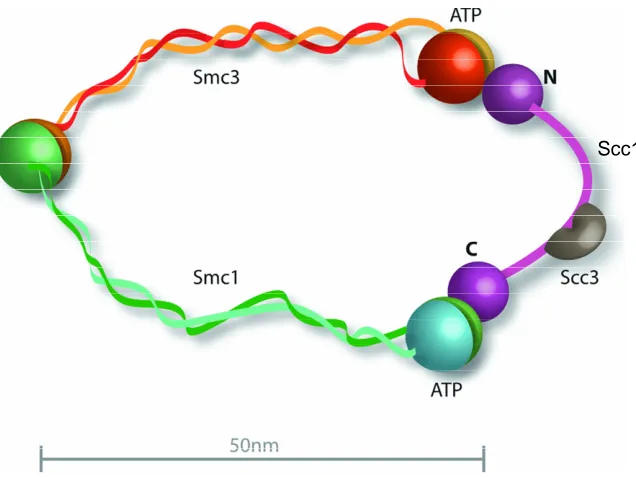
conformation connect the Walker motifs (shown in figure 2). This antiparallel
conformation was first shown by electron microscopy [79]. The Smc monomer folds back
on itself which leads to the Walker motifs situated at close proximity to each other and the
creation of hinge domain [80, 81]. Smc1 and Smc3 associate with each other at the hinge
domain resulting in the formation of a heterodimer [79] (shown in Figure 2).
The Walker A motif head domain of the Smc proteins is an ATPase domain. The
C-terminal Walker B domain of Smc contains the conserved sequence LSGG (E/Q)(K/R)
[78]. This sequence is found in the ATP binding cassettes (ABC) of ATPases [82]
(discussed later). The Walker motifs of Smc that are situated in close proximity form a
pocket that binds ATP [83]. ATP binding is important for head-head engagement and
The structure of the hinge region of Smc was first defined by the crystal structure of the
bacterium Thermotoga maritime Smc hinge domain [80]. The hinge monomers are
composed of two domains that display pseudo-two fold symmetry. The N-terminal region
of one monomer associates with the C-terminal region of the same monomer. This leads to
the formation of the antiparallel coiled-coil formation. Dimerization of the monomers
occurs through ß-sheet interactions between the monomers [80]. The interaction between
the Smc1 and 3 is independent of ATP binding [83].
Scc1 is a member of the Kleisin superfamily of proteins [84]. This family of proteins is
known to form complexes with Smc proteins [84]. TheS.cerevisae Scc1 protein contains
three domains. The N-terminal domain of Scc1 interacts with the Smc3 ATPase domain
and the C-terminal domain of Scc1 interacts with the Smc1 ATPase domain [85]. The third
domain is situated in the middle of the polypeptide chain and contains the separase
cleavage site. This domain is 180 amino acids in length and connects the N and C-terminal
domains of Scc1 [80], acting as a bridge between the two Smc subunits (shown in figure
2). Mutations in Smc1 that abolish ATP binding also disrupt the binding of Scc1 and
sister chrmatid cohesion suggesting that the binding of ATP is important in binding of
Scc1 and the function of cohesin [86]. Mutations in Smc3 that disrupt ATP binding do not
abolish Scc1 binding but also result in inactivation of cohesin function [87]. This suggests
that Scc1 binds to Smc1 first. The binding interface between the C-terminus of Scc1 and
the ATPase domain of Smc1 has been solved by X-ray crystallography [88]. The structure
shows that the two Walker domains of Smc1 bind to Scc1 and two ATP molecules are
sandwiched in between [88]. Scc1 binds to Smc1 via its winged helix motif [88]. A
winged helix motif is a compact α/β structure consisting of two wings three α-helixes and
is largely formed from β-sheets [89]. Two β sheets in the winged helix domain of Scc1
bind to the ATPase head of Smc1 [88].
Expression of a version of Scc1 that separase (a cysteine protease that cleavages Scc1 and
destroys the cohesion ring releasing the chromatids) cannot cleave shows little exchange of
cohesin or its subunits [88]. This suggests Scc1 completes the ring structure of cohesin and
locks the other components in the ring (figure 2). Electron microscopy studies on the
S.cerevisiae cohesion complex confirmed the ring shaped structure. The ring structures
consists the two Smc subunits folded up individually into rod shaped molecules of 45nm in
length that are connected at their hinge domains. This forms a V-shaped heterodimer. The
ATPase heads are then connected by Scc1 closing the ring [80] [90].
Another protein that has been shown to associate with the cohesin complex is the Scc3
protein. In vertebrate cells Scc3 exists as two closely related homologues SA1 and 2
(stromal antigen 1 and 2) [91]. These proteins associate with the cohesin complex and the
complex contains either SA1 or SA2 at one time [91]. In S.cerevisiae Scc3 was shown to
interact with Scc1 at the C-terminus but does not interact with either Smc1 or Smc3 [80].
In human cells cohesin associated with SA1 is required for telomere cohesion and cohesin
associated with SA2 is required for centromere cohesion [92]. Cells deficient in SA1 are
unable to establish or maintain cohesion between sister telomeres after DNA replication. In
SA2 depleted cells, telomere cohesion is normal but centromeric cohesion is prematurely
Scc1
Figure 2: The structure of the cohesin complex. The complex consists of 4 proteins Smc1, Smc3, Scc1 and Scc3. These proteins create a ring structure. The Smc proteins interact with each other via their hinge region and Scc1 acts as a bridge linking the ATPase head domains of the Smc proteins [93].
Loading of cohesin
The loading of cohesin onto DNA occurs at different points of the cell cycle in different
species. In lower eukaryotes cohesin is loaded onto the DNA at the end of G1 [94].
However in mammalian cells cohesin is loaded at telophase [95]. This suggest that cohesin
has other functions apart from sister chromatid cohesion (discussed later) and these
additional functions vary amongst species. In both lower and higher eukaryotes association
of cohesin and DNA becomes more stable as the cell progresses from G1to S phase [96].
The loading of cohesin requires ATP hydrolysis and the Scc2-Scc4 protein complex [97].
Scc2 is believed to interact with the cohesin complex and promote ATP hydrolysis at the
Smc heads [86]. This results in the hinge opening or Scc1 dissociation that would allow for
Cohesin was initially believed to bind to the DNA and act as an intermolecular bridge
between the DNA [98]. This theory was changed following the structural analysis of the
cohesin complex that suggested a ring shaped structure [80]. The current theory is that the
cohesin ring encircles and therefore entraps the DNA strands (Figure 3). This theory is
possible because the Smc heterodimers are 50-60nm in length and along with Scc1 is able
to encircle the DNA strand that is 10nm in diameter (Figure 2)[80]. Further evidence
supporting this theory is that disruption of the ring results in dissociation of the DNA [85,
99]. Cleavage of Scc1 by separase at the end of mitosis results in dissociation of the DNA
[85]. A further study showed that cleavage of Smc3 containing TEV cleavage sequences in
both coils of the coil-coiled domain expressed in S.cerevisiae resulted in dissociation of
the DNA from the cohesin complex [99]. This confirms that the cohesin complex encircles
the DNA instead of acting as an inter-molecular bridge. Finally, linearization of a circular
minichromosome by restriction enzyme digestion inS.cerevisiaeresulted in dissociation of
the cohesin ring from the DNA [100], suggesting the cohesin complex encircles the DNA
and does not associate with the DNA.
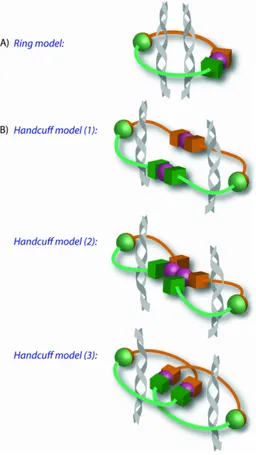
There are two models to explain how the cohesin complex encircles the DNA. The ring
model, where one cohesin ring encircles two DNA strands and the handcuff model where
two cohesin complexes are required each encircling one DNA strand and the two
complexes associate with each other (shown in Figure 3). The evidence to corroborate with
the ring model comes from data showing that interactions between multiple cohesin
complexes are undetectable by FRET analysis in S.cerevisiae [101]. Site specific
cross-linking at the three interfaces of the cohesin ring that creates a convalently closed ring was
used to show that one cohesin ring is required to encircle the DNA. Protein denaturation
Evidence to corroborate with the handcuff model includes data suggesting Scc1 interacts
with itself in a yeast-two hybrid system, which was confirmed by co-immunoprecipitation
in mammalian cells [103]. Cohesin rings are only able to encircle one strand of DNA at
silent heterochromatin regions where the DNA has a larger diameter which also suggests
that cohesin rings may multimerise to facilitate cohesion [104]. Finally the four cohesin
complex subunits have been shown to interact with themselves in a Scc3 dependent
manner. Depletion of Scc3 abolished the ability of the other three cohesin subunits to
interact with themselves [103]. This suggests Scc3 is important in creating a link between
two cohesin rings.
Cohesion establishment
Cohesion establishment is believed to occur during DNA replication and does not require
additional cohesin complexes to be loaded onto DNA [105]. Eco1 (Esco1 in humans) is a
lysine acetyltransferase and it is required for cohesion establishment [106]. Eco1
associates with numerous members of the DNA replication complex including the clamp
loader proteins Ctf18 and Elg1[107, 108]. Both of these proteins play a role in cohesion
establishment. PCNA also associates with Eco1 [109]. Therefore it is believed cohesion
establishment is linked to PCNA-dependent DNA replication. Eco1 may allow for the
dissociation and re-association of the cohesin ring as the replication fork passes, thus
facilitating cohesion establishment during DNA replication [93].
Recent evidence suggests that acetylation of cohesin is important for replication fork
processivity. Smc3 has been shown to be acetylated in vivo at two conserved lysines by
Eco1 during S phase [105, 110]. The Ctf18 subunit of the alternative RFC complex
[10]. When Ctf18 is depleted in mammalian cells replication and cohesin acetylation is
impaired [10]. Ctf18 does not acetylate Smc3 but is believed to be important in regulating
Eco1 activity [111]. Furthermore, cohesin associated proteins Wpl1 and Pds5 also regulate
cohesion establishment [112, 113]. Wpl1 and Pds5 remove cohesin from the chromosomes
during mitosis. Depletion of these proteins in mammalian cells blocks the removal of
cohesin in prophase and increases the residence time of cohesin on the chromosomes in
interphase [113]. These proteins interact with Smc3 and are removed from the complex
when it is acetylated [114].
Interestingly fork progression through the cohesin complex is necessary for establishment
as it leads to entrapment of the nascent DNA strand within the ring [105]. Therefore the
acetylation of cohesin by Eco1 may cause conformation changes to the complex that allow
for replication fork passage. The conformational change may result from the removal of
Wpl1 or Pds5 from cohesin upon acetylation [10]. It may be possible that acetylation of
Smc3 may cause dissociation of Scc1 to allow the replication fork to pass through [10].
Acetylation of Smc3 by Eco1 is important for the establishment of cohesion because
deletion of Eco1 in S.cerevisiae causes premature chromosome separation when Esp1p
(yeast homologue of separase) proteolysis is inhibited [106]. Deletion of Eco1 did not
affect the loading of Scc1 and 3 onto the chromosomes which suggests it is not required
1.4 Mitosis
ChlR1 and cohesin are important in the pairing of sister chromatids, which allows for the
equal distribution of genetic material in mitosis. Mitosis is divided into 4 main stages
prometaphase, metaphase, anaphase and telophase.
In the prometaphase stage the nucleus dissolves and the mitotic spindle microtubules
attach to the kinetochores at the centromeres (located near the centre of the chromosomes
and is the region where the sister chromatids come in close contact) of the chromosomes.
The kinetochore is a structure that containing a number protein complexes, that form
around the centromeres. The simplest kinetochore structure is found in S.cerevisiae and
contains 60 proteins in 7 complexes [115]. The complexes of the kinetochore are
conserved from yeast to humans. Two of these complexes Ndc80 and Dam1 (Duo1 And
Mps1 interacting) complexes have an important function in interacting with the
microtubules [116]. Another kinetochore protein Centromere protein A (CENP-A) a
centromeric specific conserved histone H3, is important in the recruitment and assembly of
the kinetochore complexes at the centromeres [115].
The mitotic spindle microtubules consist of polarized filaments composed of α/ß- tubulin
heterodimers arranged in a head to tail configuration within protofilaments [117]. The
microtubule polarity is due to the asymmetric arrangement of the tubulin subunits. The
minus and plus ends of the microtubule have different dynamic properties [118]. This
polarity is important in the transport of the chromosomes along the microtubule to the
poles.
The microtubules undergo repeated growth and shrinkage in various directions, searching
for kinetochores [119]. The growth and shrinkage of the microtubules is a result of the
kinetochores is not a random process in vertebrate cells as the microtubules are guided to
the kinetochores by a concentration gradient of Ran-GTP around the chromosomes [120].
Interestingly microtubules are generated by the kinetochores and these microtubules
interact with and guide spindle microtubules to the kinetochores [121]. At this stage of
mitosis the cohesin encircling the arms of the chromosomes is removed through
phosphorylation of the cohesion subunits by Polo-like kinase 1 (Plk1) [122] which will be
discussed later.
In metaphase the chromosomes are aligned on the mitotic spindles. Alignment of the
chromosomes on the spindles requires bi-orientation of the kinetochore and microtubules.
Bi-orientation is control by two mechanisms; kinetochore geometry and error correction.
Kinetochore geometry relies on a back to back position of sister kinetochores [123]. When
one kinetochore attaches to the microtubules from one spindle pole, the constraints in
geometry requires the other kinetochore faces the opposite direction and attach to the
microtubule from the opposite pole [123]. If aberrant attachments are made between the
kinetochore and microtubules the geometry mechanism cannot correct the mistake.
Tension created by correctly bi-orientated kinetochore and microtubules stabilises the
interaction between the two structures. If this tension is not present then the two structures
dissociate and a new interaction is created. This mechanism of control is termed error
correction and requires the spindle assembly checkpoint protein Aurora B kinase [124].
Defects in Aurora B or its yeast homologue Ipl1 results in the kinetochores being unable to
interact with the microtubules [125]. Aurora B localises at the centromeres in metaphase
and phosphorylates kinetochore components [126]. This weakens the interaction between
the kinetochores and microtubules [127]. This is believed to be important in the turnover
of kinetochore and microtubule interactions during bi-orientation. Once bi-orientation and
kinetochore components including Dam1 and Knl1 are dephosphorylated [128]. This
stabilises the interaction between the kinetochores and the microtubules and the cells can
progress into anaphase.
During anaphase the cohesin that encircles the chromosomes at the centromeres is cleaved
by a cysteine protease called separase, which is inhibited by the spindle assembly
checkpoint upon alignment of the chromosomes [129]. The separated sister chromosomes
are transported along the spindle microtubule to the poles. The movement of the
chromosomes towards the spindle poles is achieved by microtubule depolymerization
along with the activity of molecular motor proteins such as dynein and kinesin proteins.
Dynein associates with the kinetochores and has poleward minus end directed motility
[130]. Inhibition of dynein in drosophila embryos causes a reduction in chromatid
movement towards the poles [131]. Kinesin proteins are microtubule depolymerising
enzymes localised at the kinetochores [132]. Disruption of these proteins in vertebrate cells
causes lagging chromosomes in anaphase [133].
After the chromosomes are transported to the spindle poles during anaphase, the cell
progresses to telophase where the microtubules disappear and new nuclear envelopes form
from fragments from the parent cells nuclear envelope and portions of the endomembrane
system. The compacted chromosomes decondense back into chromatin.
1.5 Spindle assembly checkpoint
The accurate segregation of chromosomes in mitosis is controlled by the spindle assembly
checkpoint (SAC). The SAC was identified by a mutation screen inS.cerevisiae in which
poison. The screen identified a number of genes including Mad (mitotic arrest defects) 1,
2, and 3 (BubR1 in humans) and Bub (budding uninhibited by benzmidazole) 1 and 3
[134]. These genes are conserved in all eukaryotes. They are activated in prometaphase
and form the basis of the spindle assembly checkpoint (SAC) [135].
The SAC targets Cdc20, which is a co-factor of the anaphase promoting
complex/cyclosome (APC/C) [136]. The SAC negatively regulates the ability of Cdc20 to
activate the APC/C mediated poly-ubiquitination of cyclin B and securin. Hence
progression through the SAC results in proteasome-mediated degradation of cyclin B and
securin and progression to anaphase. Activation of the SAC prolongs prometaphase until
all the chromosomes have become bi-orientated between the spindle poles [136]. The SAC
monitors the interactions between the kinetochores and the spindle microtubules.
Mutations in kinetochore proteins which impair the function of the kinetochores results in
the activation of the SAC in S.cerevisiae [137]. The vertebrate homologues of the SAC
proteins were shown to accumulate at unattached kinetochores and disperse upon
microtubule attachment [138].
In humans Bub1 is present at the kinetochores from prometaphase until kinetochore
microtubule attachment in humans [139]. Bub1 is believed to recruit a number of the SAC
proteins to the kinetochores including Mad1, Mad2, BubR1, Bub3 and Cdc20 inXenopus
egg extracts [140]. Bub1 also phosphorylates Cdc20 and this inhibits Cdc20 [141].
Replacement of wild type Cdc20 with a mutant that Bub1 is unable to phosphorylate
results in mitotic exit despite spindle damage in HeLa cells [141].
The main function of the protein kinase BubR1 in regulating the SAC is to interact with
and phosphorylate the microtubule plus end directed motor CENP-E [142]. InX. laevisthe
depletion of CENP-E disrupts the SAC suggesting it directly functions in the SAC [143].
CENP-E binds to BubR1 and activates the kinase in vitro [145]. This activation is
repressed when CENP-E binds to the microtubules [145] suggesting that BubR1 is
inactivated upon kinetochore microtubule attachment. Replacement of the wildtype
CENP-E with a truncated CCENP-ENP-CENP-E that lacks the microtubule binding domain results in continued
activation of the SAC in mammalian cells [145].
The kinase Aurora B/Ipl1 is also involved in the SAC. It detects and destabilizes faulty
microtubule-kinetochore attachments [146]. This generates unattached kinetochores and
results in SAC activation [146]. There is evidence that suggests Aurora B recruits BubR1
and Mad2 to the kinetochores. Inhibition of Aurora B in mammalian cells reduces Mad2
and BubR1 localisation to the kinetochores in nocodazole treated cells [147].
There are a number of mechanisms for the inactivation of the SAC after kinetochore
attachment. Mad1 and 2 are stripped from the kinetochores by dynein that is present on
microtubules [148]. This is dependent on dynein motility along the microtubules [148].
The activated APC/C is believed to target a number of the SAC proteins for degradation.
Mps1 is a kinase involved in the SAC that localises to the kinetochores [124]. In
S.cerevisiae Mps1 is degraded at anaphase in a Cdc20 dependent manner [149].
Overexpression of Mps1 or removal of Cdc20 during anaphase results in SAC reactivation
[149].
1.6 The removal of cohesin in mitosis
At the onset of anaphase, cohesion is dissolved by the proteolytic cleavage of Scc1 by the
activated cysteine protease separase [85, 129]. Separase is activated by the degradation of
its inhibitory binding partner securin by the 26S proteasome [150]. Securin is targeted for
ligase [151]. Cdc20 act as co-activator and allows APC/C to recognise securin as a target
[151]. It is believed that Cdc20 delivers securin to APC/C [152]. Cdc20 is activated
following satisfaction of the spindle assembly checkpoint (discussed above). Securin binds
to separase at the protease active site therefore blocking the function of separase [153].
When securin is knocked out in mice, the embryonic cells are viable and undergo normal
anaphase progression [154]. This suggests there is another pathway of separase inhibition
and it has been shown that phosphorylation of serine 1226 of separase results in inhibition
of separase during metaphase [150].
Before cleavage of Scc1 by separase in anaphase, the majority of cohesin dissociates with
the chromosomes in prometaphase [122]. Polo-like kinase 1 (Plk1) is involved in this
process [122]. Plk1 is a key regulator of mitotic division [122] and a member of a family
of four related serine/threonine kinases. When Plk1 is depleted in mammalian cells the
cells fail to establish bipolar spindles [122]. Plk1 can phosphorylate Scc1 and Scc3in vitro
[155] and is required for their phosphorylation in Xenopus egg extracts [156].
Phosphorylation of Scc1 and 3 subunits results in cohesin being unable to bind to the
chromatin in the extracts [156]. Evidence suggests that the phosphorylation of Scc1 by
Plk1 can enhance the cleavage of Scc1 by separase [157]. In human cells phosphorylation
of Scc3 is important for cohesin dissociation [158] and mutations at the phosphorylation
site on Scc3 results in an inability to remove cohesin from the chromosome arms during
prometaphase [158]. This phenotype is similar in cells depleted of Plk1 [159]. Therefore
Plk1 is important for the removal of cohesin from the chromosome arms during prophase
and prometaphase.
Cohesin located at the centromeric regions of sister chromatids are protected from
phosphorylation by a protein called Shugoshin (Sgo1). Sgo1 starts to accumulate at the
edges of the chromosomes moving towards the spindle poles [161]. Sgo1 was first shown
to be involved in the process of sister chromatid cohesion in S.cerevisiae where Sgo1
deletion resulted in sensitivity to spindle destabilizing drugs and premature sister
chromatid separation [162, 163]. Depletion of Sgo1 in HeLa cells resulted in mitotic arrest
and chromosome mis-segregation [160]. Depletion of Sgo1 results in the loss of Scc1 at
the centromeres. Therefore, Sgo1 was shown to protect centromeric cohesion from
dissociation at the centromeres during prophase [160].
The serine/threonine protein phosphatase 2A (PP2A) has been shown to interact with Sgo1
[164]. This protein is important for sister chromatid cohesion as depletion of the protein
causes cohesion defects in HeLa cells [164] and premature centromere segregation in
S.cerevisiae[165]. It is believed to protect centromeric cohesin from the prophase pathway
by preventing phosphorylation of the Scc3 subunit of cohesin [166]. In vitro analysis
showed that PP2A is able to dephosphorylate the peptides of the Scc3 that are targeted by
Plk1 [166]. Furthermore PP2A is required for the recruitment of Sgo1 to the centromeric
regions [164].
The function of PP2A and Sgo1 is controlled by the kinetochore associated protein Bub1
[167]. Depletion of Bub1 prevents localisation of both PP2A and Sgo1 to the centromere
[162]. Furthermore, it has been demonstrated that the kinase activity of Bub1 restricts the
localisation of PP2A and Sgo1 to the centromere [168].
1.7 The role of ChlR1 in sister chromatid cohesion
Chl1 (Chromosome loss 1; Chl-Related1 (ChlR1) in humans) was initially discovered in a
screen for mutants of S.cerevisiae that exhibited decreased chromosome transmission
chromosome missegregation at the MATά locus on chromosome III and mutants exhibited
both loss and gain of chromosomes fragments [169]. The loss of chromosomes and
chromosome non-disjunction was 100-fold above wildtype rates [169]. In addition, the
mutant cells showed signs of cell cycle checkpoint activation leading to G2/M delay [170].
The Chl1 protein has been shown to have sequence homology to the Rad3 protein, which
is involved in nucleotide excision repair of DNA damage. The two proteins are 23%
identical and 48% similar [170]. Interestingly, regions of high homology include the ATP
binding site domain of Rad3 [170]. Mutations in Chl1 that prevent ATP binding result in
the inactivation of its function in chromosome segregation and overexpression of this
mutant interferes with chromosome transmission [171].
ChlR1 is a member of the DEAD/H family of DNA and RNA helicases. ChlR1 mRNA
and protein is expressed in dividing cells but not terminally differentiated cells or cells in
growth arrest suggesting that ChlR1 has a similar role to Chl1 in cell proliferation [172].
ChlR1 and Chl1 are 32% identical and 55% similar [173]. The expression of ChlR1 was
not present until 16 hours after the addition of serum to fibroblasts [172]. This coincides
with the entry of these cells in S phase. The expression levels increased over the next 12
hours [172]. This suggests that ChlR1 is required for S phase and mitosis. Using in vitro
transcribed and translated ChlR1 protein it has also been shown that ChlR1 binds to both
double and single stranded DNA [172].
The helicase activity of ChlR1 has been investigated using purified ChlR1 expressed in
insect cells using the baculovirus expression system [174]. The protein possesses both
ATPase and DNA helicase activities that are dependent on DNA, divalent cations and ATP
[174]. The helicase activity is abolished by a single amino acid substitution of lysine to
arginine at position 50 in the ATP binding domain of the protein [174]. ChlR1 can unwind
short single stranded DNA templates of 19 nucleotides in length [174]. Unlike other DNA
helicases, ChlR1 can translocate along single stranded DNA in both directions when the
substrates have a very long single stranded DNA region [174].
Depletion of ChlR1 resulted in pro-metaphase delay and mitotic failure. There are a higher
percentage of cells in mitosis 24 hours after depletion and there are a higher percentage of
cells with chromosome dispersed over the entire spindle [175]. This is a phenotype
associated with pro-metaphase delay. Depletion of ChlR1 also results in chromosome
mis-segregation [175]. Depletion of ChlR1 for 96 hours results in hTERT-RPE1 cells with
enlarged and fragmented nuclei indicting the cells failed to correctly segregate sister
chromatids following the delay in pro-metaphase [175]. Depletion of ChlR1 in HeLa cells
causes failure to form tight metaphase plates and after the pro-metaphase delay the DNA
decondenses without mitotic segregation [175].
ChlR1 associates with the cohesin complex and when ChlR1 is depleted the cells show
signs of abnormal sister chromatid cohesion [175]. Sister chromatid pairing was analysed
through metaphase spreads. Measurement of the distance between chromatid pairs
revealed that sister chromatids were further apart at the centromeric regions in ChlR1
depleted cells indicating abnormalities in sister chromatid cohesion [175]. These results
suggest a role for ChlR1 in chromatin condensation and organization of the DNA through
the correct assembly of cohesin onto DNA during replication [175].
There is evidence suggesting ChlR1 has a role in the spindle assembly. ChlR1 has been
shown to accumulate at the spindle poles in early mitosis where it remains until telophase
[175]. The cohesin subunits Smc1 and SA1 have also been shown to localise to the spindle
poles during mitosis [175]. This localisation is mediated by NuMA a protein required for
assembly [176]. The localisation of ChlR1 at the spindle poles and the evidence that ChlR1
associates with cohesin may indicate a role for ChlR1 in mitotic aster assembly [175].
The knockout of the mouse ChlR1 gene DDX11 resulted in lethality at the embryonic
stage of the mouse development [177]. Lethality was a result of accumulation of aneuploid
cells and placental malformation [177]. Mutant embryos were smaller in size, malformed
and exhibited sparse cellularity compared to normal or heterozygous litter mates [177].
Cells isolated from the Ddx11-/- embryos displayed a G2/M delay, increased chromosome
missegregation, decreased chromosome cohesion and increased aneuploidy [177]. These
cellular defects are displayed in mammalian cells depleted of ChlR1 with siRNA.
1.8 The role of cohesin in gene expression
Cohesin re-associates with chromosomes at the end of mitosis [95]. This re-association
occurs long before sister chromatid cohesion is established suggesting additional functions
for cohesin.
There is evidence that cohesin has a role in the control of gene expression in S.cerevisae.
The positioning of cohesin was shown to be affected by transcription with cohesin
accumulating at convergent transcripts [178]. This suggests that as the transcription
machinery moves along the DNA any cohesin it encounters is pushed along until
transcription termination. This accumulated cohesin was shown to have a role in
transcription termination in S.pombe. The cohesin is thought to act as a barrier between
coding regions to aid efficient transcriptional termination [179].
Cohesin binding sites have been discovered at heterochromatin regions, which are silent
loci with several functions from gene regulation to the protection of the integrity of
function of Sirtuins (SIR) proteins [181]. Cohesin therefore is thought to play an important
function in chromatin silencing. Cohesin is recruited to heterochromatin regions in a SIR
dependent manner [104]. Heterochromatin regions contain a number of repeated sequences
that are unstable due to recombination therefore it is believed that cohesin is recruited to
act with the SIR proteins to prevent unwanted recombination [182]. Cohesin subunits
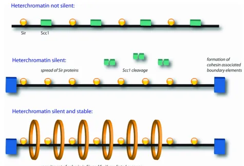
Smc1 and 3 have been found at the boundary regions of the heterochromatin and mutations
of these proteins results in disruption of the boundaries [183] (shown in Figure 4). It is
believed that cohesin helps create boundaries at the end of the heterochromatin regions to
prevent unwanted spread of the silent chromatin.
There is evidence that suggests cohesin inhibits the formation of silent chromatin.
Silencing of the heterochromatin regions is unable to take place in the presence of Scc1 in
G2/M even in the presence of SIR proteins. When Scc1 is cleaved or repressed the
heterochromatin becomes silent [184] (shown in Figure 4). This suggests that the cohesin