METABOLISM
Pitak Nasomjai
A Thesis Submitted for the Degree of PhD at the
University of St. Andrews
2010
Full metadata for this item is available in the St Andrews Digital Research Repository
at:
https://research-repository.st-andrews.ac.uk/
Please use this identifier to cite or link to this item: http://hdl.handle.net/10023/866
This item is protected by original copyright
three isomerase enzyme mechanisms in
secondary metabolism
By
Pitak Nasomjai
A thesis presented for the degree of
Doctor of Philosophy in the School of Chemistry
University of St Andrews
that it has not been submitted in any previous application for a higher degree.
I was admitted as a research student in August 2005 and as a candidate for the degree of Doctor of Philosophy in August 2006; the higher study for which this is a record was carried out in the University of St Andrews between 2005 and 2009.
Date …….………..signature of candidate ………..
I hereby certify that the candidate has fulfilled the conditions of the Resolution and Regulations appropriate for the degree of PhD in the University of St Andrews and that the candidate is qualified to submit this thesis in application for that degree.
Date …….………..signature of supervisor………..
In submitting this thesis to the University of St Andrews we understand that we are giving permission for it to be made available for use in accordance with the regulations of the University Library for the time being in force, subject to any copyright vested in the work not being affected thereby. We also understand that the title and the abstract will be published, and a copy of the work may be made and supplied to any bona fide library or research worker, that my thesis will be electronically accessible for personal or research unless exempt by award of an embargo as requested below, and that the library has the right to migrate my thesis into new electronic forms as required to ensure continued access to the thesis. We have obtained any third-party copy right permissions that may be required in order to allow such access and migration, or have requested the appropriate embargo below.
The following is an agreed request by candidate and supervisor regarding the electronic publication of this thesis:
Access to Printed copy and electronic publication of the thesis through the University of St Andrews.
Date …….………..signature of candidate ………..
First and foremost, I would like to thank my supervisor Professor David O’Hagan for his
supervision, encouragement and advice during my PhD studies.
I would also like to thank all people that were involved in my projects: Dr Darwin W. Reed
and Dr. Patrick Covello at the Plant Biotechnology Institute, Canada for “cytochrome P450
assays of fluorolittorines”; Dr. Denis Tritsch and Prof. Michel Rohmer at the Université Louis
Pasteur/Centre National de la Recherche Scientifique, France for DXR inhibition assays; Prof.
David J. Tozer at the University of Durham for DFT calculations on relative energy of
fluorobezylic intermediates and ionisation potentials and Dr. Stuart M. Cross for 5-FDRP
isomerase assays.
I am grateful to all the following people for technical support: Prof. Alexandra Slawin for
X-ray analysis, Melanja Smith and Dr Tomas Lebl for NMR spectroscopy, and Caroline
Hosburgh and Catherine Botting for mass spectrometry.
Special thanks must go to DOH members past and present: Luke, Natalie, Mayca, Matthieu,
Vincent, Thomas, David, Guillaume, Stefano, Jason Schmidberger, Gildas, Daniel, Nelly,
Deng and Daniel Smith. A very special thanks to Cosimo for passing on his knowledge.
Also, I would like to thanks all my friends: Chamnan, Walailuck, Kannika, Sujitra and Annop
for their fantastic friendships.
I am also thankful to the Royal Thai Government for financial support and the staff at the
OEA, Royal Thai embassy (London) for help.
I am deeply thankful to Assoc. Somdej and Kwanjai Kanokmedhakul without them it would
not be possible for me to reach this far.
Declaration
i
Acknowledgements
ii
Contents
iii
List of abbreviations
xiii
Abstract
xvii
1
Introduction
11.1 Carbon skeleton rearrangements in enzymology 2
1.2 Isomerisations mediated by co-enzyme B12 2
1.3 Biosynthesis of tenellin 7
1.4 Isomerisation of liquiritigenin to daidzein. Isoflavonoid biosynthesis 9
1.5 Biosynthesis of cephalosporin C 11
1.6 Biosynthesis of cholesterol 13
1.7 Biosynthesis ofmyo-inositol 14
1.8 References chapter 1 17
2
Exploring the mechanism of the rearrangement of
littorine to hyoscyamine
192.1 Introduction 20
2.3.1 Total synthesis of tropine and the structural elucidation of tropine 24
2.3.2 Biosynthesis of the tropane ring 26
2.3.2.1 The amino acid derived fragment 27
2.3.2.2 The acetate derived fragment 34
2.3.2.3 The reduction of tropinone to tropine 38
2.3.3 Biosynthesis of hyoscyamine 38
2.3.3.1 Littorine as a precursor 38
2.3.3.2 Stereochemistry of the rearrangement 41
2.4 Enzymes that mediate a molecular rearrangement 44
2.4.1 Co-enzyme B12and SAM ‘the poor man’s B12’ are not involved 44
2.4.2 The role of cytochrome P450enzymes. 46
2.5 Reactive species involved in P450transformations 49
2.6 Radical or carbocation rearrangement process 52
2.7 Computational studies 54
2.8 Fluorine as mechanistic probe 56
2.8.1 Effect of fluorine on the aromatic ring 56
2.8.2 Effect of fluorine on benzylic carbocation and radical 57
2.8.3 The use of fluorine as a mechanistic probe in tropane
alkaloid biosynthesis 57
2.9 Aims of the study 58
2.10 Results 59
2.10.1 DFT study on the relative energies ofortho-,meta-, andpara
-fluorobenzyl radicals and cations 59
2.10.3.1 Synthesis ofrac-fluorolittorines
2.10.3.1.1 Synthesis ofrac-2'-, 3'-, 4'-fluorophenyllactic acid 62
2.10.3.1.2 Synthesis ofrac-2'-, 3'-, 4'- fluorophenyllactoyl
tropines (rac-fluorolittorines) 62
2.10.3.2 The synthesis of enantio-enriched 2', 3', 4'-fluorolittorines 63
2.10.3.2.1 Resolution by preparation of (-)-menthol derivative 63
2.10.3.2.2 (1S)-(-)-Camphanoyl chloride derivative 67
2.10.3.2.3 Resolution by preparation of amides 69
2.10.3.2.4 Resolution by D-phenylalanine methyl ester
hydrochloride and (S)-phenylethylamine 71
2.10.3.2.5 Hydrolysis of enantio-enriched fluoroaryl amides 76
2.10.3.2.6 Determination of enantiomeric excess of the resolved
fluorophenyllactic acids 78
2.10.3.2.7 Preparation of enantio-enriched fluorolittorines 80
2.10.3.3 Enzyme resolution 81
2.19.3.4 Preparation of enantio-enriched (S)-[3',3'-2H2]-littorines
and (R)-[3',3'-2H2]-littorines 84
2.10.4 Bioassay results 86
2.10.4.1 Kinetic isotope effect study 86
2.10.4.2 Enzyme assays ofrac-2', 3', 4'-fluorolittorines with CYP80F1 88
2.10.4.3 Enzyme assays of enantio-enriched (R)-2', 3', 4'-fluorolittorines 90
2.10.4.4 Enzyme assays of enantio-enriched (S)-2', 3', 4'-fluorolittorines 92
2.11 Discussion 94
3
Synthesis of the ribose phosphonates
1063.1 Introduction 107
3.2 The biosynthesis of fluoroacetate and 4-fluorothreonine 107
3.3 The isomerisation of 5-FDRP to 5-FDRulP 110
3.3.1 The aldo-keto, MTR-1-P isomerase 110
3.3.2 Crystal structure of MTR-1-P isomerase fromBacilus subtilis. 111
3.3.3 5-FDRulP is an intermediate in fluorometabolite biosynthesis in
S. cattleya 112
3.4 Identification of the putative isomerase gene inS. cattleya 115 3.5 Putative mechanisms for the isomerisation of 5-FDRP to
5-FDRulP 116
35.1 Thecis-enediol and hydride shift mechanism 116
3.5.2 The phosphate migration mechanism 119
3.6 Aims of the project 120
3.7 Results and discussion 121
3.7.1 Synthesis 121
3.7.1.1 Synthetic plans 121
3.7.2 Preparation of phosphonates 123
3.7.2.1 The D-ribonic-γ-lactone route-Route A 123
3.7.2.2 The D-ribose route-Route B 127
3.7.2.3 Preparation of phostones 133
3.7.3 Bioassay results 135
4
Synthesis of potential DXR inhibitors
1414.1 Introduction 142
4.2 Biosynthesis of the isoprenoid precursors IPP and DMAPP 144
4.2.1 The mevalonic acid pathway 144
4.2.2 The MEP or DOXP pathway 145
4.3 The rearrangement of DXP to MEP 148
4.3.1 The proposed mechanism of the rearrangement of DXP to MEP 148
4.3.1.1 α-Ketol rearrangement 148
4.3.1.2 The retro-aldol/aldol rearrangement 149
4.3.1.3 Hydride/methyl shift 150
4.3.2 α-Ketol or retro-aldol/aldol rearrangement mechanism 151
4.4 The structure of the DXR enzyme 153
4.5 Inhibitors of DXR enzyme 158
4.6 Aims of the project 159
4.7 Previous study on the synthesis of potential DXR inhibitors 161
4.8 Results 163
4.8.1 Synthesis 163
4.8.1.1 Preparation of 5-hydroxy-3,4-dioxo-hexyl-phosphonate 163
4.8.1.2 Preparation of [5-13
C]-5-hydroxy-3,4-dioxo-hexyl-phosphonate 166
4.8.1.3 Preparation of [4-13C]-1-bromo-4-hydroxy-pent-2-yne 167
4.8.1.4 Preparation of [5-13C]-α-diketone 169
phosphonate 173
4.8.3 Incubation of 58a withE. coliDXR results 175
4.9 Discussion and conclusion 176
4.10 References 180
5
Chemical and biochemical experiments
1845.1 General methods 185
5.2 Synthetic experiment for Chapter 2 187
5.2.1 (RS)-o-Fluorophenyllactic acid 187
5.2.2 (RS)-m-Fluorophenyllactic acid 188
5.2.3 (RS)-p-Fluorophenyllactic acid 188
5.2.4 (rac)-o-Fluorolittorine 189
5.2.5 (rac)-m-Fluorolittorine 190
5.2.6 (rac)-p-Fluorolittorine 191
5.2.7 Methyl (RS)-p-fluorophenyllactate 192
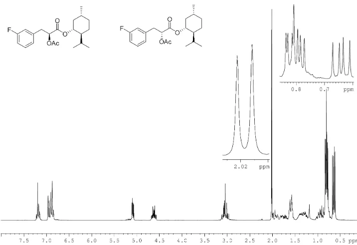
5.2.8 (-)-Menthyl (RS)-m-fluorophenyllactate 193
5.2.9 α-Acetyl-(-)-menthyl (RS)-m-fluorophenyllactate 195
5.2.10 (S)-Camphanoyl methyl (RS)-p-fluorophenyllactate 196
5.2.11 α-Acetyl-methyl-D-phenylalanyl-(RS)-p-fluorophenyllactamide 197
5.2.12 (S)-3-(2-Fluorophenyl)-2-hydroxyl-N-((S)-1-phenylethyl)
propanamide and (S)-3-(2-fluorophenyl)-2-hydroxyl-N-((S
)-1-phenylethyl)propanamide 199
phenylethyl)propanamide 201
5.2.14 (S)-3-(4-Fluorophenyl)-2-hydroxyl-N-((S)-1-phenylethyl)
propanamide and (S)-3-(4-fluorophenyl)-2-hydroxyl-N-((S
)-1-phenylethyl)propanamide 202
5.2.15 (S)-o-Fluorophenyllactatic acid 204
5.2.16 (R)-o-Fluorophenyllactatic acid 205
5.2.17 (S)-m-Fluorophenyllactatic acid 205
5.2.18 (R)-m-Fluorophenyllactatic acid 206
5.2.19 (S)-p-Fluorophenyllactatic acid 207
5.2.20 (R)-p-Fluorophenyllactatic acid 208
5.2.21 (S)-o-Fluorolittorine 209
5.2.22 (R)-o-Fluorolittorine 210
5.2.23 (S)-m-Fluorolittorine 211
5.2.24 (R)-m-Fluorolittorine 212
5.2.25 (S)-p-Fluorolittorine 213
5.2.26 (R)-p-Fluorolittorine 214
5.2.27 (2S,2'S)-[3,3-2H2]-3-Phenyl-2-hydroxyl-N-((S)-1-phenylethyl)
propanamide and (2R,2'S)-[3,3-2H2
]-3-phenyl-2-hydroxyl-N-((S)-1-phenylethyl) 215
5.2.28 (S)-[3,3-2H2]-Phenyllactic acid 216
5.2.29 (R)-[3,3-2H2]-Phenyllactic acid 217
5.2.30 (S)-[3',3'-2H2]-Littorine 218
5.2.31 (R)-[3',3'-2H2]-Littorine 219
phenylethyl)propanamide 221
5.3 Synthetic experiments for Chapter 3 223
5.3.1 2,3-O-Isopropylidene-D-ribono-1,4-lactone 223
5.3.2 5-Deoxy-5-fluoro-2,3-O-isopropylidene-D-ribono-1,4-lactone 224
5.3.3 5-Deoxy-5-fluoro-2,3-O-isopropylidene-D-ribofuranose
and (2S,3S,4S)-5-fluoro-2,3-(O-isopropylidenedioxy)pentan-1,4-diol 225
5.3.3.1 Synthesis with NaBH4 225
5.3.3.2 Synthesis with DIBAL-H 226
5.3.4 5-Deoxy-5-fluoro-1-deoxy-2,3-O
-isopropylidene-1-(dimethoxyphosphinyl)-D-ribofuranose 227
5.3.5 2,3-O-Isopropylidene-D-ribofuranose 228
5.3.6 2,3-O-Isopropylidene-5-O-trityl-D-ribofuranose 229
5.3.7 2,5-Anhydro-1-deoxy-2,3-O
-isopropylidene-1-(dimethoxyphosphinyl)-5-O-trityl-D-altritol and D-allitol 230
5.3.8 2,5-Anhydro-1-deoxy-2,3-O
-4-isopropylidene-1-(dimethoxyphosphinyl)-5-O-D-altrohexitol 232
5.3.9 2,5-Anhydro-1-deoxy-2,3-O
-4-isopropylidene-1-(dimethoxyphosphinyl)-5-O-D-allohexitol 233
5.3.10 2,5-Anhydro-1-deoxy-5-fluoro-2,3-O
-4-isopropylidene-1-(dimethoxyphosphinyl)-5-O-D-altrohexitol 234
5.3.11 2,5-Anhydro-1-deoxy-5-fluoro-2,3-O
-4-isopropylidene-1-(dimethoxyphosphinyl)-5-O-D-allohexitol 235
5.3.13 2,5-Anhydro-1-deoxy-1-phophono-D-allitol
cyclohexyl ammonium salt 237
5.3.14 2,5-Anhydro-1-deoxy-5-fluoro-1-phophono-D-altritol
cyclohexyl ammonium salt 238
5.3.15 2,5-Anhydro-1-deoxy-5-fluoro-1-phophono-D-allitol
cyclohexyl ammonium salt 239
5.3.16 Phostonic acid 240
5.3.17 5-Fluorophostonic acid 241
5.4 Synthetic experiments for Chapter 4 242
5.4.1 5-Bromo-3-pentyn-2-ol 242
5.4.2 4-(Triisopropylsilanoyloxy)-1-bromopent-2-yne 243
5.4.3 Diethyl 5-(triisopropylsilanoyloxy)-hex-3-ynyl-phosphonate 244
5.4.4 Diethyl 5-(triisopropylsilanoyloxy)-3,4-dioxohexyl-phosphonate 245
5.4.5 Deprotection of
diethyl-5-(triisopropylsilanoyloxy)-3,4-dioxohexyl-phosphonate 246
5.4.6 [1-13C]-acetaldehyde 247
5.4.7 [2-13C]-5-Bromo-3-pentyn-2-ol 247
5.4.8 [4-13C]-4-(Triisopropylsilanoyloxy)-1-bromopent-2-yne 248
5.4.9 [5-13
C]-Diethyl-5-(triisopropylsilanoyloxy)-hex-3-ynyl-phosphonate 249
5-(triisopropylsilanoyloxy)-3,4-5.4.11 Deprotection of [5- C]-diethyl
5-(triisopropylsilanoyloxy)-3,4-dioxohexyl phosphonate 250
5.5 Biochemical experiments 251
5.5.1 Bioassay of Chapter 2 251
5.5.1.1 Preparation of yeast microsomes 251
5.5.1.2 GC/MS analysis 252
5.5.1.3 CYP80F1 enzyme assays with fluorolittorines 252
5.5.1.4 CYP80F1 enzyme assays for isotope effect experiments 253
5.5.2 Bioassay of Chapter 3 254
5.5.3 Bioassay of Chapter 4 254
5.6 References 256
Appendix I 258
Ac Acetyl ACN Acetonitrile
ATP Adenosine triphosphate
br s Broad singlet
BSA N,O-bis(trimethylsilyl)acetamide
BuLi Butyl-lithium
CI Chemical ionization
CoA Coenzyme A
conc. Concentrated
d Doublet
dd Doublet of doublets
ddd Doublet of doublet of doublets
dddd Doublet of doublet of doublet of doublets
DCC Dicyclohexylcarbodiimide
DCM Dichloromethane
DIBAL-H Diisobutylaluminium hydride
DFT Density funtional theory
DMAP 4-Dimethylaminopyridine
DMAPP Dimethylallyl pyrophosphate
DMSO Dimethyl-sulfoxide
DOXP 1-Deoxy-D-xylulose 5-phosphate
de Diastereomeric excess
dec Decompose
eq. Equivalent
Et Ethyl
EtOAc Ethyl acetate
ES-MS Electrospray mass spectrometry
5'-FDA 5'-Fluoro-5'-deoxy-adenosine
5-FDR 5-Fluoro-5-deoxy-D-ribose
5-FDRP 5-Fluoro-5-deoxy-D-ribose-1-phosphate
5-FDRulP 5-Fluoro-5-deoxy-D-ribulose-1-phosphate
g Gram
GC-MS Gas chromatography mass spectrometry
h Hour
HBTU 2-(1H-Benzotriazol-1-yl)-1,1,3,3,-tetramethyluronium
hexafluorophosphate
HOBt 1-Hydroxybenzotriazole
HPLC High pressure liquid chromatography
Hz Hertz
IPP Isopentenyl pyrophosphate
IR Infrared spectroscopy
J Coupling constant
kDa Kilo Dalton
KIE Kinetic isotope effect
LDA Lithium diisopropylamide
LLD-AVC L-α-aminoadipyl-L-cysteinyl-D-valine
Me Methyl
MEP 2-C-Methylerythitol-phophate-2-phosphate
mg Milligram
min Minute
mL Millilitre
mM Millimolar
mmol Millimol
mp Melting point
MTBE Methytert-butyl ether
MTR-1-P 5-Methylthio-5-deoxy-D-ribose-1-phosphate
MVA Mevalonic acid
l Microlitres
NAD+ Nicotinamide adenine dinucleotide (oxidised form)
NADH Nicotinamide adenine dinucleotide (reduced form)
NADPH Nicotinamide adenine dinucleotide phosphate (reduced form)
NMM N-methyl morpholine
NMR Nuclear magnetic resonance
ODC Ornithine decarboxylase
PEP Phosphoenoylpyruvate
Ph Phenyl
PLP Pyridoxal phosphate
PMT Putrescine methyl transferase
PPL Porcine pancreatic lipase
Pyr Pyridine
q Quartet
qt Quintet
s Singlet
SAM S-Adenosyl-L-methionine
t Triplet
TBAF Tetrabutyl ammonium fluoride
TBTU O-(Benzotriazol-1-yl)-N,N,N',N'-tetramethyluronium tetrafluoroborate
t-BuOH tert-Butanol
TFA Trifluoroacetic acid
THF Tetrahydrofuran
TLC Thin layer chromatography
TMS Trimethylsilyl
Tris Tris(hydroxymethyl)aminoethane
TrCl Trityl chloride
This thesis is focused on an investigation of the mechanisms of three enzymatically
mediated carbon skeleton isomerisation reactions.
Chapter 1 provides an overview of some representative examples of the carbon
skeleton rearrangement reactions in enzymology.
Chapter 2 describes the preparation and use of fluorolittorines to explore the
mechanism of the rearrangement of the tropane alkaloid littorine to hyoscyamine
which is a reaction mediated by the cytochrome P450enzyme.
Chapter 3 describes the synthesis of D-ribose-1-phosphonates and the cyclic
phosphonates (phostone) that are candidate inhibitors of the enzymatic isomerisation
of 5-fluoro-5-deoxy-ribose-1-phosphate (5-FDRP) to
5-fluoro-5-deoxy-ribulose-1-phosphate (5-FDRulP), an important step in fluorometabolite biosynthesis pathway in
Steptomyces cattleya.
Chapter 4 describes the synthesis of 5-hydroxy-3,4-dioxohexylphosphonate and [5-13
C]-5-hydroxy-3,4-dioxohexylphosphonate. These compounds are proposed as
candidates for the transition state of the retro-aldol/aldol mechanism of the enzymatic
isomerisation of 1-deoxy-D-xylulose-5-phosphate (DXP) to 2-C
-methylerythitol-phophate-2-phosphate (MEP) in the biosynthesis of isopentenyl pyrophosphate (IPP)
and dimethylallyl pyrophosphate (DMAPP). The influence of pH on tautomerisation
of [5-13C]-5-hydroxy-3,4-dioxohexylphosphonate is also described.
Chapter 5 describes the general chemical and biochemical methodologies utilised in
1.1
Carbon skeleton rearrangements in enzymology
This thesis explores the mechanisms of three enzymatically mediated carbon skeleton
isomerisation reactions. These are: the littorine rearrangement to hyoscyamine; the
isomerisation of 5-fluoro-5-deoxy-ribose-1-phosphate to
5-fluoro-5-deoxy-ribulose-1-phosphate; and the isomerisation of 1-deoxy-D-xylulose-5-phophate (DXP) to 2-C
-methylerythitol-4-phosphate (MEP). These three reactions are representative of many
unique isomerisation reactions in enzymology particularly in secondary metabolism.
By way of introduction to this research some representative examples of enzymatic
carbon skeleton isomerisations are discussed in this chapter. Each individual chapter,
2-4 will then introduce details of the enzymatic mechanism under investigation.
1.2
Isomerisations mediated by co-enzyme B
12 1-3Co-enzyme B12 (1) or adenosylcobalamine is essential to almost all living organisms
but is apparently absent from plants. Co-enzyme B12 has a complex structure which
was solved using X-ray crystallography by D. C. Hodgkin in 1961. The structure
comprises a modified porphyrin ring and a central cobalt ion (Co3+). The metal ion is
covalently bonded with two axial ligands, the benzimidazole and adenosyl groups.
The key feature of co-enzyme B12 is that the Co-adenosine bond is susceptible to
homolytic cleavage to generate an adenosyl radical (2, Figure 1.1), the initiator of
co-enzyme B12 dependents reactions. This adenosyl radical participates in enzymatic
reactions such as a carbon skeleton rearrangements, 1,2-amino group shifts, and
Figure 1.1 Co-enzyme B12structure and the 5'-adenosyl radical.1
Scheme 1.1 Examples of co-enzyme B12mediated reactions.
There are four families of co-enzyme B12 dependent enzymes. These are
methyltransferases, dehalogenases, eliminases, and mutases.1 Only the mutase
(isomerase) family is described here. Around five mutase enzymes utilise co-enzyme
5'-adenosyl radical abstraction of a hydrogen atom from the substrate to form
5'-deoxy-adenosine and a substrate-derived radical. Rearrangement of the substrate-derived
radical is followed by recovery of a hydrogen atom from the methyl group of
5'-deoxy-adenosine to generate the product and regenerate the initiator. For example,
methylmalonyl-CoA mutase mediates the rearrangement of (R)-methylmalonyl-CoA
(3) to succinyl-CoA (4) a reaction which proceedsviaradical intermediates, is shown
in Scheme 1.2. This is the only mammalian B12 enzyme. The intramolecular
rearrangement process had been established by using [2-14C]-methylmalonyl-CoA.
Chemical degradation of the resultant radioactive succinyl-CoA resulted in 80% of
the radioisotope located at C-3 of succinyl-CoA, indicating that the isomerisation
involved the migration of the thioester group (Scheme 1.2).4
Scheme 1.2 Mechanism of co-enzyme B12 mediated rearrangement of
Isomerisation of the non-essential amino acid (S)-glutamate (5) to (2S,3S
)-3-methylaspartate (6) offers a unique example of a carbon skeleton isomerisation which
is mediated by the co-enzyme B12-dependent glutamate mutase. The mechanism has
been proposed to proceedviaradical intermediates and is summarised in Scheme 1.3.6
In a similar manner to the methylmalonyl-CoA rearrangement, the C-4 hydrogen atom
of glutamate is abstracted by the 5'-adenosylradical to generate the 4-glutamyl radical
(7) intermediate. Fragmentation of the glutamyl radical generates an intermediate
glycinyl radical (8) and acrylate (9), followed by a recombination of the glycinyl
radical to the other end of acrylate double bond to generate methyl aspartyl radical
(10). Unlike the isomerisation of methylmalonyl-CoA, the migrating carbon of
glutamate rearrangement is an sp3 and this prohibits formation of a cyclopropyl
radical intermediate.7, 8 This radical fragmentation-recombination mechanism was
supported by EPR studies. Incubations of2H and13C labelled glutamate substrates led
to the identification of the 4-glutamyl radical intermediate as a radical pair with
cob(II)alamin.9In addition, the glycinyl radical fragment was trapped as glycine when 14
Scheme 1.3 Proposed radical fragmentation-recombination mechanism for the
isomerisation of glutamate (5) to methylaspartate (6) using glutamate mutase.6, 9
Crystal structures of glutamate mutase bound co-enzyme B12 and glutamate from
Clostridium cochlearium have been reported.11 This revealed a radical shuttling
process from cobalt to the substrate by changing the conformation of the ribose
moiety of adenosine. The ribose was found to be present in two conformers,
C-2'-endo and C-3'-endo. The C-5' carbon of adenosine is above and points towards
co(II)balamin in the C-2'-endo conformer whereas the C-5' of adenosine is directed
towards the substrate hydrogen in the C-3'-endoconformer (Figure 1.2). Thus, the
Figure 1.2 Active site of glutamate mutase bound co-enzyme B12 and glutamate
showing; A) C-2'-endoconformer and B) C-3'-endoconformer.7, 11
1.3
Biosynthesis of tenellin (11)
12, 13The alkaloid tenellin (11) is a fungal metabolite produced by Beauvaria bassiana.
This alkaloid is known to be toxic towards mammalian erythrocytes. Structurally, it
comprises a 2-pyridone moiety and a branched alkenyl chain. The biosynthetic
precursors to tenellin are tyrosine (12) (or phenylalanine, 13), a polyketide moiety
(14), and methyl groups which are derived from L-methionine. Tenellin is
biosynthesised from a condensation of tyrosine with a β-keto polyketide intermediate
to generate a five-membered pretenellin A (15, Scheme 1.4). This intermediate then
undergoes oxidative ring expansion followed by N-hydroxylation to generate the
2-pyridone ring system of tenellin. This biosynthesis is catalysed by cytochrome P450
enzymes. Recently, the tenellin biosynthesis gene cluster has been established.13 The
cluster comprises genes tenA,tenB,tenC, andtenS. GenetenAencodes a cytochrome
P450 oxidase which is responsible for the ring expansion of tetramic acid (16) to
2-pyridone (17). Gene tenB encodes a cytochrome P450 monooxygenase which
hydroxylates the 2-pyridone nitrogen to form tenellin. Gene tenS is a hybrid
Lys32 Lys32
Arg6 Arg14 Arg6 Arg14
Glu17 Glu17
Arg10 Arg10
Gl Gl
Ado Ado
Glu33 Glu33
B12 B12
polyketide synthase-nonribosomal peptide synthase (PKS-NRPS) and it is involved in
the biosynthesis of pretenellin-A (15).
Scheme 1.4 Tenellin 11 is biosynthesised from tyrosine 12 (or phenylalanine 13)
by condensation with a polyketide intermediate, e.g.14.
Three mechanisms have been proposed for this oxidative ring expansion reaction and
these are summarised in Scheme 1.5. The first mechanism (route A) involves
oxidation of16to a quinomethide intermediate 18, followed by a ring expansion and
tautomerisation to generate 2-pyridone17. The second mechanism (route B) involves
formation of a benzylic alcohol (21) via a benzylic radical (20), followed by ring
expansion to form an imine19. The third proposed mechanism (route C) proceeds via
the generation of a benzylic radical (20), followed by the formation of cyclopropyloxy
radical 22, which then undergoes ring expansion to generate radical23 and then loss
of a hydrogen atom to give 2-pyridone17.
At present, the most likely mechanism is route C. Route A cannot account for the
demonstrated that prototenillin-D (21, route B) did not convert to 2-pyridones when
incubated with cell-free extracts ofB. bassiana.
O N H O HO 16 R = O N O O H O R R O HO N O O R O O N O HO H O R HO O N H O HO R O HO N H O O R O A B -H -H O N H O HO 15 R HO HO N H OH O R O O N H O HO R O 17 19 18 20 21 22 23 C
Scheme 1.5 Proposed mechanisms A-C for the rearrangement of tetramic acid16to
2-pyridone17.13
1.4
Isomerisation of liquiritigenin (24) to daidzein (25).
14, 15Isoflavone biosynthesis.
Isoflavones are a subclass of the flavone natural products. They have a general C6-C3
-C6structure with a phenyl ring at C3 of a pyrone ring. The isoflavones originate from
higher plants, and particularly the Legumeminosae/Fabaceae. These plants produce
isoflavones as defence substances against phytopathogenic microorganisms.
The isoflavone skeleton arisesviathe direct rearrangement of flavones. A cytochrome
P450 dependent enzyme catalyses this rearrangement, an enzyme which requires
NADPH and molecular oxygen. The plant Pueraria lobata, expresses a cytochrome
P450 dependant isoflavone synthase which catalyses the conversion of the flavone,
liquiritigenin (24) to the isoflavone daidzein (25). The process is thought to proceed
via a radical intermediate. Abstraction of C-3 hydrogen atom, is followed by a
1,2-aryl migration to generate 2,7,4'-trihydroxyisoflavanone (26) as shown in Scheme 1.6.
A key feature of this transformation is an “oxygen rebound” process, in which the
rearranged radical intermediate is quenched by an hydroxyl radical from Fe(IV)-OH to
generate 2,7,4'-trihydroxyflavone. The mechanism was deduced after an oxygen-18
investigation on the origin of the oxygen atoms at 2-OH of26and the C-4 carbonyl of
25. Incubation of liquiritigenin (24) with cytochrome P450 under 18O2 resulted in the
detection of 2,7,4'-trihydroxyisoflavanone (26) carrying 2-18OH. Oxygen-18 was not
incorporated into the C4 carbonyl of 26. Also an incubation of the cytochrome P450
enzyme with [4-18O]-liquiritigenin showed a retention of the ratio of 16O:18O in the
product, [4-18O]-daidzein, relative to starting material. Thus the C4 carbonyl is not
Scheme 1.6 Isomerisation of the flavonoid liquiritigenin 24 to the isoflavone
daidzein25.14, 15
1.5
Biosynthesis of cephalosporin C(27)
3Another example of an enzymatically mediated isomerisation by means of ring
enlargement following from tenellin, can be found in the biosynthesis of
cephalosporin C (27) from penicillin N (35). Penicillins (28) and cepharosporins (29)
are important antibiotics produced by fungi such as the Penicillium and
Cephalosporium spp, respectively. These cyclic tripeptides are derived from the
amino acid precursors L-valine (30), L-cysteine (31), and L-α-aminoadipate (32). It is
noteworthy that the configuration of L-valine is inverted to D-valine during the
biosynthesis and that the configuration of L-α-aminoadipate is inverted to
D-α-aminoadipic acid in the cepharosporins. It has been demonstrated that the
L-α-aminoadipyl-L-cysteinyl-D-valine (LLD-ACV, 33) is the only stereoisomer that is
converted to the β-lactam antibiotics.16
A summary of the biosynthesis of
cephalosporin C is shown in Scheme 1.7. The intermediate LLD-ACV tripeptide is
acid moiety to D-α-aminoadipic acid, to form penicillin N (35). In order to convert
penicillin N to cephalosporin C, the five-membered thiazolidine ring is rearranged to
generate the six-membered dihydrothiazine ring of the cephalosporins. This ring
enlargement also proceedsviaa radical rearrangement and is mediated by an iron-oxo
species (L4FeIV-O•) as illustrated in Scheme 1.7.
1.6
Cholesterol biosynthesis
17Steroid biosynthesis offers one of the most elaborate examples of a molecular
rearrangement in enzymology. Although diverse in structure, all steroids are derived
from the straight chain triterpene precursor, squalene. cholesterol is the principal
animal steroid, a constituent of cell membranes, and it has been found in all animal
tissues. Cholesterol is derived from a metabolite of lanosterol which itself originates
from squalene. The cyclisation of squalene starts with an oxidation of the terminal
double bond initiated by an epoxidase to generate squalene oxide (36, Scheme 1.8).
This epoxidation reaction requires NADH and molecular oxygen. In the event,
squalene adopts a chair-boat-chair-boat conformation on the enzyme surface. The
cascade cyclisation is then catalysed by the terpene cyclases to generate the
protosteryl cabocation (37). The rearrangement starts by an elimination of H+ at C-9
followed by a series of Wagner-Meerwein 1,2-shifts to generate lanosterol (38). This
steroid is then modified by a series of subsequent transformations to generate
O Me Me Me Me Me Me Me Me Enz-AH
chair-boat-chair boat conformer
HO Me Me Me Me Me Me Me Me H H H HO Me Me Me Me H Me Me H Me Me Me Me Me HO H H H rearrangement squalene O2/NADPH
36
37
38 39
9
H
Scheme 1.8 Steroid cholesterol39derived from rearrangement of lanosterol38.17
1.7
Biosynthesis of L-myo-inositol
18, 19An example of a carbohydrate ring isomerisation is demonstrated during the
biosynthesis ofmyo-inositol (40). This naturally occurring cyclitol carbohydrate is the
most important stereoisomer of all nine possible inositol stereoisomers. It is a
precursor of all inositol containing compounds such as inositol phosphates and cell
wall polysaccharides. Inositol metabolism plays an important role in growth
regulation, signal transduction, and membrane biogenesis. Myo-inositol is
biosynthesised fromD-glucose-6-phosphate (41). The transformation is catalysed by a
NAD-dependent glucose-6-phosphate-1-L-myo-inositol-1-phosphate cyclase. The C-5
initiates an aldol condensation of C-6 to C-1 to generate cyclic ketone 44. NADH
reduction of the C-5 carbonyl generates myo-inositol-3-phosphate (43).
Dephosphorylation of43then generatesmyo-inositol.19
Scheme 1.9 Conversion of D-glucose-6-phosphate (43) tomyo-inositol (40).18
In summary, these carbon skeleton isomerisation reactions in enzymology are unusual
in mechanisms. Particularly, unusual are the carbon skeleton rearrangements via
radicals involving co-enzyme B12 and cytochrome P450 enzymes. This thesis is
focused on investigations of the mechanism of three remarkable isomerisation
reactions in secondary metabolism. The isomerisation of the alkaloid littorine (45) to
hyoscyamine (46) which is catalysed by a plant cytochrome P450 enzyme is discussed
in Chapter 2. The isomerisation of 5-fluoro-5-deoxy-ribose-1-phosphate (47) to
Chapter 3. The isomerisation 1-deoxy-D-xylulose-5-phophate (DXP, 49) to 2-C
-methylerythitol-phophate-4-phosphate (MEP, 50) an enzyme reaction of the new
biosynthetic pathway to isoprenoid precursors is discussed in Chapter 4.
O O Me N OH O Me N OH O O F O OH OH 47 48 isomerase O F O HO OH P O -O -O P O O -O -(EC 5.3.1.23) cytochrome P450 enzyme OH OH O O P O O O 49 DXP reductoisomerase (DXR) 50 HO OH O OH P O O O 45 46
Scheme 1.10 Three isomerisation reactions of secondary metabolism which
1.8
References
1. W. Buckel, C. Kratky and B. T. Golding,Chem. Eur. J., 2006,12, 352-362.
2. T. Bugg, An introduction to enzyme and coenzyme chemistry, Blackwell
Science, 1997.
3. K. B. G. Torssell, Natural product chemistry: A mechanistic, biosynthetic and
ecological approach, 2 edn., Apotekarsocieteten, Stockholm, 1997.
4. H. Eggerer, P. Overath, F. Lynen and E. R. Stadtman, J. Am. Chem. Soc.,
1960,82, 2643-2644.
5. S. Patterson and D. O'Hagan,Phytochem., 2002,61, 323-329.
6. W. Buckel and B. T. Golding,Chem. Soc. Rev., 1996,26, 329-337.
7. K. Gruber and C. Krathy,Curr. Opin. Chem. Biol., 2000,6, 598-603.
8. R. Bunerjee,Chem. Rev., 2003,103, 2083-2094.
9. H. Bothe, D. J. Darley, S. P. J. Albracht, G. J. Gerfen, B. T. Golding and W.
Buckel,Biochemistry, 1998,37, 4105-4113.
10. H.-W. Chih and E. N. G. Marsh,J. Am. Chem. Soc., 2000,122, 10732-10733.
11. K. Gruber, R. Reitzer and C. Krathy, Angew. Chem. Int. Ed., 2001,40,
3377-3380.
12. K. L. Eley, L. M. Halo, Z. Song, H. Powles, R. J. Cox, A. M. Bailey, C. M.
Lazarus and T. J. Simpson,ChemBioChem., 2007, 8, 289-297.
13. L. M. Halo, M. N. Heneghan, A. A. Yakasai, Z. Song, K. Williams, A. M.
Bailey, R. J. Cox, C. M. Lazarus and T. J. Simpson,J. Am. Chem. Soc., 2008,
130, 17988-17996.
14. M. F. Hashim, T. Hakamatsuka, Y. Ebizuka and U. Sankawa, FEBS, 1990,
15. T. Hakamatsuka, M. F. Hashim, Y. Ebizuka and U. Sankawa, Tetrahedron,
1991,47, 5969-5978.
16. G. Bahadur, J. E. Baldwin, T. Wan and M. Jung, J. Chem. Soc. Commun.,
1981, 1146-1147.
17. J. Mann, Chemical aspects of biosynthesis, Oxford University Press, Oxford,
1994.
18. D. E. Kiely and W. R. Sherman,J. Am. Chem. Soc., 1975,97, 6810-6814.
Exploring the mechanism of the
rearrangement of littorine to
2.1
Introduction
Alkaloids are one of the most diverse groups of secondary metabolites which are not
only widely distributed in plants but also to a lesser extent in microorganisms and
animals. Within the plant kingdom, 21,120 alkaloids have been identified so far.1 As
the name implies, alkali-like, the alkaloids are basic in solution, and contain at least
one nitrogen atom in their structure (mostly heterocyclic). However, amino acids,
peptides, nucleotides, nucleic acids, and amino acids are excluded.2 Due to their
pharmacological properties alkaloids have found extensive use as medicinal agents
such as local anaesthetics and stimulants; e.g. cocaine 51, caffeine 52, the analgesic
morphine53, and the anti-malaria agent, quinine54.
Figure 2.1 Selected medicinal alkaloids: cocaine51, caffeine52, morphine53and
quinine54.
2.2
The tropane alkaloids
Structurally, the tropane alkaloids possess an 8-azabicyclo[3.2.1]octane (nortropane,
55) skeleton which forms an ester bond with the tropic acid (56) moiety.
Hyoscyamine46, atropine58, and scopolamine (or hyoscine59)have emerged as the
Figure 2.2 The core structure of the tropane alkaloids; nortropane55, tropic acid
56and some medically important tropane alkaloids; hyoscyamine46, atropine58, and
scopolamine (or hyoscine59).3
When (-)-hyoscyamine is extracted from plants into solvents it racemises to
(±)-hyoscyamine, or atropine. The name atropine derives from Greek mythology, the god
Atropos, one of the three Fates, who cut the thread of life. Pharmacologically, atropine
and hyoscyamine act competitively as antagonists of acetylcholine (60) at the
muscarinic receptors. Interestingly, the superimposition of atropine and acetylcholine
reveals a structural similarity. Atropine comprises an ester and amine, which is
protonated at physiological pH.4
Figure 2.3 The superimposition of atropine 57 (ionised at physiological pH) and
In low doses, atropine is used in ophthalmology to dilate the pupil (mydriatis) of the
eyes prior to examination of the retina. It is also used pre-operatively to reduce mucus
secretions during anesthesia and to treat bradyarrhythmias.5 The tropane alkaloids are
also used to reduce sweating, bronchial secretions, and to dilate the bronchi. Atropine
sulfate combined with obidoxime is used in combination as an injection to counteract
organophosphate insecticide poisoning and nerve gases. At higher doses, the tropanes
produce hallucinogenic/aphrodisiac side effects. To minimize the hallucinogenic side
effects, the quaternary salt of atropine, ipratopium 61, and the atropine analogue,
atropine methonitrate 62 and amprotropine 63 are currently used clinically.
Scopolamine presents a similar therapeutic profile to atropine and hyoscyamine. Its
hydrobromide salt is used for its anti-depressant activity, although it causes delirium
and mydriasis and in higher doses can be fatal.
Figure 2.4 Quaternary salt of atropine; ipratropium 61, atropine methonitrate 62
and amprotropine63.
Plant-containing tropane alkaloids are commonly found in three major genera. These
are Atropa, Datura, and Hyoscyamus, of the Solanaceae family (potato family).
distributed in the whole plants and particularly the roots, leaves and berries. Atropine
is present as a trace tropane alkaloid. Solanacea plants have long been associated with
ritualistic, mystery and early medical procedures. They were essentially sources of
poison, hallucinogens, and medicine in ancient and medieval times. Cleopatra
systematically tested the poison of Atropa belladonna (the deadly nightshade) and
Hyoscyamus niger (henbane) on her slaves in search for the best poison for suicide,
although she chose the asp venom in the end. The wives of Roman emperors, Livia
(wife of Augustus) and Agrippina (wife of Claudius) poisoned their husbands with the
juice of deadly nightshade.6 Although it is poisonous, this juice was wildly used by
ladies during Renaissance Italy to exaggerate the size of their eyes by dilating their
pupils, hence making them more attractive to the opposite sex. The name, Atropa
belladonna derives from this well known effect, “bella donna” meaning beautiful
lady. Datura stramonium, known as ‘thorn apple’ or Jimson weed, is commonly
distributed in Europe, North America and Asia Minor. The seeds and berries of thorn
apple are particularly poisonous as they contain hyoscyamine and scopolamine at
toxic levels. Ingestion of a few berries can be lethal. The seeds and plant extracts of
thorn apple were used for a wide variety for medicinal purposes, including the
treatment of mania, epilepsy, melancholy, rheumatism, convulsion, and madness.7 It
was once believed that smoke from Jimson weed could relieve the respiratory
symptoms of asthma and the Spanish sold it in the form of herbal cigarettes during the
nineteenth century.6, 8 Cancer cures and motion sicknesses were reported.9 The
Columbian Indians used Datura species for infanticide by smearing extracts on the
nipple of the mother. The Mexican Indian rain priests chewed the roots to
communicate with the spirits of the dead to intercede with the gods for rain. Extracts
others believe it possible to obtain supernatural powers through drug-induced visions.7
The Solanaceas plants have been closely associated with magic and witchcraft.
Deadly nightshade, henbane, and mandrake (Mandragora officinarumm) were used in
witches’ brews in the Middle Age. There is little doubt that the tropane alkaloids in
the brew induced hallucination and produced what had been described as animal
behaviour as well as experiencing a fanciful flying and inducing a frenzied dancing.
In particular, mandrake has much associated folklore. Upon hearing the terrible shriek
when uprooting the mandrake the collector would die.6To avoid the fate the collector
used dogs to uproot mandrake and plugged their ears with wax. Due to the Y-shaped
root, mandrake has also been associated with enhancement of fertility.
2.3
Biosynthesis of tropane alkaloids
2.3.1 Total synthesis of tropine and the structural elucidation of tropine
The synthesis and elucidation of the tropine structure was very much on the agenda in
late nineteenth century organic chemistry. In 1863 Kraut obtained atropic acid and
tropine (64) after boiling atropine with BaSO4, and later Lossen showed that the initial
product of the hydrolysis atropic acid, was tropic acid. The reverse synthesis was
demonstrated by Ladenburg a year later, when upon treatment of tropine and atropic
acid under dry hydrochloric acid conditions, he generated atropine. However, the
structure was still unsettled.10-12 It was not until 1903 that the first total synthesis of
tropine was successfully performed by Robert Martin Willstätter, and as a
consequence he successfully elucidated the structure of tropine.12 He is also credited
for the first synthesis of cocaine, another tropane alkaloid. In 1915 Willstätter
received the Nobel Prize in chemistry for his work on pigments and especially
O
Br NMe2
NMe2 NMe2 Br Br Me2 N Br Br Me N Me N Br Me N OH Me N Me N OH O
64 65 64a
Me2
N Br
Scheme 2.1 Summary of Willstätter’s first total synthesis of tropine.12
In 1917 Robert Robinson published a one pot synthesis of tropinone (65) a reaction
which has become a synthesis classic.13 The possibility that tropine might result from
the condensation of succindialdehyde (66) and methylamine (67) occurred during
discussions between Robinson and Lapworth.13-15Robinson then demonstrated that by
adding succindialdehyde to an aqueous solution of methylamine (67) and acetone he
could obtain tropinone. The yield was improved significantly when the calcium salt of
direct route to tropinone led Robinson to speculate that the in vivo biosynthesis of
tropinone might parallel this chemistry.14
Scheme 2.2 Robert Robinson’s 1917 classic one-pot synthesis of tropinone.13, 16
2.3.2 Biosynthesis of the tropane ring
The biosynthesis of the tropane ring arises from two origins. The C4N pyrrolidine,
carbon atoms 1, 5, 6, 7 have an amino acid origin and the C3 ketone fragment
comprising carbon atoms 2, 3, and 4 derive from acetate. There were two hypotheses
at that time on the biosynthesis of the tropane nucleus. The Robinson hypothesis
second was presented by Mortimer based on a previous hypothesis by Dawson which
suggested tryptophan as the biosynthetic precursor of the entire tropane ring via
known biosynthetic intermediates to nicotinic acid.17 However, Robinson suggested
that this was less likely as it would require the oxidative fission between C-1 and C-2
of compound71. Also it was known that piperidine alkaloids are related to the amino
acid lysine, thus in the same way, these pyrrolidine alkaloids would be related to
ornithine.18 N H NH3 O O 69 70 NH2 O NH3
O O 71 NH2 O O NH2 O O HO Me N O 72 65
Scheme 2.3 Dawson’s incorrect biosynthesis hypothesis of tropinone (65) from
tryptophan (69).
2.3.2.1 The amino acid derived fragment
The amino acids argentine (73) and ornithine (74) had been suggested precursors for
the biosynthesis of the pyrrolidine moiety of the tropane skeleton by Robinson, after
his 1917 in vitro synthesis.14 Initial studies revealed that the feeding of
content to a greater extent than other amino acids. Also ornithine was detected in the
pressed juice of A. belladonna sprouts, supporting this hypothesis.19, 20 Leete’s
radiolabelled experiments by feeding (RS)-[2-14C]-ornithine (74a) to mature D.
stramonium plants revealed that the radioactivity was located at C-1.21-24
Complementary feeding experiments were also carried out by Bothner-By.
Presumably, feeding of sodium [1-14C]-acetate (75a) to root culture ofD. stramonium
produced the [5-14C]-ornithine (74b) which then biotransformed to hyoscyamine.
Chemical degradation of hyoscyamine revealed that radioactivity was found to be
located at C-5 of the pyrrolidine moiety of hyoscyamine (46a, 46b).25
Scheme 2.4 Leete’s feeding experiment demonstrated the regiospecific
incorporation of radioactivity into C-1 and C-5 of the pyrrolidine ring of hyoscyamine
(46a, 46b).24 This was also supported by Bothner-By’s feeding experiments of
sodium [1-14C]-acetate (75a).25
Other intermediates in the biosynthesis of the tropane alkaloids have been revealed.
indicated N-methylputrescine as a precursor of the tropinone ring.26 Also
administration of DL-[5-14C]- and DL-[5-3H]-ornithine (74d,74e)27to A. belladonna
supported the involvement of N-methylornithine (76) as a biosynthetic precursor of
the pyrrolidine moiety, and 76 was also identified as a natural metabolite by
isolation.27 N-Methylputrescine (77) had been found to be an intermediate of the
tropanes in Datura metel,28 whilst radioactive 4-methylaminobutanal(78a) had been
detected inDaturaplants which were fed with [2-14C]-ornithine (74a).29
Leete’s early proposal of the biosynthetic pathway is shown in Scheme 2.5.26 The
amino acid ornithine (74) is first methylated, followed by decarboxylation to yield N
-methylputrescine (77). Oxidative deamination would then deliver
4-aminobutylaldehyde 78, a substrate for cyclisation to N-methylpyrrolinium salt (79).
Condensation of79with an acetate derived fragment (see below) would then generate
tropinone65. It is noteworthy that intermediates after76 in this biosynthetic proposal
are asymmetric.
Leete’s proposal required ornithine and other intermediates to become incorporated in
an asymmetric manner into the pyrrolidine moiety of the tropane ring. However,
symmetrical incorporations of radiolabelled substrates into the tropane alkaloids in
Datura plants30, Hyoscyamus niger31, H. niger,32 and Erthroxylon coca33 have been
reported. The doubly labelled [1,4-14C]-putrescine (80a) feeding experiment with D.
mentel incorporated equally into C-1 and C-5 of the pyrrolidine moiety.34
Additionally, feeding experiments of [2-13C,2H3]-acetate (75b) to root cultures of D.
stramoniumconfirmed the symmetrical incorporation into hyoscyamine (46d,46e) as
shown in Scheme 2.7.35
Scheme 2.6 Feeding experiment demonstrating the regiospecific incorporation of
[1,4-14C]-putrescine (80a) into C-1 and C-5 of hyoscyamine (46c).34
Scheme 2.7 Feeding of [2-13C, 2H3]-acetate (75b) into root cultures of D.
stramoniumrevealed the symmetrical incorporation into C-6 and C-7 of hyoscyamine
These apparent inconsistent results from feeding experiments were rationalised.12
Pyrrolinium (79) may tautomerise in vivo to result in a symmetrical incorporation.
This was shown when deuterium in [2-2H]-N-methylpyrrolinium chloride (79a) was
incorporated into both C-1 and C-5 of the tropane ring of 7β-hydroxytropine (81a,
81b) after feeding experiments to D. stramonium.12, 36 However, in a contradictory
study, radioactivity was only located at C-2' of nicotine (82a) whenNicotina tabacum
was fed with N-methyl-[2-14C]-pyrrolidinium chloride (79b), inconsistent with an in
vivotautomerisation.37
Scheme 2.8 Tautomerism in N-methylpyrrolidinium (79) salt would lead to the
observed symmetrical labelling.36
Scheme 2.9 [14C]-Feeding experiments in Nicotiana tabacum showed asymmetric
labelling into nicotine (82a).37
Alternatively, Leete has proposed38the involvement of PLP in the biosynthesis of the
biosynthesis of hyoscyamine support this hypothesis (Scheme 2.10). Decarboxylation
of ornithine by ornithine decarboxylase (ODC, EC 4.1.1.17) will generate Schiff base
(84). Hydrolysis of84 would give the aminoaldehyde (85), followed by methylation
to generate 4-N-methylaminobutanal (78). Tautomerisation of 84 to Schiff base 86
followed by hydrolysis would however result in a loss of the symmetry in the
labelling of putrescine (80). Methylation mediated by putrescine N-methyltransterase
(PMT, EC 2.1.1.53)32, 39 would then form N-methylaminobutyraldehyde (78),
followed by cyclisation to generate79. Recent investigations have shownS
-adenosyl-L-methionine acts as a methyl donor for PMT.40 This was proven by the
administrating [2H3-methyl]-methionine to transformed root cultures of D.
stramonium.35
H2N
NH2
H2N NHMe H2N O
O NH3 O NHMe ODC N OH PO CH N O O H2N
N H OH PO CH N H2N
N OH PO
CH N H2N
O NH2 PMT MPO N Me X 74 83 84 85 86 80 77 78 79
Scheme 2.10 Proposed involvement of PLP dependent ornithine decarboxylase in
L-ornithine is an accepted precursor for the biosynthesis of tropane alkaloids and
more recent studies suggest a role for arginine (87) since ornithine and arginine are
metabolically inconvertibleviathe urea cycle. An investigation withH. albus, feeding
with L-[2,3-3H3]-arginine (87a), found that radioactivity in N-methylputrescine
became incorporated into hyoscyamine.41 Tracer studies in Datura cultures feeding
DL-[5-14C]-ornithine (74a), L-[U-14C]-arginine (87b), [U-14C]-agmatine (88a), all
showed incorporations into hyoscyamine. Interestingly, hyoscyamine production was
substantially inhibited by the arginine-decarboxylase inhibitor,
DL-α-difluoromethylarginine (89), but not by the corresponding ornithine decarboxylase
inhibitor, DL-α-difluoromethylornithine (90). This suggests that arginine is the more
direct precursor of tropine biosynthesis by decarboxylation to agmatine88.39
H2N
NH3 O O N H NH3 O O NH2
H2N
urea cycle
N H NH2
H2N NH2
74 87 88 Me N O 15
Scheme 2.11 Ornithine (74) and arginine (87) are interconvertableviathe urea cycle
Figure 2.5 DL-α-difluoromethylarginine (89), an arginine decarboxylase inhibitor
2.3.2.2 The acetate derived fragment
It is obvious that the biosynthesis of the tropane alkaloids requires the condensation of
N-methylpyrrolinium (79) with an acetate derived unit. Either acetone, acetate (75) or
acetoacetate (91) have been discussed as candidate intermediates in Robinson’s
hypothesis.14 Early studies involving radiolabelling experiments by feeding sodium
[1-14C]-acetate (75a), sodium [2-14C]-acetate (75c), and [2-14C]-ornithine (74a)
pointed to the involvement of the alkaloid hygrine (92) as an intermediate in tropane
alkaloid biosynthesis.42-44 In addition, feeding experiments with (RS)-[N-methyl -14
C,2'-14C]-hygrine (92a), (2R)-[2'-14C]-hygrine (92b) and (2S)-[2'-14C]-hygrine (92c)
with D. innoxia, H. niger, and A. belladonna all resulted in radioactive
hyoscyamine.43, 45, 46 However, there were inconsistencies in stable isotope labelling
studies indicating that hygrine may not be involved hyoscyamine biosynthesis. This
was revealed by the feeding of [1,2,3,4-13C]-acetoacetate (91a) which showed a
similar labelling pattern to feeding sodium [1,2-13C2]-acetate (75d).36, 47Additionally,
the incorporation of (RS)-[2',3'-13C2]-hygrine (92d) into hyoscyamine and
scopolamine D. innoxia was very low.48 Ethyl (RS)-[2,3-13C2,3-14
C]-4-(1-methyl-2-pyrrolidinyl)-3-oxobutanone (95a) was successfully incorporated into scopolamine
and hyoscyamine at high levels in both D. innoxia48 and D. stramonium.49
Presumably, the ethyl ester was hydrolysed in vivo and was incorporated as its free
acid or perhaps as a co-enzyme A ester (95b). Interestingly, the feeding, by
hydroponics to D. innoxia roots showed no preference for (R)- or (S)- 95 into
scopolamine (59). Also the possibility that (RS)-[2,3-13C2,3-14
C]-4-(1-methyl-2-pyrrolidinyl)-3-oxobutanone (95a) could undergo decarboxylation to (RS )-[2',3'-13
C2,2'-14C]-hygrine (92e) was ruled out, since labelled (RS)-[2',3'-13C2]-hygrine (92d)
Pyrrolinium salt (79) reacts consecutively with two acetate (or acetyl-CoA,75e) units
to form thioester (95b), as suggested by Hemscheidt and Spenser.36 Cyclisation via
the pyrrolinium salt (96) forms the thioester of 2-carboxy-3-tropinone (97), followed
by hydrolysis and decarboxylation to deliver tropinone (65).
Scheme 2.12 Proposed biosynthesis of tropinone via the consecutive incorporation
of acetyl-CoA (75e). Hygrine is not an intermediate.48, 49
Richard Robins and co-workers49 re-evaluated the intermediacy of ethyl (RS )-[2,3-13
C2,3-14C]-4-(1-methyl-2-pyrrolidinyl)-3-oxobutanone (95a) in transformed root
cultures of D. stramonium. The results confirmed that hygrine (92) was not
incorporated into hyoscyamine. There was no preference for the (R)- over the (S
)-isomer of (RS)-[2,3-13C2,3-14C]-4-(1-methyl-2-pyrrolidinyl)-3-oxobutanone (95a) or
its thioester (95b). However, a feeding experiment with sodium [1,2-13C2]-acetate
(75d) suggested the involvement of an intact C3 unit because this gave an
incorporation of label at C-2 and C-4 in hyoscyamine, but there was also a significant
not become incorporated into the tropane ring, so a stepwise process as shown in
Scheme 2.12, is not supported by this experiment.
Scheme 2.13 Re-evaluation of β-ketoester 95ain tropane biosynthesis.49
Alternatively, there has been a suggestion that 1,3-acetonedicarboxylic acid (68)
could be a precursor to the acetate derived fragment of the tropane ring.12, 36, 50, 51A
closely related biosynthesis had been explored for lycopodine (99) in Lycopodium
tristachyum. Feeding of [1,2,3,4-13C4]-1,3-acetonedicarboxylate (68a) resulted in the
incorporation of an intact C3 unit into two separate fragments (Scheme 2.14).52 The
condensation of piperidinium ion (100) with acetonedicarboxylate would generate
intermediates 101 and 102 which would then condense to generate lycopodine. A
condensation of the pyrrolinium salt (79) with acetondicarboxylate (68) for tropane
biosynthesis would mirror this observation (Scheme 2.15), however this hypothesis
Scheme 2.14 The incorporation of [1,2,3,4-13C]-acetonedicarboxylate (68a) into
lycopodine (99a) inL. trischyum.52
Scheme 2.15 Proposed incorporation of acetonedicarboxylate (68) into tropinone
2.3.2.3 The reduction of tropinone to tropine
[Methyl-14C]-Tropinone is an established precursor to tropine.53 Two NADPH
dependent enzymes have been isolated and purified from transformed root cultures of
D. stramonium. Tropinone reductase I converts tropinone to tropine, whereas
tropinone reductase II generates pseudotropine (ψ-tropine, ).54
The structures of TR-II
has been solved by X-ray structure analysis.55
Scheme 2.16 The reduction of tropinone by tropane reductases (TR I and II).54
2.3.3 Biosynthesis of hyoscyamine
2.3.3.1 Littorine as a precursor
Robinson recognised that tropic acid is an isomer of the phenylpropanoic acid
skeleton and he proposed that tropic acid may originate by a rearrangement of the
phenylpropanoid skeleton.56 Several researchers have shown that the
phenylpropionate moiety of littorine (45) is derived from phenylalanine (13).57-59
Further feeding experiments by administrating [1,3-13C2]-phenylalanine (13a) to D.
innoxiahave demonstrated the incorporation of13C into C-1 and C-2 of the tropic acid
coupling.60 Remarkably this experiment demonstrated an intramolecular
rearrangement where the two isotopes become contiguous in the resultant tropate.
Scheme 2.17 Leete’s feeding of [1,3-13C2]-phenylalanine (13a) proved an
intramolecular rearrangement in the biotransformation of the tropate ester.60
It is known that phenylalanine is metabolically interconvertable in vivo with
phenyllactate (103) presumablyviaphenylpyruvate (104), so the role of phenyllactate
in the biosynthesis of the tropane alkaloids then attracted attention. Phenyllactate was
shown to incorporate into tropic acid in D. sanguinea61 and a radiolabelled study
showed that the 3H:14C ratio from feeding of (RS)-phenyl-[1-14C,2-3H]-lactate (103a)
remained essentially constant in the recovered hyoscyamine and scopolamine.62
Further, feeding experiments with phenyl-[1,3-13C2]-lactic acid (103b)63 in D.
stramoniumresulted in the similar spin-spin couplings by13C-NMR at C-1 and C-2 of
the tropic acid recovered by hydrolysis of hyoscyamine and scopolamine. These
findings clearly suggested that phenyllactate is an obligatory intermediate in the
biosynthesis of tropic acid and that the rearrangement of the side chain takes place at
the phenyllactate level, not the phenylpyruvate level.62 Feeding experiments
supplementingD. stramoniumtransformed root cultures with sodium (R,S
)-phenyl-[2-13
C,2-2H]-lactate (103c) resulted in a substantial retention of13C-D attached to C-3' of
H]-phenyllactates (R-103d, S-103d) showed that only the (R)-isomer resulted in
hyoscyamine containing a dual labelled 13C-D (28.9%) indicating that the bond
remained intact during the biosynthesis (Scheme 2.18). For the (S)-isomer the
deuterium was lost, indicating this bond had been broken during hyoscyamine
formation. Presumably, (S)-phenyllactate is converted in vivo to (R)-phenyllactic acid
perhapsvia phenylpuruvate, prior to incorporation into hyoscyamine biosynthesis. 65, 66 O O NH3 O O O O O OH D -D O O D HO -D O O H HO OH O RO OH O RO D
13b 104a (S)-103d
R = tropinyl
46i 46j
(R)-103d
=13C
(R)-103
Scheme 2.18 Anin vivointerconversion between (R)- [2-13C,2H] and (S)- [2-13C,2
H]-phenyllactate (R-103d,S-103d)viaphenylpyruvate (104a) resulted in a loss of 13C-D
The most direct precursor of tropane biosynthesis emerged when the quintuply
labelled of littorine, (RS)-phenyl-[1,3-13C2]-lactoyl-[methyl-2H3]-tropine (45a), was
fed to transformed root culturesD. stramonium. This revealed an intact incorporation
of the quintuply labelled precursor into hyoscyamine.67, 68 Additionally, the
intramolecular rearrangement was again confirmed by NMR with 13C spin-spin
coupling due to the adjacent enriched C-1' and C-2' carbons of hyoscyamine (46k).
These experiments clearly suggest that littorine is the true substrate for rearrangement
to hyoscyamine.
Scheme 2.19 The feeding of (RS)-phenyl-[1,3-13C2]-lactoyl-[methyl-2H3]-tropine
(45a) to D. stramonium resulted in the quintuply labelled hyoscyamines (46k)
securing littorine (45) as a direct precursor to hyoscyamine (46).67, 68
2.3.3.2 Stereochemistry of the rearrangement
The studies described above have shown the intramolecular rearrangement of
phenylalanine to tropane alkaloids.60, 69, 70 In order to establish the stereochemical
course of the rearrangement of littorine (45) to hyoscyamine (46), isotopic labelling
experiments were carried out inD. stramonium. Initially, the migration centre at C-3
C,3-2
H1,phenyl-2H5]-phenyllactate (103e) carrying deuterium at the 3-pro-S site, and
separately (2R, 3S)-[2-13C,3-2H1]-phenyllactate (103f), carrying deuterium at the
3-pro-R site. These experiments revealed that the 3-pro-Rdeuterium was lost during
the transformation and that the 3-pro-Sdeuterium was retained. The resultant tropate
ester has the (S)-configuration so it was deduced that there is an overall inversion of
configuration at C-3 of (R)-phenyllactate during the rearrangement.71
Scheme 2.20 Exploring the stereochemistry of C-3 of phenyllactic acid during its
conversion to tropic acid inD. stramonium.71
The stereochemical course at C-2 of littorine as it progressed to C-3 of hyoscyamine
was established by feeding (RS)-[2-3H]-phenyllactate (103g) to generate radiolabelled
hyoscyamine (46m) with tritium located at C-2.72 After in vivo isomerisation the
radiolabelled hyoscyamine was isolated and the resultant configuration of the C-T
bond was revealed by oxidation to acetic acid and then chiral acetic acid analysis. The
resultant [2H,3H]-acetic acid (108) had the (R)-configuration (Scheme 2.21). Thus, by
hyoscyamine. Clearly, again, there is an overall inversion of stereochemistry in going
from littorine to hyoscyamine at this migratory terminus. This was consistent with a
previous study by Haslam which indicated inversion of configuration at C-2'.73 A
summary of the stereochemical course of the rearrangement is shown in Scheme 2.22.
Scheme 2.21 Incorporation of (RS)-[2-3H]-phenyllactate (103g) to D. stramonium
and the chemical manipulation of hyoscyamine (46m) to chiral acetic acid (108).72
Scheme 2.22 Summary of the stereochemical course of the rearrangement of littorine