ORIGINAL RESEARCH
PEDIATRICS
Early White Matter Changes in Childhood Multiple Sclerosis:
A Diffusion Tensor Imaging Study
A. Blaschek, D. Keeser, S. Mu¨ller, I.K. Koerte, A. Sebastian Schro¨der, W. Mu¨ller-Felber, F. Heinen, and B. Ertl-Wagner
EBM 2
ABSTRACT
BACKGROUND AND PURPOSE: Loss of integrity in nonlesional white matter occurs as a fundamental feature of multiple sclerosis in adults. The purpose of our study was to evaluate DTI-derived measures of white matter microstructure in children with MS compared with age- and sex-matched controls by using tract-based spatial statistics.
MATERIALS AND METHODS: Fourteen consecutive pediatric patients with MS (11 female/3 male; mean age, 15.1⫾1.6 years; age range, 12–17 years) and age- and sex-matched healthy subjects (11 female/3 male; mean age, 14.8⫾1.7 years) were included in the study. After we obtained DTI sequences, data processing was performed by using tract-based spatial statistics.
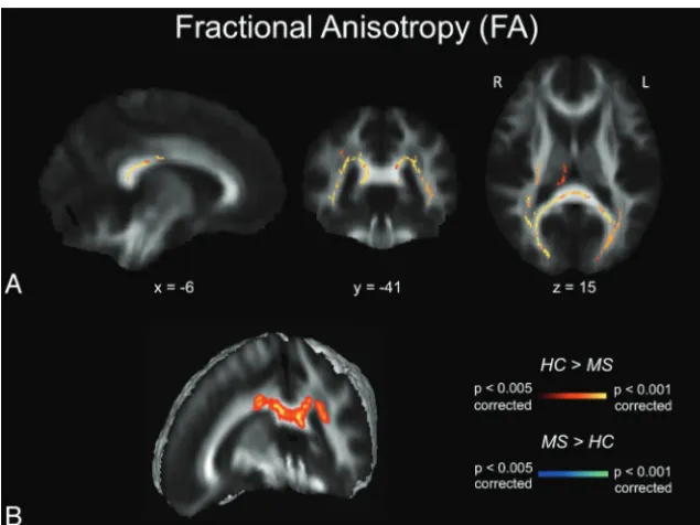
RESULTS:Compared with healthy age- and sex-matched controls, children with multiple sclerosis showed a global decrease in mean fractional anisotropy (Pⱕ.001), with a concomitant increase in mean (P⬍.001), radial (P⬍.05), and axial diffusivity (P⬍.001). The most pronounced fractional anisotropy value decrease in patients with MS was found in the splenium of the corpus callosum (P⬍.001). An additional decrease in fractional anisotropy was identified in the right temporal and right and left parietal regions (P⬍.001). Fractional anisotropy of the white matter skeleton was related to disease duration and may, therefore, serve as a diagnostic marker.
CONCLUSIONS: The microstructure of white matter is altered early in the disease course in childhood multiple sclerosis.
ABBREVIATIONS:AD⫽axial diffusivity; FA⫽fractional anisotropy; MD⫽mean diffusivity; RD⫽radial diffusivity; TBSS⫽tract-based spatial statistics
M
S most commonly occurs in adults in their late 20s and 30s, but early onset in childhood and adolescence is increasingly being diagnosed, with up to 10% of MS cases manifesting before adulthood. Conventional MR imaging has become an essential part of diagnostic decision making in MS. On MR imaging, chil-dren tend to have fewer white matter lesions and lessenhance-ment with gadolinium-based contrast agents.1Lesion load has been shown to correlate moderately with clinical outcome in lon-gitudinal studies in children and adults.2
In addition to the above-mentioned localized lesions, white matter microstructure is known to be altered in the macroscopi-cally normal-appearing white matter.3 Advanced MR imaging techniques such as DTI allow the evaluation of the microstructure of the cerebral white matter by detecting subtle changes in the magnitude and direction of water diffusion. White matter dam-age is mainly reflected by a decrease in fractional anisotropy (FA) and an increase in mean diffusivity (MD). MD consists of axial diffusivity (AD) and radial diffusivity (RD), measuring diffusivity parallel and perpendicular to the main axis of white matter tracts, respectively.4Both parameters are purported to indicate myelin and axonal injury.5Recently, DTI studies in adult patients with MS demonstrated axonal and myelin injury occurring much ear-lier in the course of the disease than previously assumed.6,7
Physiologic myelination is known to expand well into early adult-hood in conjunction with cognitive, behavioral, emotional, and mo-tor development.8-10Any pathology interfering with this process of myelination is likely to affect white matter integrity as has been shown in hypoxic-ischemic encephalopathy, former premature
in-Received November 15, 2012; accepted after revision January 3, 2013.
From the Department of Pediatric Neurology and Developmental Medicine (A.B., A.S.S., W.M.-F., F.H.), Hauner Children’s Hospital, Ludwig-Maximilian-University, Munich, Germany; Department of Psychiatry and Psychotherapy (D.K.) and Insti-tute for Clinical Radiology (D.K., S.M., I.K.K., B.E.-W.), Ludwig-Maximilian University, Munich, Germany; and Psychiatry Neuroimaging Laboratory (I.K.K.), Brigham and Women’s Hospital, Harvard Medical School, Boston, Massachusetts.
A. Blaschek and D. Keeser contributed equally to the manuscript. F. Heinen and B. Ertl-Wagner shared senior authorship.
This work was supported by a research grant from Merck Serono. Inga Koerte was supported by the Else Kro¨ner-Fresenius Stiftung, Bad Homburg, Germany. Please address correspondence to Astrid Blaschek, MD, Department of Pediatric Neurology and Developmental Medicine, Hauner Children’s Hospital, Ludwig-Maximilian-University Munich, Lindwurmstraße 4, 80337 Munich, Germany; e-mail: [email protected]
Indicates article with supplemental on-line tables. Indicates article with supplemental on-line figure.
EBM
fants, and normal-pressure hydrocephalus.11The onset of childhood MS occurs within this vulnerable period of central nervous system maturation. To date, only a few MR imaging studies have investi-gated white matter microstructure in children with MS. These stud-ies demonstrated decreased FA12or slightly increased mean diffusiv-ity13in the macroscopically unaffected white matter by using either summary measures of the entire brain or a region-of-interest ap-proach. Most recently, a study assessing major white matter tracts in children with MS compared with children with a single demyelinat-ing event showed a decrease in FA with a concomitant increase in MD in patients with MS only.14,15
Tract-based spatial statistics (TBSS) allows a coregistration of likely white matter tracts to analyze multisubject diffusion tensor data. For this purpose, subject data are projected on a mean FA skeleton, which refers to the center of all tracts common to both groups, before applying voxelwise between-group comparisons.16 This technique permits an observer-independent voxelwise anal-ysis of the main white matter tracts with a sensitivity to intergroup differences, considering less across-subject FA variability.16
The aim of our study was to evaluate DTI-derived measures of white matter microstructure in children with MS compared with age- and sex-matched controls by using TBSS.
MATERIALS AND METHODS
SubjectsThis study was approved by the local institutional review board. Oral and written informed consent was obtained from all partic-ipants and their legal guardians.
Fourteen consecutive pediatric patients with MS (11 female/3 male; mean age, 15.1⫾1.6 years; age range, 12–17 years) were included in the study. Inclusion criteria were an age younger than 18 years and a definite diagnosis of MS with a relapsing-remitting course. Exclusion criteria consisted of MR imaging–related con-traindications (eg, cardiac pacemakers, ferromagnetic implants, or claustrophobia). All patients were clinically evaluated on the basis of the Kurtzke Expanded Disability Status Scale.
MR imaging of age- and sex-matched healthy subjects (11 fe-male/3 male; mean age, 14.8⫾1.7 years) was included from a local data base of healthy volunteers; all volunteers had been im-aged with the identical MR imaging protocol on the same MR imaging scanner (Magnetom Verio; Siemens, Erlangen, Ger-many). All volunteers underwent MR imaging for scientific pur-poses only. Inclusion criteria were an age match to a patient in-cluded in the MS cohort of⫾6 months and a sex match to the respective patient. Exclusion criteria were any history of chronic or ongoing medical conditions, intake of medication, a history of traumatic brain injury (including mild traumatic brain injury), a tory of headache disorders, a history of learning disorders, any his-tory of other neurologic or psychiatric disorders, and structural brain abnormalities on conventional MR imaging sequences.
Clinical Tests
As part of the clinical work-up, selected elements of standardized tests were available for 13 of the 14 patients. Two parts of the Multiple Sclerosis Functional Composite score were adminis-tered: the right- and left-hand Nine-Hole Peg Test, to measure upper extremity fine motor skills, and the Timed 25-Foot Walk
Test, to assess the lower extremities. All values were compared with normative data.17,18All clinical scores were converted toz scores. Thezscore indicates the deviation from the mean popu-lation score. Azscore lying outside the 95% normal distribution is considered abnormal. Screening of cognitive function was per-formed with 2 subtests of widely used cognitive tests in 11 pa-tients: the Trail-Making Test (subtests A and B)19and the German Leistungspru¨fungssystem (subtest 5) for verbal fluency.20 All raw values were age-corrected for each patient according to Helmstaedter et al,21to receive an age-independent measure.
MR Imaging Acquisition
MR imaging of the brain was performed on a 3T scanner (Magnetom Verio; Siemens Healthcare, Erlangen, Germany) by using a 12-ele-ment phased-array head coil for both the patient cohort and the healthy control subjects. The following structural sequences were ac-quired for all subjects: 3D magnetization-prepared rapid acquisition of gradient echo: TR, 11 ms; TE, 4.76 ms; FOV, 250 mm; voxel size, 1⫻1⫻1 mm3; iPAT (Siemens syngo software) acceleration factor, 2; sagittal sections covering the entire brain, 160; and FLAIR: TR, 94 ms; TE, 7000 ms; FOV, 220 mm; voxel size, 0.9⫻0.9⫻3 mm3; distance factor, 10%; fat saturation; axial sections, covering the entire brain, 45. A DTI sequence with 20 independent diffusion directions and 3 averages was applied with the following parameters:b⫽0 and 1000 s/mm2; matrix size, 128⫻128 mm2; FOV, 230⫻230 mm2. The resulting voxel size was 1.8⫻1.8⫻4.0 mm3. Thirty-six transverse sections were acquired.
Image Analysis and Postprocessing
Evaluation of Lesions. All structural sequences were visually as-sessed and graded by a board-certified neuroradiologist with⬎10 years’ experience in MR imaging of the brain.
DTI Analysis. Image data processing was performed by using the TBSS approach implemented in FMRIB Software Library 4.19 (FSL; http://www.fmrib.ox.ac.uk/fsl). Images were corrected for eddy cur-rents due to changing gradient fields and head motion.22Brain masks were created by using the Brain Extraction Tool in FSL.22FA and mean, axial, and radial diffusivity were calculated for each voxel. FA data of all subjects were aligned to a common space by using the FMRIB58 FA standard space with nonlinear registration.16A mean FA image and a mean FA skeleton were created, which corresponded to the centers of all tracts common to the group (On-line Fig 1). The white matter skeleton is, therefore, a representation of white matter tract geometry, and fiber bundle centers are represented in the mean skeleton. The threshold of the mean FA skeleton white matter mask (shown as a green underlay) was set to an FA value between 0.2 and 0.8 to exclude voxels that consisted of gray matter or CSF. The voxel size was set to 1⫻1⫻1 mm Montreal Neurological Institute space. The obtained binary skeleton mask determined all subsequent pro-cessing steps.
permuta-tions).23We used threshold-free cluster enhancement to avoid choosing an arbitrary initial cluster-forming threshold. This method provides a voxelwise significance (Pvalue) that is cor-rected for multiple comparisons.24TheP values are fully cor-rected for multiple comparisons across voxels but only for each white matter mask and only as 1-tailedPvalues. Because we used 4 different white matter masks and investigated between-group increases and between-between-group decreases, a P ⬍ .05 equaled an adjustedP⬍.00625 [0.05 / (4⫻2)], aPvalue⬍.01 corresponded to an adjusted P⬍ .00125, aP ⬍.005 corre-sponded to an adjustedP⬍ .000625, and aP ⬍.001 corre-sponded to an adjustedP⬍.000125. We considered aP⬍.05 statistically significant.
The same procedure was applied to MD, RD, and AD data.
Clinical Correlations
Within the patient group, clinical results were correlated with mean FA values and of 2 ROIs, which displayed the most significant group differences in the TBSS analysis. The defined ROIs had a diameter of 20 mm around the peak voxel on the FA skeleton. For all analyses,P
values⬍.05 were considered statistically significant.
Statistical Analysis
All clinical scores were calculated aszscores. A z score lying out-side the 95% normal distribution is conventionally conout-sidered abnormal.
The Pearson product moment correlation coefficient (r) was calculated for the correlation between clinical data and mean in-dividual FA values. Correlations were calculated by using the Sta-tistical Package for the Social Sciences, Version 19 (SPSS, Chicago,
Illinois), and aPvalue⬍.05 was consid-ered statistically significant.
RESULTS
Demographic Data, Disease, and MR Imaging Characteristics Fourteen consecutive pediatric patients with MS and age- and sex-matched healthy subjects were included in the study (On-line Table 1). Disease duration in pa-tients ranged between 2 and 57 months (median, 16.5 months). Patients had a low level of neurologic impairment measured by the Kurtzke Expanded Disability Status Scale (mean, 0.75⫾1.2) and the Timed 25-Foot Walk Test (3.73⫾1.3 seconds), consistent with the relatively short disease duration. Cognitive screening with the Trail-Making Test A (mean, 5.9⫾0.5), Trail-Making Test B (mean, 5.72⫾0.6), and the Verbal Fluency test (mean, 4.18⫾ 0.6) showed no differences in normative data (z scores ⬍ 2).21 Unusually high completion times (zscore⬎2) were only observed for the Nine-Hole Peg Test (dominant hand mean, 19.5 ⫾2.6 sec-onds; nondominant hand mean, 19.7⫾ 1.3 seconds) compared with normative data.17Mean lesion load for all patients was low in supratentorial (3.25⫾0.96) and infratentorial (2.5⫾0.76) regions.
Diffusion Parameters in TBSS Analysis
Mean FA values of the white matter skeleton were significantly decreased in patients with MS compared with age- and sex-matched controls (P⬍.005). MD (P⬍.001), RD (P⬍.001), and AD (P⬍.05) values were increased in patients with MS, with changes in RD being more extensive than those in AD. Detailed results of TBSS analysis can be found in On-line Table 2. The most pronounced decrease in FA was found in the splenium of the corpus callosum (P⬍.001) (Fig 1). This result remained stable after excluding 3 patients with lesions within the splenium and surrounding regions (P⬍.001). An additional reduction of FA values was identified in the right temporal and right and left pa-rietal regions (Fig 1A). We did not detect any FA value increase in the patient group compared with the control group.
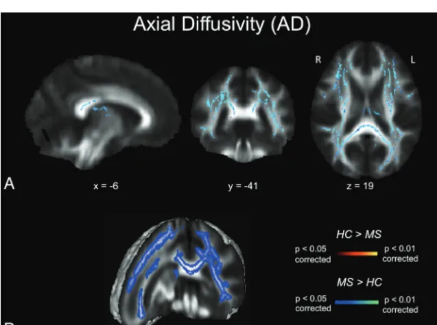
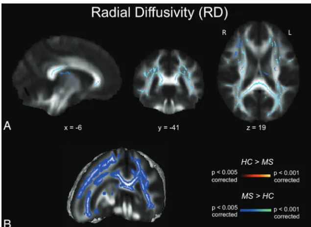
Diffusivity maps revealed a widespread increase in MD in most white matter tract regions, including the corpus callosum, superior longitudinal fasciculus, fornix, corona radiata, corticospinal tract, and uncinate fasciculus (Fig 2). Of the 2 components contributing to MD, AD increase (Fig 3) was less widespread than RD increase (Fig 4). Neither the anterior or posterior corpus callosum nor the internal capsules exhibited any AD alterations (Fig 3). The anterior internal capsule did not show any RD alterations. We did not detect any significant MD, RD, or AD decreases in the patient group.
Correlation between FA and Clinical Scores
We found a statistically significant correlation between mean skeletal FA and disease duration (r⫽ ⫺0.57;P⬍.01), right temporal (r⫽
[image:3.594.56.374.45.283.2]⫺0.55;P ⬍.05), and left parietal (r⫽ ⫺0.54;P⬍.05) regions. No significant correlations were found for the Kurtzke Expanded Disability Status Scale, annual relapse rate, number of total relapses, lesion load, Nine-Hole Peg Test, Timed 25-Foot Walk Test, or cognitive screening with FA values.
DISCUSSION
We investigated the white matter micro-structure in children with MS. TBSS analysis revealed significantly decreased FA values along with a widespread in-crease in MD, RD, and AD in patients with MS. These findings provide evi-dence for the influence of MS in nonle-sional macroscopically unaffected WM even at an early stage of the disease. Po-tential causes of these alterations are di-verse and include direct disease effects such as axonal injury and demyelination or indirect effects such as protracted or inhibited maturation.
Our findings are in accordance with previously published data on the micro-structure of white matter in childhood MS.12,13Two recent studies of Vishwas et al14,15demonstrated higher MD and lower mean FA values in all white matter pathways in pediatric patients with MS compared with controls. However, no changes were observed in 27 patients with a single demyelinating event,15 from which the authors concluded that there might be a window of opportunity to prevent further damage between the period of a single demyelination and the diagnosis of MS. This is in accordance with the significant negative correlation between mean FA of the white matter skeleton and disease duration that we observed in our study. In adults, con-flicting data on the correlation of FA mea-sures with the annual relapse rate, number of total relapses, and lesion load exist. Dis-ease durations of⬎3 years have been re-ported to negatively correlate with FA,25,26 whereas in clinically isolated syndrome, no correlation was reported.6
It has been proposed that global changes in white matter occur to a lesser degree in childhood at the beginning of the disease compared with adult-onset MS.6However, early changes in childhood MS are known to be characterized by pathologic MR imaging findings of gray FIG 2. MD maps.A, TBSS MD results for healthy subjects compared with patients with MS.
Significant clusters of increased MD values in patients with MS compared with healthy controls are shown as correspondingPvalues in red-orange (scale ranging from red to yellow for the comparison HC⬎MS and scale ranging from blue to light blue for the comparison MS⬎HC) and have been thresholded atP⬍.005 for between-group comparisons (corrected for multiple comparisons).B, 3D visualization of significantly (P⬍.01, corrected for multiple comparisons) different white matter clusters between the 2 groups. Note that the results are thickened for visual purposes only. L indicates left; R, right (radiologic convention); HC, healthy controls.
[image:4.594.55.374.45.279.2] [image:4.594.56.375.404.641.2]and white matter structures that are generally more subtle compared with MR imaging findings in adult MS.12,13,27This difference may explain why patients with early-onset MS tend to have a longer du-ration of illness before reaching a state of irreversible disability.28 Considering the fact that myelination continues well into adoles-cence, it is conceivable that MS may interfere with this process of physiologic myelination in addition to the ongoing demyelination by the disease process itself.8,29Longitudinal assessment might be a use-ful tool for gaining insight into the dynamics of the disease process, especially because established markers for disease activity such as le-sion load correlate only moderately with clinical outcome in longitu-dinal studies in adults and children.2
The spatial extent and distribution of increase in diffusivity indi-ces (MD/RD/AD) was far more widespread than the decrease in FA values in our patient cohort. Changes in diffusivity parameters are indicators of mainly extracellular diffusion and might be related to axon or myelin damage. Water diffusion in the white matter of the brain is highly sensitive to alterations in the microstructural integrity of cellular membranes; thus, water diffusion is a sensitive marker for early differences in tissue properties related to neuroinflammation, for example.4,5An increase in RD is proposed to reflect myelin de-struction as demonstrated in mouse models or human postmortem brain studies.30-32The relevance of AD as a marker of axonal damage is less well-understood. In our study, AD was increased but to a lesser extent than RD; these findings are in accordance with those in earlier studies in adults with MS.10,25In contrast, Lin et al33showed a trend toward lower AD in the normal-appearing pyramidal tract in adult patients with MS. Recent pathologic studies in degenerative neuro-logic diseases indicate a topographic concordance between WM loss and AD changes in Huntington disease or Friedreich ataxia.34,35
Ro-sas et al34suggested that the increase in AD might be due to reduced axonal diameters, resulting in increased extra-axonal space. Thus, interpretation of this diffusivity value is still controversial, with most stud-ies in MS pointing toward an increase in AD. On the other hand, RD plays a pre-dominant role in detecting subtle patho-logic damage from very early on in MS, making it a potential marker for monitor-ing changes in the course of the disease.
In addition to the already-known global decrease in skeletal FA,12patients with MS displayed a regional FA reduc-tion in the splenium of the corpus callo-sum and in the right temporal and left and right parietal regions. Reduction in FA was most profound in the splenium. These changes in FA may serve as an early indicator of MS white matter pa-thology in childhood MS. Our results are in accordance with DTI studies in adult-onset MS, which report that the earliest changes in white matter tracts occurred in the corpus callosum.6A fo-cal FA decrease in the splenium of the corpus callosum has furthermore been described in patients with isolated optic neuritis.36Our results suggest that the corpus callosum is one of the most affected regions in early-onset MS. It may, therefore, be a potential target region for the early detection of disease processes by MR imaging. Special attention was drawn to this region by Hagmann et al,37who combined modern MR imaging techniques with network anal-yses. This publication presented evidence for the existence of a structural core of highly interconnected brain regions, located primarily in the posterior medial and parietal cortex. The sp-lenium of the corpus callosum has been proposed as an integral part of this core, interconnecting both hemispheres.
In our relatively small patient cohort, we observed a significant negative correlation between mean FA of the white matter skele-ton and disease duration, while no correlation was found for other clinical parameters. In adults, conflicting data on the correlation of FA measures with annual relapse rate, number of total relapses, and lesion load exist. Disease duration of⬎3 years is reported to negatively correlate with FA,25,26whereas in clinically isolated syndrome, no correlation was reported.6
Study Limitations
There are several limitations to our study that need to be taken into account when interpreting the data. First, the sample sizes in our patient and control cohorts are relatively small due to the rare nature of MS in children. Nevertheless, the alterations in diffusivity param-eters were highly significant and appear robust in light of the tests applied. In addition, the MR images of the age- and sex-matched healthy controls were recruited from a local data base, in which the clinical assessment did not include the Nine-Hole Peg Test and the Timed 25-Foot Walk Test. The study-related MR imaging protocol and the MR imaging scanner were identical in both cohorts, though. FIG 4. RD maps.A, TBSS RD results for healthy subjects compared with patients with MS.
[image:5.594.55.373.45.277.2]CONCLUSIONS
Our study demonstrates the white matter microstructure of non-lesional tissue to be affected in childhood MS, even in early stages of the disease. Decreased FA values and increased diffusion pa-rameters may indicate impaired myelination. The observed de-crease in FA in the white matter correlated with disease duration.
ACKNOWLEDGMENTS
We thank all participants in this study.
Disclosures: Astrid Blaschek—RELATED: Grant:Research grant from Merck Serono GmbH;*UNRELATED: Payment for Lectures (including service on speaker bureaus):
Speaker honoraria from Biogen Idec GmbH, Merck Serono GmbH, and Bayer Schering GmbH;*Travel/Accommodations/Meeting Expenses Unrelated to Activities Listed:
Bayer Schering GmbH, Merck Serono GmbH. Sophia Mueller—UNRELATED: Travel/ Accommodations/Meeting Expenses Unrelated to Activities Listed:I received a travel grant from the GlaxoSmithKline Foundation (2012) to travel to the Annual Meeting of the Organization for Human Brain Mapping. I presented work that is unrelated to the sub-mitted study. Wolfgang Mu¨ller-Felber—UNRELATED: Board Membership:Pompe reg-istry board. Florian Heinen—RELATED: Grant:Merck Serono GmbH;*Consulting Fee or Honorarium:Speakers honorarium from Meck Serono; UNRELATED:Payment for Lec-tures (including service on speaker bureaus):Merz Pharma, Pfizer Deutschland GmbH, and Allergan GmbH. Birgit Ertl-Wagner—RELATED: Grant:German Research Council;
UNRELATED: Board Membership:Philips Healthcare, Bracco, Springer Medical Publisher;
Consultancy:Munich Medical International, Philips Healthcare;Grants/Grants Pending:
Eli Lilly,* Genentech,* Geurbet,* Merck Serono;*Payment for Lectures (including service on speaker bureaus):Siemens, Bayer Schering;Payment for Manuscript Preparation:
Siemens, Springer Medical Publisher, Thieme Medical Publisher, Bracco; Royalties:
Springer Medical Publisher, Thieme Medical Publisher;Payment for Development of Educational Presentations:Siemens, Bracco, Springer, Thieme;Stock/Stock Options:
Siemens (stock owned by spouse);Travel/Accommodations/Meeting Expenses Unre-lated to Activities Listed:Siemens.*Money paid to institution.
REFERENCES
1. Banwell B, Shroff M, Ness JM, et al.MRI features of pediatric mul-tiple sclerosis.Neurology2007;68(16 suppl 2):S46 –53
2. Mikaeloff Y, Adamsbaum C, Husson B, et al.MRI prognostic factors for relapse after acute CNS inflammatory demyelination in child-hood.Brain2004;127(pt 9):1942– 47
3. van der Valk P, Amor S.Preactive lesions in multiple sclerosis.Curr Opin Neurol2009;22:207–13
4. Pierpaoli C, Basser PJ.Toward a quantitative assessment of diffu-sion anisotropy.Magn Reson Med1996;36:893–906
5. Song SK, Sun SW, Ju WK, et al.Diffusion tensor imaging detects and differentiates axon and myelin degeneration in mouse optic nerve after retinal ischemia.Neuroimage2003;20:1714 –22
6. Raz E, Cercignani M, Sbardella E, et al.Clinically isolated syndrome suggestive of multiple sclerosis: voxelwise regional investigation of white and gray matter.Radiology2010;254:227–34
7. Bodini B, Cercignani M, Khaleeli Z, et al.Corpus callosum damage pre-dicts disability progression and cognitive dysfunction in primary-progressive MS after five years.Hum Brain Mapp2013;34:1163–72 8. Barnea-Goraly N, Menon V, Eckert M, et al.White matter
develop-ment during childhood and adolescence: a cross-sectional diffu-sion tensor imaging study.Cereb Cortex2005;15:1848 –54 9. Koerte I, Heinen F, Fuchs T, et al.Anisotropy of callosal motor fibers
in combination with transcranial magnetic stimulation in the course of motor development.Invest Radiol2009;44:279 – 84 10. Roosendaal SD, Geurts JJ, Vrenken H, et al.Regional DTI differences
in multiple sclerosis patients.Neuroimage2009;44:1397– 403 11. Utsunomiya H.Diffusion MRI abnormalities in pediatric
neurolog-ical disorders.Brain Dev2011;33:235– 42
12. Tortorella P, Rocca MA, Mezzapesa DM, et al.MRI quantification of gray and white matter damage in patients with early-onset multiple sclerosis.J Neurol2006;253:903– 07
13. Mezzapesa DM, Rocca MA, Falini A, et al.A preliminary diffusion tensor and magnetization transfer magnetic resonance imaging study of early-onset multiple sclerosis.Arch Neurol2004;61:366 – 68 14. Vishwas MS, Chitnis T, Pienaar R, et al.Tract-based analysis of
cal-losal, projection, and association pathways in pediatric patients with multiple sclerosis: a preliminary study.AJNR Am J Neuroradiol 2010;31:121–28
15. Vishwas MS, Healy BC, Pienaar R, et al.Diffusion tensor analysis of pediatric multiple sclerosis and clinically isolated syndromes.
AJNR Am J Neuroradiol2013;34:417–23
16. Smith SM, Jenkinson M, Johansen-Berg H, et al.Tract-based spatial statistics: voxelwise analysis of multi-subject diffusion data. Neuro-image2006;31:1487–505
17. Poole JL, Burtner PA, Torres TA, et al.Measuring dexterity in chil-dren using the nine-hole peg test.J Hand Ther2005;18:348 –51 18. Nieuwenhuis MM, Van Tongeren H, Sorensen PS, et al.The six spot
step test: a new measurement for walking ability in multiple sclero-sis.Mult Scler2006;12:495–500
19. Reitan RM, Wolfson D.The trail making test as an initial screening procedure for neuropsychological impairment in older children.
Arch Clin Neuropsychol2004;19:281– 88
20. Horn W. Leistungspru¨fungssystem Manual. Go¨ttingen, Germany: Hogrefe; 1983
21. Helmstaedter C, Schoof K, Rossmann T, et al.Introduction and first validation of EpiTrack Junior, a screening tool for the assessment of cognitive side effects of antiepileptic medication on attention and executive functions in children and adolescents with epilepsy. Epi-lepsy Behav2010;19:55– 64
22. Smith SM.Fast robust automated brain extraction.Hum Brain Mapp2002;17:143–55
23. Nichols TE, Holmes AP.Nonparametric permutation tests for func-tional neuroimaging: a primer with examples.Hum Brain Mapp 2002;15:1–25
24. Smith SM, Nichols TE.Threshold-free cluster enhancement: ad-dressing problems of smoothing, threshold dependence and locali-sation in cluster inference.Neuroimage2009;44:83–98
25. Liu Y, Duan Y, He Y, et al.Whole brain white matter changes re-vealed by multiple diffusion metrics in multiple sclerosis: a TBSS study.Eur J Radiol2012;81:2826 –32
26. Yu HJ, Christodoulou C, Bhise V, et al.Multiple white matter tract abnormalities underlie cognitive impairment in RRMS. Neuroim-age2012;59:3713–22
27. Bala´ssy C, Bernert G, Wober-Bingol C, et al.Long-term MRI obser-vations of childhood-onset relapsing-remitting multiple sclerosis.
Neuropediatrics2001;32:28 –37
28. Renoux C, Vukusic S, Mikaeloff Y, et al.Natural history of multiple sclerosis with childhood onset.N Engl J Med2007;356:2603–13 29. Wilke M, Krageloh-Mann I, Holland SK.Global and local
develop-ment of gray and white matter volume in normal children and ad-olescents.Exp Brain Res2007;178:296 –307
30. Song SK, Yoshino J, Le TQ, et al.Demyelination increases radial diffu-sivity in corpus callosum of mouse brain.Neuroimage2005;26: 132– 40 31. Schmierer K, Wheeler-Kingshott CA, Tozer DJ, et al.Quantitative magnetic resonance of postmortem multiple sclerosis brain before and after fixation.Magn Reson Med2008;59:268 –77
32. Zollinger LV, Kim TH, Hill K, et al.Using diffusion tensor imaging and immunofluorescent assay to evaluate the pathology of multiple sclerosis.J Magn Reson Imaging2011;33:557– 64
33. Lin F, Yu C, Jiang T, et al.Diffusion tensor tractography-based group mapping of the pyramidal tract in relapsing-remitting mul-tiple sclerosis patients.AJNR Am J Neuroradiol2007;28:278 – 82 34. Rosas HD, Lee SY, Bender AC, et al.Altered white matter
micro-structure in the corpus callosum in Huntington’s disease: implica-tions for cortical “disconnection.”Neuroimage2010;49:2995–3004 35. Pagani E, Ginestroni A, Della Nave R, et al.Assessment of brain white
matter fiber bundle atrophy in patients with Friedreich ataxia. Ra-diology2010;255:882– 89
36. Bester M, Heesen C, Schippling S, et al.Early anisotropy changes in the corpus callosum of patients with optic neuritis.Neuroradiology 2008;50:549 –57