ORIGINAL RESEARCH
PEDIATRICS
Asymmetric Meckel Cave Enlargement: A Potential Marker of
PHACES Syndrome
XJ.N. Wright andX V. Wycoco
ABSTRACT
BACKGROUND AND PURPOSE: PHACES syndrome is a complex of morphologic abnormalities of unknown cause and includes posterior fossa abnormalities; head and neck infantile hemangiomas; arterial, cardiac, and eye anomalies; and sternal or abdominal wall defects. Accurate identification of the syndrome is important for optimal treatment. The purpose of this study was to investigate the incidence of asymmetric Meckel cave enlargement, a potential novel imaging marker, in a population of patients referred for evaluation of possible PHACES syndrome.
MATERIALS AND METHODS: Eighty-five patients referred for neuroimaging evaluation of possible PHACES syndrome were identified and stratified on the basis of their ultimate clinical PHACES diagnosis categorization into PHACES, possible PHACES, or not PHACES. MR imaging studies were subsequently reviewed for the presence or absence of unilateral Meckel cave enlargement, with the reviewer blinded to the ultimate PHACES syndrome categorization.
RESULTS:Twenty-five of 85 patients (29%) were ultimately categorized as having PHACES or possible PHACES according to consensus guidelines. Asymmetric Meckel cave enlargement was present in 76% (19/25) of these patients and in 82% (19/23) of only those patients with definite PHACES. This finding was present in none of the 60 patients determined not to have PHACES syndrome. In 7/19 patients (37%) with this finding, subtle MR imaging abnormalities consistent with PHACES were missed on the initial MR imaging interpretation.
CONCLUSIONS: Asymmetric Meckel cave enlargement was a common feature of patients with PHACES in our cohort and may serve as a novel imaging marker. Increased awareness of this imaging feature has the potential to increase the diagnostic accuracy of PHACES.
ABBREVIATIONS:IAC⫽internal auditory canal; PHACES⫽posterior fossa abnormalities; head and neck infantile hemangiomas; arterial, cardiac and eye anomalies; and sternal or abdominal wall defects
P
HACES syndrome (Online Mendelian Inheritance in Man No. 606519; omim.org) is a complex of morphologic malities of unknown cause and includes posterior fossa abnor-malities; head and neck infantile hemangiomas, often in a seg-mental distribution; arterial, cardiac, and eye anomalies; and sternal or abdominal wall defects.1Accurate recognition of thesyndrome is important to identify the potentially increased risk associated with the treatment of infantile hemangiomas in the setting of underlying arterial or cardiac pathology,2,3as well as to
initiate careful surveillance and aggressive intervention for poten-tial speech and language delays related to posterior fossa
anoma-lies.4,5There is increasing consensus that the stigmata of PHACES
may be subtler than previously thought, and new diagnostic cri-teria are being considered. We have observed that asymmetric Meckel cave enlargement is a frequent neuroimaging finding in patients with PHACES syndrome and may serve as an easily rec-ognizable marker that can improve diagnostic sensitivity.
MATERIALS AND METHODS
Following Seattle Children’s Hospital institutional review board approval, we retrospectively identified 93 patients referred for MR imaging of the head and neck due to concern for PHACES syn-drome between 1994 and 2016, based on the presence or history of large (⬎5 cm) or segmental head and neck hemangiomas (n⫽92) or sternal clefting (n⫽1). Imaging protocols varied but generally included contrast-enhanced MR imaging of the brain and time-of-flight MRA of the head and neck. In all cases, a coronal fluid-sensitive sequence (T2-weighted, STIR, or steady-state free pre-cession [balanced fast-field echo, FIESTA, or CISS]) was included in the protocol. Five patients were excluded due to lesions other
Received October 6, 2016; accepted after revision January 17, 2017.
From the Department of Radiology (J.N.W.), University of Washington and Seattle Children’s Hospital, Seattle, Washington; and Department of Neurological Inter-vention and Imaging (V.W.), Alterna Wellness Center, Nedlands, Western Australia. Please address correspondence to Jason N. Wright, MD, M/S MA.7.220, Seattle Children’s Hospital, PO Box 5371, Seattle, WA 98105; e-mail: jnixon@uw.edu
Indicates article with supplemental on-line table.
than hemangiomas identified on MR imaging, and 3 additional patients were excluded due to lack of available clinical records.
We then stratified the remaining 85 patients on the basis of their ultimate clinical PHACES diagnosis categorization within the published 2009 consensus criteria into PHACES, possible PHACES, or not PHACES.1Current diagnostic criteria are as
previ-ously published by Metry et al.1Work-up included a combination of
dermatologic evaluation, ophthalmologic examination, cardiology consultation including echocardiography, and structural MR imag-ing evaluation of the head and neck.
We subsequently reviewed all MR imaging studies for the pres-ence or abspres-ence of asymmetric unilateral Meckel cave enlarge-ment, blinded to the ultimate PHACES syndrome categorization. Particular attention was paid to the coronal fluid-sensitive se-quences at the level of the sella turcica, which highlighted the comparative volumes of the bilateral Meckel caves. “Presence” was defined as obvious subjective asymmetric enlargement based on visual assessment, with an estimated volume ratio of approxi-mately 1:1.5 used as a cutoff. Our goal was to evaluate the utility of this novel imaging finding in a clinically applicable manner re-quiring no advanced morphometric analysis for implementation. Minimal asymmetry was not considered sufficient for positivity.
RESULTS
Of the 85 included patients evaluated by MR imaging for suspi-cion of PHACES syndrome, 25 patients (29%) were ultimately categorized as having PHACES (n⫽23) or possible PHACES (n⫽2) according to consensus guidelines. Mean and median ages for the PHACES cohort were 20 and 4 months, respectively. Eighty-four percent were female, in line with prior published re-ports.6Summary statistics for all patients are presented in the Table.
All patients with PHACES presented with large or segmental head or neck hemangiomas, excepting 1 patient (patient 4) cate-gorized as having possible PHACES, who was evaluated for sternal clefting noted at birth and who subsequently developed a left fa-cial segment 3– distribution hemangioma of⬍5 cm. Additional diagnostic criteria for PHACES present in each patient are pro-vided in the On-line Table.
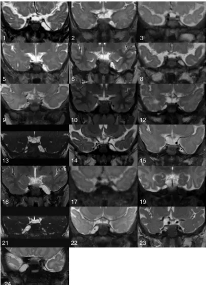
Asymmetric Meckel cave enlargement was present in 76% (19/ 25) of all patients with PHACES (Fig 1); the finding was present in 82% (19/23) of patients with definite PHACES. Patients with Meckel cave involvement were more likely to have a facial seg-ment 1 or 2 distribution of facial hemangiomas, while patients
without were more likely to have a facial segment 3 or cervicotho-racic distribution, though overlap occurred in both directions. Five patients (patients 2, 5, 11, 20, and 23) demonstrated internal auditory canal (IAC) hemangiomas, and 1 patient (patient 23) had a Meckel cave hemangioma, all associated with ipsilateral Meckel cave enlargement.
Of the patients with unilateral Meckel cave enlargement, all enlargements were ipsilateral to the facial or head and neck hem-angioma. Sixty-eight percent (13/19) had associated ipsilateral cerebellar hypoplasia, and 58% (11/19) had dysplastic cerebellar clefting associated with the hypoplasia. This finding is compared with 60% (15/25) with ipsilateral cerebellar hypoplasia in all pa-tients with PHACES, and 48% (12/25) with associated dysplastic clefting. In all cases, the cerebellar hemispheric abnormalities were ipsilateral to both the hemangioma and the asymmetrically enlarged Meckel cave.
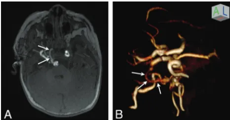
Eighty-nine percent (17/19) of patients with Meckel cave en-largement had craniocervical vascular anomalies, most with arte-rial dysplasia. This finding is comparable with 84% (21/25) noted in all patients with PHACES in our cohort. When present, arterial abnormalities were always ipsilateral to the side of the facial hem-angioma and Meckel cave enlargement, though additional con-tralateral abnormalities occurred in a large minority of cases (41%) (Fig 2).
Major cardiac or arch anomalies were present in 21% (4/19) of patients with asymmetric Meckel cave enlargement, compared with a slightly higher 28% (7/25) of all patients with PHACES in our cohort.
Excepting strabismus related to lid or orbital involvement by fa-cial hemangiomas, ocular anomalies described in PHACES were not identified in any patient on imaging or fundoscopic examination.
Of note, in 7 of the 19 patients (37%) with asymmetric Meckel cave enlargement, there were subtle MR imaging abnormalities of the posterior fossa, intracranial vessels, or aortic arch and cervical vessels that were missed on the initial MR imaging interpretation (patients 1, 2, 3, 6, 8, 14, and 17). One of these patients (patient 3) was nevertheless diagnosed with PHACES prospectively on the basis of cardiac abnormalities identified on echocardiography. The 6 additional patients were not prospectively identified and were only retrospectively diagnosed with PHACES during our review. One of these patients (patient 1) was initially imaged be-fore the original description of PHACES by Frieden et al in 1996.7
Sixty of the 85 patients (71%) evaluated by MR imaging were Cohort summary data and statistics
All PHACES Definite PHACES Possible PHACES Not PHACES
No. (% total) 25 (29) 23 (27) 2 (2) 60 (71)
Female (No.) (% category) 21 (84) 19 (82) 2 (100) 50 (83)
Age (mean⫾SD) 20⫾39 mo 22⫾40 mo 1⫾0.5 mo 10⫾17 mo
Age (range) (median) 5 days to 14 yr (4 mo) 5 days to 14 yr (5 mo) 1–2 mo (1.5 mo) 1 mo to 7 yr (4 mo)
Meckel cave enlargement (No.) (% category) 19 (76) 19 (82) 0 (0) 0 (0)
Posterior fossa anomalies (No.) (% category) 15 (60) 15 (65) 0 (0) 0 (0)
Facial hemangioma (No.) (% category) 24 (96) 23 (100) 1 (50) 60 (100)
Arterial anomalies (No.) (% category) 21 (84) 20 (90) 1 (50) 0 (0)
Cardiac anomalies (No.) (% category) 13 (52) 12 (52) 1 (50) 0 (0)
Eye anomalies (No.) (% category) 0 (0) 0 (0) 0 (0) 0 (0)
Sternal or midline abdominal anomalies (No.) (% category)
[image:2.594.57.531.55.192.2]ultimately determined not to have PHACES syndrome. Of these patients, asymmetric ipsilateral Meckel cave enlargement was not identified in any patient.
DISCUSSION
[image:3.594.81.504.45.628.2]have fallen under the rubric of PHACES, a neurocutaneous syn-drome of uncertain etiology. In our series, 29% of patients re-ferred for MR imaging evaluation for suspicion of PHACES were ultimately diagnosed with PHACES or possible PHACES per con-sensus guidelines, in line with the overall prevalence reported in prior studies.1,6
Thorough and accurate neuroimaging evaluation is one of the mainstays of complete evaluation for such patients, with MR im-aging of the brain and MRA of the head and neck recommended in all cases. In addition to evaluating the distribution and extent of proliferative phase infantile hemangiomas, these studies may re-veal abnormalities of the posterior fossa, cervicocranial arteries, globes, or aortic arch and great vessels. These findings are impor-tant for optimal risk assessment before initiation ofblocker therapy for hemangiomas,2,3as well as to cue
neurodevelopmen-tal surveillance for possible associated speech and language delays related to posterior fossa anomalies.4,5
In this study, we have demonstrated that the finding of asym-metric Meckel cave enlargement ipsilateral to the facial hemangi-oma is a common feature of PHACES, present in 76% of patients. This imaging finding can serve as a useful and conspicuous marker for the syndrome. When applied as an independent diag-nostic criterion to our full patient cohort, the finding demon-strated a sensitivity of 76%, a specificity of 100%, a positive pre-dictive value of 100%, a negative prepre-dictive value of 91%, and an accuracy of 93% in predicting a clinical diagnosis of PHACES or possible PHACES syndrome.
In no case was asymmetric Meckel cave enlargement an iso-lated intracranial finding of PHACES. However, greater than one-third of our patients with Meckel cave enlargement had additional subtle stigmata of PHACES that were missed on the initial evalu-ation. These may have been more easily or accurately identified with a heightened pretest probability for PHACES associated with asymmetric Meckel cave enlargement. Thus, recognition of this finding could increase the diagnostic sensitivity of neuroimaging for PHACES syndrome. If these additional intracranial findings had been prospectively made in the setting of asymmetric Meckel
cave enlargement, the diagnostic sensitivity of MR imaging/MRA for PHACES would have increased by 24% in our cohort.
Meckel cave enlargement as a finding in PHACES syndrome has only rarely been described in the literature to date, to our knowledge, and is not an abnormality that is currently included in the consensus criteria for PHACES. Given the incidence of the finding in our cohort, this omission likely reflects under-report-ing. Oza et al8described the finding of unilateral Meckel cave
prominence in 3 of the 16 patients in their series. The finding was subsequently described in 2 additional case reports.9,10Unilateral
Meckel cave enlargement is also demonstrated in Figs 6 and 7 in the textVascular Lesions of the Head and Neck: Diagnosis and Man-agementby Persky et al,11Figs 1 and 2 from the article by Judd et
al,12and Fig 5 from the article by Meltzer et al.13Furthermore,
some authors have described an association of arachnoid cysts and PHACES,1,2,6,8,12,14and an asymmetric Meckel cave could
conceivably be confused with a middle cranial fossa arachnoid cyst, as was the case in one of our patients (patient 21).
The etiology of unilateral Meckel cave enlargement ipsilateral to the facial hemangioma and posterior fossa anomalies in PHACES syndrome is not definitely known. One plausible expla-nation derives from the theory that PHACES is caused by aberrant or deficient migration of the cephalic neural crest in a metameric distribution.15Neural crest cells and paraxial mesoderm derived
from the rhombencephalic metamere contribute to the formation of the skull base; trigeminal nerve ganglia; and facial bones, soft tissues, and blood vessels. Meckel cave enlargement may therefore represent a component of skull base dysplasia secondary to a postzygotic mutation or early prenatal insult in this territory. A similar theory was advanced to explain the relatively high inci-dence of enlarged IACs seen in PHACES syndrome observed by Meltzer et al.13
Alternatively, unilateral enlargement of Meckel cave may be the result of direct expansion secondary to an extant or previously involuted Meckel cave hemangioma. Judd et al12reported on the
association between PHACES and intracranial hemangiomas. Most commonly described in the cerebellopontine angle or IAC, hemangiomas centered in or extending to Meckel cave both have been described in the literature12,13and noted in our series. One
of the patients described by Judd et al demonstrated an enlarged Meckel cave containing a hemangioma on initial imaging (Fig 2C
in Judd et al), which then progressed to isolated Meckel cave en-largement following hemangioma involution (Fig 2Ein Judd et al), supporting a mechanical etiology of the enlargement.
Metzler et al13similarly raised the possibility of a causal
asso-ciation between IAC hemangioma and IAC enlargement. This was based on the increased prevalence of IAC hemangiomas within the enlarged IACs in children when imaged at younger than 1 year of age, compared with children older than 1 year of age at initial imaging, in whom the hemangiomas were presumed to have previously involuted. Given that Meckel cave hemangioma was present in only 1 of 19 patients with Meckel cave enlargement in our cohort, with a median age of 4 months at imaging, it is unlikely that mechanical enlargement can adequately explain the etiology of this finding in all patients in our cohort.
Limitations of our study included the retrospective nature of the analysis and the relatively small sample size. Also, a subset of FIG 2. A persistent right trigeminal artery in a 1-year-old girl (patient
[image:4.594.53.286.47.169.2]patients with PHACES may possibly present without obvious cu-taneous stigmata. These patients would likely have been missed by our diagnostic algorithm, and any data regarding the prevalence of asymmetric Meckel cave enlargement may not be applicable to this population of patients.
CONCLUSIONS
Asymmetric Meckel cave enlargement was a common feature of patients with PHACES and possible PHACES in our cohort and may serve as a conspicuous marker for the syndrome. Increased awareness of this imaging feature has the potential to increase the diagnostic accuracy of the neuroimaging evaluation for PHACES in the setting of large or segmental facial hemangiomas.
REFERENCES
1. Metry D, Heyer G, Hess C, et al; PHACE Syndrome Research Confer-ence.Consensus Statement on Diagnostic Criteria for PHACE Syn-drome.Pediatrics2009;124:1447–56CrossRef Medline
2. Siegel DH, Tefft KA, Kelly T, et al.Stroke in children with posterior fossa brain malformations, hemangiomas, arterial anomalies, co-arctation of the aorta and cardiac defects, and eye abnormalities (PHACE) syndrome: a systematic review of the literature.Stroke
2012;43:1672–74CrossRef Medline
3. Metry D, Frieden IJ, Hess C, et al.Propranolol use in PHACE syn-drome with cervical and intracranial arterial anomalies: collective experience in 32 infants.Pediatr Dermatol2013;30:71– 89CrossRef Medline
4. Tangtiphaiboontana J, Hess CP, Bayer M, et al.Neurodevelopmental abnormalities in children with PHACE syndrome.J Child Neurol
2013;28:608 –14CrossRef Medline
5. Brosig CL, Siegel DH, Haggstrom AN, et al.Neurodevelopmental
outcomes in children with PHACE syndrome.Pediatr Dermatol
2016;33:415–23CrossRef Medline
6. Metry DW, Haggstrom AN, Drolet BA, et al.A prospective study of PHACE syndrome in infantile hemangiomas: demographic fea-tures, clinical findings, and complications.Am J Med Genet A2006; 140:975– 86Medline
7. Frieden IJ, Reese V, Cohen D.PHACE syndrome: the association of posterior fossa brain malformations, hemangiomas, arterial anom-alies, coarctation of the aorta and cardiac defects, and eye abnor-malities.Arch Dermatol1996;132:307–11Medline
8. Oza VS, Wang E, Berenstein A, et al.PHACES association: a neuro-radiologic review of 17 patients.AJNR Am J Neuroradiol2008;29: 807–13CrossRef Medline
9. Arora SS, Plato BM, Sattenberg RJ, et al.Adult presentation of PHACES syndrome.Interv Neuroradiol2011;17:137– 46CrossRef Medline
10. Rosmaninho A, Machado S, Bastos-Leite AJ, et al.PHACE syndrome: a new case report.Eur J Dermatol2011;21:289 –90CrossRef Medline
11. Persky MS, Waner M, Blei F, et al.Vascular Lesions of the Head and Neck: Diagnosis and Management.New York: Thieme; 2015:chap 6 12. Judd CD, Chapman PR, Koch B, et al.Intracranial infantile
heman-giomas associated with PHACE syndrome.AJNR Am J Neuroradiol
2007;28:25–29Medline
13. Meltzer DE, Robson CD, Blei F, et al.Enlargement of the internal auditory canal and associated posterior fossa anomalies in PHACES association. AJNR Am J Neuroradiol2015;36:2159 – 62
CrossRef Medline
14. Hartemink DA, Chiu YE, Drolet BA, et al.PHACES syndrome: a review. Int J Pediatr Otorhinolaryngol 2009;73:181– 87 CrossRef Medline
15. Krings T, Geibprasert S, Luo CB, et al.Segmental neurovascular syndromes in children.Neuroimaging Clin N Am2007;17:245–58