warwick.ac.uk/lib-publications
A Thesis Submitted for the Degree of PhD at the University of Warwick
Permanent WRAP URL:
http://wrap.warwick.ac.uk/109991
Copyright and reuse:
This thesis is made available online and is protected by original copyright.
Please scroll down to view the document itself.
Please refer to the repository record for this item for information to help you to cite it.
Our policy information is available from the repository home page.
T H E B R I T I S H L I B R A R Y D O C U M EN T
SUPPLY
CENTRE
TITLE
STUDIES OS THE ENTRY OF RIC I N SUBUNITS INTO CELLSA U TH O R
C.rr J O « C l . » * « «IN STITU TIO N
and DATE
Un i v e r s i t y of Warwick, 1 1 1 8Attention is drawn to the fact that the copyright of
this thesis rests with its author.
This copy of the thesis has been supplied on condition
that anyone who consults it is understood to recognise
that its copyright rests with its author and that no
information derived from it m ay be published without
the author’s prior written consent.
T T ems
1 2| 1 S| 1 4 1 s ! 6
T H E B R I T I S H L I B R A R Y D O C U M E N T SUPPLY C E N T R E Boston Spa. Wetherby West Yorkshire
United Kingdom R ED U CTIO N X _______
21
STUDIES ON THE ENTRY OF RICIN SUBUNITS INTO CELLS
Gary James Clements B.Sc. (Hons) Brunei
A thesis submitted for the degree of
Doctor of Philosophy
Department of Biological Sciences,
University of Warwick,
Coventry,v
U.K.
For Mum, Dad and Karen.
'All things are poison and nothing is without poison.
The dose alone decides ...."
CONTENTS
Section Title Peee
List of Tables 111
List of Figures Iv
Acknowledgements xl
Declaration xlli
Abbreviations xlv
Chapter I : Introduction 1
1:1:1 Overview 1
1:1:2 Structure of Rlcln 3
1:1:3 Biological Activities of Rlcln Subunits 4
1:1:4 The Cytotoxic Nature of Rlcln 5
1:2:1 The Structure, Evolution and Function of
Ricin B Chain 7
1:2:2 Structure, Function and Evolution of
Ricin A Chain 11
1:3:1 Internalization and Intracellular
Trafficking of Ricin 17
1:4:1 A Comparison of Structure and Function
Between Ricin and Diphtheria Toxin 21
1:4:2 Cell Surface Binding 22
1:4:3 Endocytosis and Intracellular Routing 23
1:4:4 Transport to the Cytosol 24
1:4:5 Enzymatic Inactivation of Protein Synthesis
by Ricin and Diphtheria Toxin 24
1:4:6 Concluding Remarks 26
Section £fl&£
Chapter 2 : Materials and Methods 28
2:0 Materials 28
2:1:1 Purification of Castor Bean Lectins 30
2:1:2 Affinity Chromatography of Castor
Bean Lectins using Sepharose 4B 30
2:1:3 Separation of Castor Bean Lectins Using
Gel Filtration Upon Sephacryl S-200 31
2:2:1 Purification of Rlcin Subunits 31
2:2:2 Reduction of Interchain Disulphide Bond 31
2:2:3 Separation of Reduced Ricin Subunits
Using Chromatofocusing 32
2:2:4 Purification of Rlcin A Chain by Affinity
Chromatography Using Aslalofetuin 32
2:2:5 Storage of Ricin and Ricin Subunits 33
2:3:1 Sodium Dodecyl Sulphate - Polyacrylamide
Gel Electrophoresis : Mini Gel System 33
35
Page
Section Title £&&£
2:3:3 Sodlua Dodecyl Sulphate - Polyacrylamide
Gel Electrophoresis : 20 cm x 20 cm System 35
2:4 Acrylamide Gel Staining Techniques 37
2:4:1 Silver Staining Polyacrylamide Protein Gels 37
2:4:2 Coomassie Blue Staining 37
2:4:3 Drying of SDS-PAGE Gels 38
2:4:4 Fluorography and Autoradiography 38
2:5 Sample Preparation for SDS-PAGE 39
2:5:1 Trichloroacetic A c i d Protein Precipitation 39
2:5:2 Acetone Protein Precipitation 39
2:5:3 Treatment of Proteins with Endo
0-N-acetylglucosamlnldase H 39
2:6 Western Blotting 40
2:6:1 Biotin-Streptavldln Hethod 40
Section Title Efl&e
2:6:3 Developing Using 4 Chloro-1-Napthol 41
2:7 Electroelution 41
2:7:1 Preparative SDS-PAGE 41
2:7:2 Electroelution of Excised Protein 42
2:8:1 Spectroscopic Determination of Protein
Concentration 42
2:9 Biological Activity Assay Techniques 43
2:9:1 Cell Cytotoxicity Assay 43
2:9:2 Toxin Preparation for Cell Cytotoxicity Assay 43
2:10 Cell-Free Toxicity Assay 44
2:10:1 Preparation of Rabbit Reticulocyte Lysate 44
2:10:2 Preparation of Reaction M i x 45
2:10:3 Cell-Free Toxicity Assay Usi n g Non-Nuclease
Treated Rabbit Reticulocyte Lysate 46
2:11:1 Incubation of Eukaryotic Ribosomes with Toxin 47
2:11:2 Kirby Buffer 48
2:11:3 Determination of RNA Concentrations 48
2:11:4 Treatment of RNA with Aniline 49
2:11:5 Electrophoresis of RNA Samples 49
Section Title P.flgÇ
2:12 Protein-Protein Conjugation Using
Heterobifunctional Thiol Linkers 50
2:12:1 Protein Derlvatisatlon Using
N-succlnlmidyl- 3-(2-pyrldyldithio)
proprlonate (SPDP) 50
2:12:2 Determination of the Extent of
Derlvatisation with SPDP 51
2:12:3 Protein Derivatisatlon Using 2-iminothiolane 52
2:12:4 Determination of Derivatisatlon with
2 -imlnothlolane 54
2:13 Preparation of Polyclonal Antibodies
Section Title Page
2:13:1 Innoculura Preparation 55
2:13:2 Preparation of Antisera 56
2:14:1 Growth and Maintenance of E. coll Strains 56
2:14:2 Bacterial Strains 57
2:15:1 Preparation of Competent Cells 57
2:16:1 Transformation of Competent Cells 58
2:17 Extraction and Purification of Plasmid DNA 58
2:17:1 Mini Preparation of Plasmid DNA Using the
Boiling Method 58
2:17:2 Mini Preparation of Plasmid DNA Using the
Alkaline Lysis Method 59
2:17:3 Large-Scale Preparation of Plasmid DNA
Using the Boiling Method 59
2:17:4 Equilibrium Density Gradient Centrifugation 60
2:18 Preparation of DNA from the Single-Stranded
2:18:1
2:18:2
2:19:1
2 :2 0
2:20:1
2:2 0 :2
2:21:1
2:22:1
2:23 2:23:1 2:23:2 2:23:3 2:23:4 Section
Preparation of Single-Stranded M13 DNA
Preparation of RF DNA from M13
Use of Restriction Endonucleases
Isolation of DNA Fragments
Isolation of DNA Fragments U s ing Low Melting
Point Agarose
Isolation of DNA Fragments U s ing DE81 Paper
Ligation of DNA Fragments
Treatment of Linearized Plasmid DNA with
Alkaline Phosphatase
Oligonucleotide Site-Directed Mutagenesis
The Mutagenic Oligonucleotide
Oligonucleotide Phosphorylation
Priming of Single-Stranded M13 Template
Using Mutagenic Oligonucleotide
Section Title Page
2:23:5 Transformation of 71:18 Mut L E. coli 70
2:23:6 Colony Hybridization Using Mutagenic
Oligonucleotide 71
2:23:7 Plaque Purification 72
2:24 Dideoxy Chain Termination DNA Sequencing 73
2:24:1 Dideoxy Chain Termination DNA Sequencing
-The Annealing Reaction 74
2:24:2 Dideoxy Chain Termination DNA Sequencing
-The Sequencing Reaction 74
2:24:3 Gel Electrophoresis 75
2:24:4 25% Formamide Buffer Gradient Gels 76
2:24:5 Autoradiography 77
2:25 In vitro Transcription and Translation
of the Mutated Ricin A Chain Clone (A Stop) 77
2:25:1 Cloning into pSP64ABam from M13mpl9 78
Section Title Ea*£
2:25:3
2:25:4
2:25:5
2:26
2:26:1
2:26:2
2:26:3
2:26:4
2:26:5
2:26:6
Translation of SP6 Transcripts In a
Wheatgerm Lysate System 83
Translation of SP6 Transcripts In a Rabbit
Reticulocyte Lysate System 84
Toxicity Analysis of Rabbit Reticulocyte
Translation Products 84
Expression of Truncated Ricin A Chain In
E. coll 85
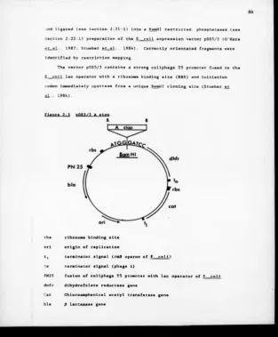
Construction of pDS5/3 A Stop 85
Transformation and Induction of Expressed
A Stop 87
Scaled-Up Expression of Truncated A Chain
and Sample Preparation 87
Purification of Truncated A Chain Using
Chromatofocuslng 88
Determination of Ricln A Chain-Specific
Activity In Chromatofocused Fractions 88
Use of Blue Sepharose Affinity
Section Title Page
Chapter 3 90
3:1:1 Purification of lectins from Ricinus communis
Seeds 90
3:2 Purification of Ricin Subunits 97
3:2:1 Chromatofocusing and SDS-PAGE Analysis 97
3:2:2 A Chain Purification : Removal of
Contaminating B Chain by Affinity
Chromatography 101
3:2:3 Cytotoxicity Assay : Analysis of Purified
Subunits 103
3:2:4 Cell-Free Ass a y : Analysis of Purified
Subunits 109
3:2:5 Assessment of Ribosome Modifying Activity
of Purified Ricin B Chain 111
3:2:6 Quantitation of Ricin A Chain Contamination
in Ricin B Chain Preparation 113
3:3 Discussion 115
Section £ U £
3:3:2 Purification of Ricin Subunits 117
3:3:2:1 Reduction of the Interchain Disulphide Bond 117
3:3:2:2 Separation of the Reduced Ricin Subunits 118
3:3:2:3:1 Analysis of Subunit Purity by SDS-PAGE 119
3:3:2:3:2 Analysis of Ricin A Chain in a Cytotoxicity
Assay 120
3:3:2:3:3 Analysis of Ri cin A Chain in a Cell-Free
Assay 122
3:3:2:3:4 Analysis of Ricin B Chain in a Cytotoxicity
Assay 122
3:3:2:3:5 Analysis of Ricin B Chain in a Cell-Free
Assay 123
3:3:2:3:6 Galactose-Binding Activity of Purified
Ricin B Chain 123
3:3:2:3:7 Activity of Reassociated Subunits 124
Chapter 4 126
4:1 Introduction of Thiol Linkers into
Gelonin Using Heterobifunctional Reagents 126
4:1:1 Derivatisation of Gelonin Using SPDP 126
4:1:2 Derivatisation of Gelonin Using 2 -IT 130
4:2 Determination of Ribosome Inactivating
Activity of Derivatised Gelonin 134
4:3 Reassociation Experiment : Comparison
of Ricin B Chain/A Chain and Ricin B
Chain/Derivatised Gelonin in a
Cytotoxicity Assay 136
4:4 SDS-PAGE Analysis of Derivatised Gelonin/
Ricin B Chain Conjugate 139
4:5 The Use of Chromatofocusing in the
Purification of Ricin B Chain-Gelonin
Conjugates 145
4:6 SDS-PAGE Analysis of Chromatofocused
Section Title Page
4:7 Section 4:8 4:8:1 4:8:2 4:8:3 4:8:4
Use of Blue Sepharose CL 6B Affinity
Chromatography to Purify Ricin B Chain-
Gelonln Conjugate
Analysis of the Biological Activity of
Partially Purified Conjugate Ricin-B
Chain-Gelonin
Cell-Free Assay : To Determine the Ribosome
Inactivating Activity of the Partially
Purified Conjugate
RNA Modification Assay : To Determine a
Ricin A Chain-Type Modification with a
Ricin B Chain-Gelonin Conjugate
Analysis of the Galactose Binding Activity
of Ricin B-Gelonln Conjugate
Cytotoxicity Assay : Analysis of the Activity
of Partially Purified Ricin B Chain-
Gelonin Conjugate Preparations
Section
5:1
5:2
5:3
5:4
5:5
5:6
5:7
Chapter 5 178
Introduction of a Premature Translation
Termination Codon Into Rlcln A Chain cDNA
b y Oligonucleotide Site-Directed
Mutagenesis 178
Colony Hybridization Using the Mutagenic
Oligonucleotide as a Probe 178
Analysis of Mutated DNA Sequence Encoding
for Rlcln A Chain 182
Analysis of pUC8RA Stop by Limited
Restriction Endonuclease Mapping 185
Analysis of pSP64ABam A Stop by
Limited Restriction Mapping 188
Analysis of pSP64ABam A Stop Transcripts
in a Uheatgerm Lysate System 191
Analysis of the Toxicity of Ricin A Chain
Transcripts in a Rabbit Reticulocyte
Lysate Translocation System 193
Analysis of pDS5/3 A Stop by Limited
Restriction Endonuclease Mapping
Title Page
Section Page
5:8:1 Western Blot Analysis of Transformed
E. coll Cells U pon Induction 197
5:9 Partial Purification of Ricin A Stop by
Chromatofocusing 200
5:10 Determination of Ricin A Chain-Specific
Activity in Chromatofocused Fractions 209
5:11 Evaluation of Affinity Chromatography
Using Blue Sepharose as a Method for the
Purification of Ricin A Stop 211
5:12 Discussion 215
Chapter 6 223
6:1 General Discussion 223
The potent cytotoxin ricin, obtained from the seeds of the castor
oil plant Ricinus c o mmunis, is composed of two polypeptide subunits
linked by a single disulphide bond. The binding of this molecule to the
surface of eukaryotic cells is mediated v i a the sugar-binding activity
of the B subunit. The exact nature of the ricin receptor(s) on the cell
surface is unclear, but is most probably some glycoprotein or glycolipld
containing exposed galactosyl residues. The ricin molecule becomes
internalized by the cell and, by an unclear mechanism, ricin A chain
escapes its endocytic vesicle entering the cell cytoplasm where it
enzymatically and irreversibly inhibits eukaryotic ribosomes, bringing
about the cessation of protein synthesis.
The toxicity of ricin A chain-containing immunotoxins can in many
model systems be enhanced by the subsequent addition of ricin B chain.
This apparent ricin B chain-mediated potentiation of cytotoxicity
suggests that this subunit has some role in the mechanlsm(s)
facilitating the entry o f ricin A chain into the cell cytosol.
The w ork presented in this thesis has attempted to examine the
potential of ricin B chain as a carrier of proteins into cells other
than ricin A chain. In this example ricin A chain has been replaced
with the type I ribosome inactivating protein, gelonln.
Further to these studies, preliminary work considering the possible
Importance of the hydrophobic C-terminus o f ricin A chain in the
ii
demonstrated that it is possible to delete at least 30 amino acid
residues from the C-terminus of the A chain and retain full ribosomal
inactivation activity as judged by in vitro analysis. This truncated
form of ricin A chain has been expressed in an E. coli expression system
and a soluble and active recombinant protein has been partially
purified. The implications of this work and possible future analysis of
Ill
Lift at
lifciai
Table T i tle Pace
1:1 Lethal Doses of Ricin 2
4:1 Analysis of Galactose Binding Activity of
Ricin B Chain-Gelonin Conjugate 162
5:1 Limited Restriction Endonuclease Mapping
o f pUC8RA Stop 187
5:2 Limited Restriction Endonuclease Mapping of
pSP64ABam A "Stop" 190
5:3 Limited Restriction Endonuclease Mapping of
Ltst pf Figur**
Figure Title Page
2:1 BamHI Fragment Encoding Rlcln A C h ain from
pRICA 68
2:2 Cloning of Bglll/EcoRI A Stop Fragment from
M13mpl9 R A Stop Into pUC8RA 80
2:3 Cloning of ££mHI Fragment from pUC8 RA Stop
Into pSP64ABam HI 81
2:4 pSP64ABam A Stop 83
2:5 pDS5/3 A Stop 86
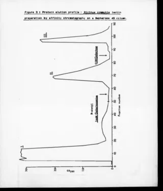
3:1 Protein Elution Profile : Ricinus communis
Lectin Preparation by Affinity Chromatography
Using Sepharose 4B 91
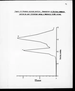
3:2 Protein Elution Profile : Separation of
Rlclnus communis Lectins by Gel Filtration
Using a Sephacryl S-200 Column 93
3:3 SDS-PAGE Analysis o f Fractions Taken from
Sephacryl S-200 G e l Filtration 95
3:4 Cytotoxicity Assay : Inhibition of Protein
Synthesis by P u r i f i e d Ricin Concentrations 96
3:5 Protein Elution Pro f i l e : Purification o f Ricin
Subunits by Chromatofocusing 98
3:6 SDS-PAGE Analysis o f Chromatofocused Ricin
B Chain 100
3:7 SDS - PAGE Analysis o f Endo H Treated Ric i n and
Ricin Subunits 102
3:8 Cytotoxicity Assay : Inhibition of Protein
Synthesis by R i c i n A Chain at Different
Stages of Purification 105
3:9 Cytotoxicity Assay : Inhibition of Protein
Synthesis by Pu r i f i e d Ricin B Chain 106
3:10 Cytotoxicity Assay : Inhibition of Protein
Synthesis by Reassociated Ricin Subunits 107
Vi
3:11 SDS-PAGE Analysis of Ricin, Purified Rlcln
Subunits and Reassociated Ricin Subunits
Under Denaturing, Non-Reducing Conditions 108
3:12 Cell-Free Assay : Inhibition of Protein
Synthesis by Purified R i c i n Subunits 110
3:13 RNA Modification Assay : Analysis of Ricin B
Chain Dilutions for R i c i n A Chain Specific
Activity 112
3:14 RNA Modification Assay : Determination of the
Lowest A-Chain Concentration to Produce
Modification of 28S R N A 114
4:1 Protein Elution Profile : Separation of
Derivatlsed Gelonln f r o m Unreacted 2-
iminothiolane 131
4:2 Cell-Free Assay : Inhibition of Protein
Synthesis by Gelonln, Gelonln SPDP and
Gelonin 2 -IT 135
4:3 Cytotoxicity Assay : Inhibition of Protein
Synthesis by the Type I RIP Gelonln 137
vii
4:4 Cytotoxicity Assay : Inhibition of Protein
Synthesis In Vero Cells b y Dilutions of
Gelonin 2 -IT Mixed with P u rified Ricin B
Chain (1 x 10*» M) 138
4:5 SDS-PAGE Analysis of Rlcin B Chain-Gelonin
Reaction Mixture 141
4:6 U e s t e m Blot Analysis of the Electroeluted
Ricln B Chain-Gelonin Conjugate 142
4:7 Protein Elution Profile : Partial Purification
of Ricln B Chain-Gelonin Conjugate by
Chromatofocusing 147
4:8 SDS-PAGE Analysis of Chromatofocused Ricln
B Chain-Gelonin Conjugate Material 150
4:9 Protein Elution Profile : Partial Purification
of Ricln B Chain-Gelonin Conjugate by Affinity
Chromatography Using Blue Sepharose CL 6B 152
4:10 SDS-PAGE Analysis of Protein Fractions from
Blue Sepharose CL 6B Af f i n i t y Chromatography
Step 154
vili
4:11 Cell-Free Assay : Inhibition of Pro t e i n
Synthesis by Samples from Different Stages of
Purification of the Ricin B Chaln-Gelonin
Conjugate 157
4:12 RNA Modification Assay : Analysis o f Purified
Ricin B Chain-Gelonin Conjugate Fractions
for RNA Modification Activity 158
4:13 Cytotoxicity Assay : Inhibition b y Partially
Purfied Ricin B Chain-Gelonin Conjugate 164
5:1 Hydrophoblcity Plot of the C-Terminus of Ricin
A Chain 179
5:2 Colony Hybridization Screen of Mutagenised
Clones Probed with the Radiolabelled
Mutagenic Oligonucleotide 181
5:3 DNA Sequence Analysis of Mutagenised Ricin A
Chain Clones on Standard Buffer Gradient Gels 183
Figure Title Page
5:4 Analysis of Mutated Ricin A Chain D N A sequence
ix
Figure H t l » Page
5:5 Restriction Endonuclease Mapping of pUC8 RA
"Stop" 186
5:6 Restriction Endonuclease Mapping of p S P64Bam
A "Stop" 189
5:7 Analysis of Ricin A Chain Transcripts in a
Wheatgerm in vitro Translation System 192
5:8 Analysis of Ricin A Chain Transcripts in a
Rabbit Reticulocyte in vitro Translation
System 194
5:9 Restriction Endonuclease Mapping of pDS5/3
A "Stop" 196
5:10 Expression of Ricin A "Stop* in E. coli :
Analysis of Whole Cell Products by
Western Blotting 198
5:11 Protein Elution Profile : Partial Purification
of Recombinant Truncated Ricin A Chain by
Page
5:12 Purification of Recombinant A "Stop" I :
Western Blot Analysis of Chromatofocused
Fractions 204
5:13 Purification of Recombinant A "Stop" II :
Western Blot Analysis of Chromatofocused
Samples 206
5:14 RNA Modification Assay : Analysis of Partially
Purified Recombinant Ricin A "Stop" 210
5:15 Protein Elution Profile : Partial Purification
Truncated Ricin A Chain by Affinity
Chromatography 212
5:16 RNA Modification Assay : Evaluation of Blue
Sepharose Affinity Chromatography in the
Purification of Recombinant Truncated
Figure Title
xi
Acknowledgements
I would like to thank Lynne Roberts, my supervisor for her help and
advice throughout this project. In particular I am indebted to Lynne
for her prompt and constructive criticism of this thesis. Thanks also
to Andy Vright, my Industrial Supervisor, for his help and encouragement
during my three years.
I am also grateful for the assistance offered to me from members of
the ricin group, past and present. In no particular order I would like
to thank Drs. Bernle Prior for her help and advice particularly at the
beginning of the project; Mary O'Hare for help with chromatofocusing and
recombinant A chain expression; Robert Spooner for teaching me the
necessary molecular techniques, in particular DNA sequencing;
Peter Richardson for help with the ricin B chain binding assays,
Angelica Gebhardt for good cell lines and listening to my ideas;
Jane Gould for advice on RNA assays and photography and Khalid Hussain
for more molecular techniques. Special thanks also to
Dr. Martin Hartley for teaching me the ribosome modification assay he
developed, to Professor Mike Lord for the many useful conversations and
to Dr. Liz Jones for reticulocyte lysates.
I thank my fellow postgraduate students, again both past and
present, who have been such good company over the past three years. In
particular, Mike May for his assistance with in vitro lysate systems a n d
xii
I especially wish to acknowledge the drug targeting group of the
Institute of Cancer Research for their help with protein derivatisation
and their gifts of gelonin. I am grateful to A lan Cumber,
Geoff Parnell, Tony Forrester and Eddie Wawrzynezak from this group.
O n the thesis production side I would like to thank
Professor Mike Lord for use of his computer and Mrs. Carol Howes for her
tremendous efforts in preparing and typing this thesis.
For my Mum and Dad, an opportunity to finally thank them for their
support throughout my education and providing me with opportunities
which, without their efforts would not have b een possible. Special
thanks to the Wilkinsons (Karen's family) for their help and hospitality
over the last three years. Also thanks to Dave Black and
Steve Carpenter for being good training partners.
Finally a very special thank you to Karen whose support has been
continuous and invaluable throughout.
I acknowledge financial support of the SERC in the form of a CASE
Studentship and the provision of help and materials from ICI
xlli
Declaration
All the results presented In this thesis were obtained by the
author and have not been used In any previous application for a degree.
All sources of Information have been specifically acknowledged by means
o f reference.
Afafeiâzlaslgni
ADP adenosine diphosphate
APS ammonium persulphate
Bis N'N'-methylene-bisacrylamide
BSA bovine serum albumin
CIP calf intestinal alkaline phosphatase
CsCl caesium chloride
dH20 distilled water
DNA deoxyribonucleic acid
dNTPs deoxyribonucleoside triphosphates
DTT dithiothreitol
E. coli Escherichia coll
EDTA diamlnoethanetetra-acetic acid disodlum salt
Endo H endoglucosaminidase H
FCA Freunds complete adjuvant
FI CA Freunds incomplete adjuvant
Fuc fucose
GlcNac N-acetylgalactosamine
HAc acetic acid
HCI hydrochloric acid
IPTG isopropyl-0-D-thio galactopyranoside
Kd Kilodalton
M molar
Man mannose
NAD nicotinamide adenine dinucleotide
X V
PBS phosphate buffered saline
PMSF phenylmethylsulphonylfluoride
OD optical density
PVP polyvinylpyrollidone
RCAj Rtcinvg çpnawnte agglutinin
rATP riho adenosine triphosphate
R F replicative fora
RNA ribonucleic acid
RNAase ribonucléase
SDS sodium dodecyl sulphate
S P6 Sglrognella typMffvrivn phage
SPDP N - s uceinimidy1-3-(2-pyridyIdithio) proprionate
TCA trichloroacetic acid
TEMED NNN'N'-tetramethylethylenediamine
Tris HCl tris (hydroxymethyl) aminomethane hydrochloride
v / v volume/volume
w / v weight/volume
X-gal 5-bromo-4-chloro-3-indolyl 0 galactose
Li. Intrgflvctlgn
1:1:1 Overview.
The protein rlcln Is an extremely potent cytotoxin occurring with a
number of other seed storage proteins within protein b ody organelles found
In the seeds of the castor oil plant Rlclnus communis (Tulley and Beevers,
1976; Youle and Huang, 1976). The toxic and medical properties associated
w ith the seeds of this plant have b een realised since ancient times; their
use b e ing recorded In classical Greek and Sanskrit medicine. More
recently, in the nineteenth century, Paul Ehrlich established some of the
basic principles o f immunology using crude rlcin preparations obtained from
castor beans (Ehrlich, 1891). Ehrlich Is of course the originator of the
term "magic bu l l e t " , a term used to describe reagents designed for the
targeted destruction of diseased cells. Nowadays this term frequently
describes the construction of rlcin-antlbody conjugates, commonly referred
to as immunotoxins.
A n historical perspective of the scientific work relating to ricln has
b een made by Balint (1974) in his extensive review. In summary, Stlllmark
In 1887 was the first person to name the toxic component from the castor
oil seed, rlcin. However, its activity had been noted as early as 1878 by
Rltthausen and later confirmed by Dixon In 1887. The first detailed
description of r l cin poisoning In man was published in 1899 by Muller.
Early purification procedures were improved by Osborne (1905) and later by
Robert (1913) who introduced an ammonium sulphate precipitation stage. The
major breakthrough however did not occur until 1965 w hen affinity
chromatography u s ing sepharose was first achieved by Dlrheimer. This
technique now forms the basis for most present day methods of ricin
Ricin is a potent toxin towards eukaryotic cells. It has been
estimated that 1 K g of the purified toxin w o uld be lethal to 3.6 million
people, which equates to a lethal dose of 0.27 mg per person, (Balint,
1974). Sensitivity to the toxin varies from species to species; the horse
and the guinea p i g being the most sensitive. Indeed Ehrlich has calculated
that a single g ram of ricin w o uld be enough to kill l.S million guinea
pigs. At the other extreme, the domestic hen is the most resistant of the
higher animals tested, and the frog apparently has an outstanding
resistance to the toxin (Balint, 1974). Table 1:1 describes the
sensitivity of different species to ricin intoxication (after Olsnes and
P i h l , 1982).
Table 1:1
Animal Lethal dose 50%
Mice 2.7 ¿tg/Kg body weight
Rabbit *50 ng/Kg
Dog *1.25 pg/Kg
Human *0.15 /ig/Kg
*intravenous administration o f abrin, a toxin closely related to ricin.
Inevitably throughout the ages ricin has b e e n exploited for criminal
purposes, with possibly the m ost famous case b e ing the assassination of the
Bulgarian expatriate Georgy Mar k o v on Waterloo Bridge in London in 1978.
Markov was shot in the 1 « 8 w i t h what is believed to be a ricin impregnated
pellet, fired from a modified umbrella.
The use of ricin for medicinal purposes is, thankfully, far more
[image:38.356.14.347.10.396.2]their potential as specific anti-tumour agents whereby the toxins are
targeted to cancerous cells b y being coupled to a suitable cell binding
monoclonal antibody. These so-called lmmunotoxlns have achieved reasonable
efficacy in whole cell and animal model systems, but as yet only a limited
response in clinical tri a l s .
1 ; 1 ; 2 S t r w c of
Risln-Ricln is a glycosylated, heterodimerlc protein w ith a n estimated
molecular weight of 62,057 daltons (Olsnes and Fihl, 1982). It is composed
of two subunits termed A chain and B chain. Both are N-glycosylated and
have molecular weights of 30,625 daltons and 31,432 daltons respectively.
The complete amino acid sequence of ricin A chain (Funatsu et a l ., 1978)
and ricin B chain (Funatsu et a l ., 1979) has been established by peptide
analysis and also deduced from the nucleotide sequence o f cDNA clones
coding for preproricin (Lamb et a l ., 1985). These latter studies showed
preproricln to be composed of a 35 amino acid N-terminal sequence preceding
the A chain (267 amino acids), which is joined to the B chain (262 amino
acids) by a 12 amino acid linker region. In the mature protein the signal
sequence and linker are removed by specific processing enzymes. The two
subunits are covalently linked by a single disulphide b o n d between cysteine
residues at position 259 in the A chain and position 4 in the B chain.
However, it is apparent from X-ray crystallographic analysis of ricin that
a more complex Interaction occurs between the two subunits (Montfort £_£
a l .. 1987). In addition to the interchain disulphide bond, ricin B chain
contains 4 intrachain disulphide bonds. In contrast ricin A chain has no
intrachain disulphide linkages. Both subunits possess two N-glycosylation
sites, both of which are occupied in the B chain by high mannose type
oligosaccharides (Foxwell et a l ., 1985). Ricin A chain may exist in two
4
occupied with a complex oligosaccharide containing xylose and fucose, or as
the A, species in which the second N-glycosylation site is also occupied.
A more detailed account of the oligosaccharide side chains of the ricin
subunits is considered in the discussion section of Chapter 3.
1 ;1 ;3 p io io g ic a i A c tiv itie s ,
vt
R ic in subunjt$.
The structure and function of the respective subunits will be
considered in more detail elsewhere. However, each subunit may be
characterised through distinct biological activities. Ricin A chain, the
effectomer, is k no w n to enzymatically inactivate eukaryotic ribosomes
resulting in a cessation of protein synthesis in the Intoxicated cell.
However, to inactivate the ribosomes ricin A chain must first become
internalized into the cell, and then translocated into the cell cytoplasm.
The biological activities of ricin B chain are believed to facilitate these
processes. Ricin B chain, the haptomer, has a well-defined sugar-binding
o r lectin activity, exhibiting a specificity for galactosyl residues. This
activity of the B chain mediates the binding of the w h ole toxin to the
surfaces of cells, probably to galactosyl residues w h ich occur o n exposed
glycoproteins and glycolipids on the cell surface. No specific receptors
h ave been identified for ricin, rather its binding appears to be
opportunistic towards a range of cell-surface molecules. The number of
bin d i n g sites is therefore enormous. He La cells for example, possess 3 x
1 0 T potential binding sites for ricin (Olsnes and Pihl, 1982). Together
therefore, the two subunits constitute an extremely potent toxin which can
bind, through the B chain, to any one of millions of sugar residues on a
w i d e range of different cell types and become internalised resulting in the
inactivation of susceptible ribosomes.
In addition to its sugar-binding role there exists indirect evidence
5
cytosol. This effect has b een observed in studies in w h ich the cytotoxic
activities of ricin A chain-containing immunotoxins has b een potentiated in
the presence o f ricin B chain (Youle and Neville, 1982; McIntosh et a l ..
1983). This effect is apparently not a result o f the binding of ricin B
chain to galactosyl residues on the cell surface because it has also been
demonstrated in the presence of high lactose concentrations which would
ensure blockage of the binding sites.
l i l i A Th? C v to tp g U <?f Rl<?ln.
The cytotoxic activity of ricin on eukaryotic cells may be considered
in three stages. Stage I involves binding of the whole toxin to the cell
surface. This is mediated through the sugar-binding sites of the B
subunit. That ricin cytotoxicity can be abolished by the presence of free
lactose (100 m M ) , suggests that (a) binding to the cell surface is a
prerequisite for intoxication and (b) binding mediated through the lectin
activity of ricin B chain represents the predominant means by which ricin
interacts with (and ultimately kills) the cells. It remains unclear
however, as to whether particular cell surface sugar residues on particular
glycoproteins or glycolipids are more important than others with respect to
the next stage, of toxin internalisation and ultimate delivery of ricin A
chain into the cytosol. Studies by Simmons et a l . (1986) have suggested
that the mannose-rich oligosaccharide side chains of ricin may have some,
possibly minor, role in the internalisation of ricin.
Stage II is concerned with the internalisation of ricin and
translocation of ricin A chain into the cytosol. As it is of particular
interest to the subject of this thesis, it has been considered in a
separate section later in the Introduction.
Stage III involves the inactivation of eukaryotic ribosomes, leading
synthesis is brought about by the enzymatic inactivation o f the large (60S)
ribosomal subunit b y ricin A chain (Olsnes and Pihl. 1976; Sperti et a l . .
1973). Cawley et a l . (1978) have shown this inactivation to be catalytic
in nature, requiring no cofactors. Kinetic experiments h ave indicated that
a single ricin A chain molecule can inactivate salt-washed ribosomes at a
rate of 1500 per minute (Km - 1-2 x 10*T M ) , (Olsnes Et ftl- , 1975). A
more detailed account of ribosome inactivation has been given in the later
section, Ricin A chain : Structure, Function and Evolution.
1 = 1:5 aiflimthaaia <?f
Ricin and the related castor b ean lectin, RCA^, are s ynthesised
simultaneously in the endosperm cells of maturing castor b e a n seeds
(Gifford et a l .. 1982). This synthesis occurs during and after testa
formation when the organelles are being rapidly formed (Roberts a n d Lord,
198 1 a ) . The structural and functional homology between the two lectin
species would suggest a similar biosynthetic pathway, indeed it seems
likely that duplication of an ancestral gene may have evolved into the
genes encoding for these two lectins. Work b y Butterworth and L o r d (1983)
has shown that both the A and B subunits of ricin and the A' and B'
subunits of RCAj are derived from large precursors. The ricin precursor,
preproricln, is composed of a 35 amino acid N-terminal signal peptide
preceding the A chain (267 amino a c i d s ) , which in turn is linked to the B
chain (262 amino acids) by a 12 amino acid linker region (Lamb et al ■ ,
1985). The N-terminal signal peptide is cleaved during cotranslational
translocation of the nascent precursor across the endoplasmic reticulum
membranes at which stage core glycosylation occurs and the disulphide bonds
are formed (Roberts and Lord, 1981b). The glycosylated p r e cursor is
then apparently directed via the Golgi to vesicles which fuse w i t h the
sequence. Within the protein bodies the precursor is processed to the
mature form of ricin. Details surrounding these final processing events
are presently being investigated.
The synthesis of ricin as a precursor molecule, its concomitant
segregation within the endomembrane system and its transport to protein
bodies prior to processing prevents enzymatic inactivation of castor bean
endosperm ribosomes by ricin A chain. Thus if ever exposed to the
ribosomes as a result of inefficient translocation, ricin A chain occurs as
a precursor which is known to be biologically inactive (Lynne Roberts,
Personal Communication).
1:2 = 1 The s tr v c w r * . EvglvUen tn4 Fwnctipp 9 f E lg in g chain.
Ricin B chain is a lectin w ith an affinity for galactosyl residues.
Earlier work by Zentz et a l . (1978) and Houston and Dooley, (1982)
established that ricin B chain binds two sugar molecules in a non-
cooperative manner. These two authors disagree however as to the binding
affinities of each of these sites; Zentz et a l . . suggesting that the s u gar
binding domains can be differentiated as a low and a high affinity binding
domain, whilst Houston and Dooley maintain that the binding affinities
of each site are more or less e q u a l .
Villafranca and Robertus (1981), have shown that ricin B chain is a
gene duplication product, showing approximately 32% homology at the amino
acid level between the two halves of the molecule. Each half of this
molecule contains a sugar-binding domain. Work by Mise et a l . (1986) , has
implicated the amino acid residue tyrosine 248 in the "strong” galactose
binding site, and an undetermined tryptophan residue in the weak site
(Hatakeyama et a l .. 1986). Each sugar-binding domain is proposed to
contain two disulphide loops which show significant homology with residues
8
Dlctvostellum dlscoideum (Robertus and Ready, 1984). These same authors
have suggested that this peptide was an ancient galactose b inding unit from
w h ich rlcln B chain has evolved. More recently X-ray crystallographic
analysis, revealing the three-dimensional structure of ricln at a
resolution of 2.8 A, has confirmed that rlcln B chlan Is composed of two
globular regions, which although separate, share Identical folding
topologies. Each will bind a lactose molecule (Montfort et a l . . 1987). It
Is Interesting to note however, that each sugar molecule binds to different
sites within each domain.
A more detailed analysis of each domain, described b y Rutenber et a l .
(1987), shows that each homologous domain can be divided into four peptide
sub-domains. These authors have named these peptides A- , a-, fi- and y-sub-
domalns. The A-peptide of domain 1 (peptide 1A, residues 1-16) is
homologous to the A peptide of domains 2 (peptide 2A, residues 136-150).
These two peptides are however not related to the other sub-domains within
the molecule. At this point It Is interesting to note that the 1A sub-
doma i n forms the N-terminal extension of ricln B chain w h ich interacts with
the carboxyl-terminal domain of ricin A chain (Montfort et a l . ■ 1987). The
remaining sub-domains, the a, fi and y peptides are homologous with each
other. These sub-domains, which comprise the main body o f each domain have
undergone considerable divergence but, despite this they still demonstrate
a statistically significant homology.
In the model presented by Rutenber et a l . (1987) the two sugar-binding
domains, which are linked by the 2A peptide, each bind a lactose molecule
in a non-homologous fashion at the domain level. However, at the sub-
dom a i n level, the binding of lactose in each respective domain, to peptides
la and 2 y Is homologous (Rutenber et a l .. 1987). At both binding sites (in
sub-domains la and 2y) the lactose molecule is believed to lie In a pocket
formed on one side by a kink In the peptide chain and o n the other side an
9
conserved in b o t h sub-domains, and che aromatic side chains belong to
tryptophan residue 37 in la and tyrosine residue 248 in 27. These side
chains however, serve only as a flat binding surface and apparently do not
specifically interact with the sugar hydroxyl groups (Montfort et al ■ ,
1987). S pecific non-covalent interactions in the fora of hydrogen bonds do
occur b e t w e e n the sugar and amino acid residues Asn 46 and Asn 2SS. These
binding residues are only conserved in the sub-domains which still b ind the
sugar and h ave therefore been considered as prime targets for modification
in attempts to abolish the sugar-binding activity of ricin B chain. Other
highly co n s e r v e d residues are believed to fulfil structural roles.
Rut e n b e r et a l . (1987), speculate that ricin B chain has evolved from
an ancient galactose-binding peptide of approximately 40 residues which
resembles the la domain found presently in the molecule. They further
speculate that the ancient molecule existed as a trimer and that f rom gene
duplication a n d fusion a afii molecule resembling a modern day B chain
domain evolved. Such a fusion is thought to have abolished binding in the
P subunit, leaving only two binding sites in the a and 7 sub-domains. The
addition o f the A peptide is expected to have stabilized the structure to
form a XafJ-y u n i t which by a further duplication event would form the
structure (,XaP~i)3 , resembling modern ricin B chain. This final duplication
event is thought to have blocked access of galactose to the 1A unit. Sub
domain 2a appears to have also lost its binding residues at some point.
This leaves two sugar-binding domains, la and 2 7 whi c h are 75 A apart.
Rutenber et a l . (1987) speculate that the selection of two sugar-binding
sites this distance apart reflects a requirement for cell surface binding
and the triggering of endocytosis. The primitive "galactose-fold"
speculated b y these workers is consistent with structural analysis o f the
lectin di s c o i d l n I, extracted from the slime mould Dlstvostellum
discoldeura. fueling the speculation that ricin B chain has evolved from a
10
Throughout this analysis of the structure and function of ricin B
chain it is interesting to note that no obvious candidate for a membrane
penetrating domain, s uch as those observed in diphtheria toxin (Greenfield
et a l . , 1987) and Pseudomonas aeruginosa exotoxin A (Allured et a l . , 1986)
has b e e n identified. Despite this, ricin B chain apparently possesses some
ability to potentiate the cytotoxic activity of certain ricin A chain-
containing immunotoxins (Youle and Neville, 1982; McIntosh et a l .. 1983).
This activity, as m e n tioned earlier, is apparently independent of the
sugar-binding activity of ricin B chain.
The possibility that ricin B chain interacts directly with lipid
membranes has been examined by a number of workers (Ishida et a l . . 1983;
Utsumi et a l .. 1984). In their experiments Ishida et a l . . 1983 used the
Newcastle Disease Vir u s (NDV) as the target membrane for ricin and ricin
subunits. The membrane of NDV is rich in the ganglioside GMj which
provides an avid b i n d i n g site for ricin and ricin B chain. In the case of
both whole ricin and r i c i n B chain, the presence of galactose significantly
reduced membrane interaction. In contrast the binding of ricin A chain to
the virus membrane envelope was unaffected by the presence of galactose.
These workers conclude that both subunits of ricin have the inherent
ability to penetrate the lipid bllayer.
In similar experiments Utsumi et a l . (1984) considered the interaction
of ricin and ricin subunits with dipalmitoylphosphatidylcholine (DPPC)
vesicles. They o b served no association between these vesicles and the
unreduced toxin, but considerable interaction when the purified ricin
subunits were presented to the vesicles separately. In fact, their
experiments indicated that ricin B chain demonstrated the greater lipid-
protein association o f the 2 subunits. However, these same authors
observed a significantly greater perturbation of the DPPC bilayer after
incubations with ric i n A chain as compared with either whole ricin or ricin
strong associations with lipid membranes, only ricin A chain was able to
achieve full penetration. T his finding is in agreement with the
speculation that the hydrophobic C-terminal region of the A chain is
important in membrane penetration and entry of ricin A chain into the cell,
OJchlda et a l .. 1980).
If, as has been suggested by these results, ricin B chain does not
actively participate in membrane penetration, then the observed
potentiation of A chain immunotoxins cytotoxic activity may be the result
of some indirect or cooperative activity. Possibly ricin B chain acts to
protect the A chain from proteolytic degradation, or perhaps being
associated with ricin B chain acts to direct ricin A chain to an
appropriate intracellular compartment for translocation into the cytosol.
Observations b y Montfort et a l . (1987) and Lewis and Y o u l e , (1986) have
suggested that the ricin subunits form an intimate association. In
particular Montfort et a l . (1987) have speculated from their analysis of
the three-dimensional structure of ricin, that the carboxyl-terminus of
ricin A chain inserts between the two sugar-binding domains of the B chain.
Possibly this association acts to maintain the hydrophobic C-terminus of
the A chain in a protected conformation until intracellular conditions are
optimum in facilitating subunit dissociation and the subsequent exposure of
this protected region. For the mean time these suggestions remain untested
speculation.
1 ;2
¡2
s*r*»?tvnrt.
Function
Evglutlw
9 f R icin A
Chain-The X-ray crystallographic analysis of ricin carried out by Montfort
et a l . (1987) has divided ricin A chain into three arbitary domains. The
first, comprising 117 amino-terminal residues, forms a flat domain at the
base of the A chain, dominated by a five-stranded /3-sheet structure. The
This dom a i n is located above, and slightly to the left of the first domain.
The t h i r d domain, comprising residues 211-267 forms a compact disc-like
domain which, as well as interacting w i t h the first two domains of the A
chain, interacts with the ricin B chain. As mentioned in the previous
section, this interaction results in the insertion of this C-terminal
domain between the two sugar-binding domains of ricin B chain. The A chain
is a m o r e globular protein than the B chain. It is approximately 55 A
long, 4 5 A wide and 35 A thick. Of this, the C-terminal disc-like domain
comprises of a region 25-30 A across a n d 15-20 A thick. In addition to its
strong interaction w i t h ricin B chain, this C-terminal domain represents a
significantly hydrophobic region of the molecule which has been implicated
in membrane associations (Uchida et a l . . 1980).
R i c i n A chain is an enzyme whose substrate is 28S rRNA. Recently,
w ork b y Endo et a l . (1987) has demonstrated a specific N-glycosidase
a ctivity associated with the A chain. The location of the active site
however, remains unclear. Montfort et a l . (1987) have identified a
p u tative active site within the A c h a i n structure, but readily acknowledge
the n e e d to carry out further studies. Interestingly this cleft, formed at
the interface between the three d o m a i n s , would be exposed to the aqueous
m e dia in the unreduced holotoxin, suggesting that u pon reduction and
dissociation from the B chain, ricin A chain undergoes some degree of
conformational change to form the active molecule.
R i cin A chain inhibits protein synthesis by inactivating the function
of the 60S subunit o f eukaryotic ribosomes, (Sperti et a l . . 1973). This
activity appears to be confined to eukaryotic ribosomes only. Ribosomes
extracted from prokaryotic sources are totally insensitive when incubated
w ith concentrations of ricin that result in complete inhibition in
eukaryotic systems (Greco et a l .. 1974). Ribosomes extracted from higher
plants have also b e e n shown to be susceptible to ricin inactivation,
13
translation system was some 23,000 times higher than the concentration
giving the same effect on mammalian ribosomes, (Harley and Beevers, 1982).
These findings are in agreement with the result described in Figure 5:7 in
which ricin A chain raRNA translated in a wheatgerm in vitro system does not
apparently inactivate the ribosomes in that system. In contrast, when the
same RNA is translated in a rabbit reticulocyte lysate system complete
ribosome inactivation occurs within the duration of the experiment (see
Figure 5:8). In addition the ribosomes from the protozoan, Tetrahvmena
pvrlformis have also been reported to be resistant to ricin (Wilde et a l . ,
1979).
As mentioned earlier in this chapter, a single molecule of ricin A
chain can inactivate a large number of ribosomes. Olsnes et a l . (1975),
have estimated that a single A chain molecule can inactivate 1500 salt-
washed ribosomes per minute, which translates to a Km value between 1-2 x
10*T M. The use of salt-washed ribosomes in these kinetic studies is
important, since pre-bound elongation-factor 2 (EF-2) which is removed in
the salt wash, has been shown to protect ribosomes against ricin
inactivation. This suggests that ricin A chain may bind at or very close
to the normal EF-2 binding site. Protection o f ribosomes is also seen with
amino acyl-tRNA (Fernandez-Puentes et a l .. 1976). Furthermore ricin A
chain treated ribosomes are apparently unable to bind EF-1, thereby
blocking the binding of amino acyl-tRNA. All this data suggests that the
action of ricin A chain is to somehow modify the binding sites for these
accessory factors on the ribosomes. It is possible that EF-2 may bind to
the same site or a site close to the EF-1 binding site on the ribosome.
Interestingly ricin A chain has been shown to demonstrate some amino acid
homology with hamster EF-2. The deletion of a contiguous stretch of six
homologous residues from the ricin A chain sequence results in a molecule
which displays no activity when tested in a ricin A chain sensitive in
[image:49.356.14.349.6.389.2]14
whether this loss o f activity has resulted from the inability of an
otherwise active r i cin A chain molecule to bind to the ribosomes, or the
consequence of malfolding.
Evidence that ricin A chain might bring about ribosomal inactivation
by inducing some subtle conformational change in the ribosome comes from
the observations of Cawley et a l . (1979) who were able to protect and even
rescue ribosomes from the effects of ricin A chain. In their experiments,
they found that hi g h concentrations of M g 2+ ions reduced A chain
inactivation and have speculated that this may be the result of an induced
conformational change. More recently, work by Teroa et a l . (1988) has
demonstrated that the labelling of the ribosomal protein L-14 with either
*H or 1* C labelled N-ethylmaleimide was reduced after treatment of the
ribosome with ricin. This result suggests that ricin may alter the
conformation of the ribosome in the vicinity of that protein. The same
authors have speculated that these conformational changes are related to
the specific N-glycosidase activity which is characteristic of ricin A
chain action o n eukaryotic ribosomes.
This N-glycosidase activity, discovered by Endo et a l . (1987) produces
an apparently minor, but highly specific, modification of the 28S rRNA.
More precisely it catalyses the removal of an adenine residue from position
4324 in a highly conserved region of rat liver 28S rRNA. The ribosomal RNA
backbone is not cleaved by the action of the A chain, but the
phosphodlester linkages on either side of this residue become
hypersensitive to cleavage using reagents such as aniline at low pH. This
ability to cleave ricin A chain modified rRNA using simple chemicals is
exploited in the RNA modification assay described in Section 2:11. Endo
has also demonstrated that ricin A chain can catalytically modify naked
rRNA extracted from both eukaryotic and prokaryotic ribosomes. This
suggests that susceptible prokaryotic rRNA is somehow protected by the
15
able to deduce che minimum requiremenc for che subscrace of ricin A chain
Co be rRNA in a seem loop sCrucCure, having Che sequence GA GA within Che
loop (Endo, Y . , abstract from the International Symposium on immunotoxins,
June 9-11, 1988, Sheraton University Centre, Durham, North Carolina,
U.S.A.).
Considered together, this data would support a model in which ricin A
chain binds at or near to the binding site for EF-2 on the ribosome. Ricin
A chain then catalyses the removal of the adenine residue at position 4324
in the 28S rRNA. This adenine residue is located within a ribonucleotide
sequence GA GA, situated in a stem/loop structure. This modification of
28S rRNA has b een associated with conformational changes in the 60S
ribosomal subunit (Terao et a l . , 1988) which are thought to result in
inactivation of the whole ribosome. Interestingly this region of 28SrRNA,
modified by the action of r i cin A chain, is h i g h l y conserved in the large
rRNA of both prokaryotic and eukaryotic ribosomes. The observation by Endo
(Symposium abstract) that prokaryotic rRNA, w h e n purified from associated
ribosomal proteins, is also modified by ricin A chain suggests that
prokaryotic ribosomes form a conformation which somehow protects this
susceptible region.
Although these recent developments have contributed considerably to
our understanding of ribosomal Inactivation by ricin A chain, much work
remains before the precise mechanism can be defined. Perhaps the greatest
benefit to be gained from the efforts of Endo a n d co-workers is that it has
focused the research on ricin A chain activity, which prior to this h a d
consisted of an examination o f a number of putative A chain associated
phenomena. In order to avoid unnecessary confusion, these earlier
observations which led to speculation regarding the mode of action o f ricin
A chain, such as dephosphorylation (Houston, 1978) or inhibition of
Recently a number of authors have identified ribosomal RNA
modification activity, identical to that found with ricin A chain, in a
variety of other ribosome inactivating proteins, for example barley toxin,
(Endo et a l .. 1988(c)), viscumin, a type II RIP from mistletoe (Endo at
a l ., 1988(a)) gelonin, saporin, momordin, pokeweed antiviral proteins (PAP,
PAP-II and PAP-S) (Endo et a l .. 1988(b)), recombinant ricin A chain,
tritln, trichosanthin and dianthins (Stirpe et a l ., 1988) . All these RIPs
come from plant sources although from species that are taxonomically
unrelated. The source and data describing the toxicity of these and many
other RIPs have been extensively reviewed by Stirpe and Barbieri (1986).
This diversity of RIP sources suggests that these proteins, and indeed
ricin A chain, may have evolved from some primitive common ancestor.
Furthermore, the observation that the cytotoxln from the bacterium Shigella
dvsenteriae (Shiga toxin) also catalytically inactivates ribosomes in a
ricin A chain-like fashion supports this possibility (Endo and Tsurugi,
1987). The biology and biochemistry of Shiga and Shiga-like toxins have
been reviewed by O'Brien and Holmes (1987), and their nucleotide sequence
determined and compared with ricin (Calderwood et a l . , 1987). These
workers conclude that the A subunit of Shiga-like toxin is homologous with
the A chain of ricin. Analysis of the amino-terminal sequences of RIPs
extracted from Phytolacca americana (PAP, PAPII, PAP-S), Phytolacca
dodecandra (dodecandrin) and the A chains of ricin and modeccln, has
indicated that all these proteins appear to be related (Ready et a l .,
1984). These same authors suggest that these proteins have diverged from a
common ancestor, in some cases fusing with the genes for sugar-binding
proteins, the predecessor to the modern B chains (type II RIPs). In other
cases they remain as single chain molecules (type I RIPs). This type of
analysis, together with the discovery of a common rRNA modification
activity and the widespread, divergent occurrence of these proteins goes a
The occurrence of hydrophobic regions w i t h i n the ricin A chain
sequence, in particular the significance of the hydrophobic C-terminal
portion of this molecule have been considered briefly in the previous
section. The possibility that this region may be involved in membrane
interactions and its association with ricin B chain are considered more
fully in the discussion section of Chapters 5 a n d 6.
1:3:1 In«?erp,aUsatlan-..»nd Intrac^Hy;»t TrgfflgfclhR of Ricin.
For ricin A chain to bring about cessation of protein synthesis in
eukaryotic cells it must gain access to its intracellular target, the 60S
ribosomal subunit located in the cell cytosol. Access to this environment
requires that ricin is internalized by the cell and that ricin A chain at
least, is able to translocate the intracellular membrane and enter the
cytosol.
The binding of ricin to the cell surface is mediated via sugar-binding
sites within the B chain. As mentioned earlier, ricin is able to bind to a
variety of cell surface molecules bearing terminal galactosyl residues,
although it is unclear as to whether all these binding events result in the
internalization of the toxin. Methods for detecting ricin on the surface
of exposed cells, such as the use of horse-radish peroxidase (hrp)
conjugates, or the use of imrounoperoxidase cytochemistry, have indicated
that ricin binds evenly over the cell surface, including regions of coated
and uncoated pits. This is in contrast with similar studies carried out on
transferrin-horse radish peroxidase complexes w h ich are detected within or
close to coated pit structures. This suggests that the receptors for
transferrin are located only within this region (Sandvig et a l .. 1987).
Interestingly, Shigella toxin - hrp, a toxin w ith an apparently identical
ribosome inactivating activity to ricin binds to cells in a similar manner
preparation). It is important however, in any analysis of these protein
labelling studies to determine the extent of any chemical modifications on
the protein, induced by the presence of the label itself. For instance van
Deurs et a l . (1985) have reported that ricin-gold and polyvalent ricin-hrp
could not be removed from the cell surface by lactose, whereas monovalent
ricin-hrp could. If chemically altered, this begs the question - how
physiological is the system under study? Such studies are further hampered
b y the apparently slow uptake of ricin by cells and the evidence that some
internalized toxin becomes recycled to the cell surface (Sandvig and
Ols n e s , 1979). The apparent multiplicity of possible e n try routes for
bound ricin is a further indicator of the opportunistic nature of the cell
binding step.
To date there is no firm understanding of the mechanism(s) by which
ricin becomes internalized. Despite the limitations of the immunogold
conjugate approach considered earlier, the association o f ricin into coated
pit regions, observed using this technique, suggests t hat at least some
ricin becomes internalized by this route, i.e. the s tandard endocytic
pathway. Equally this does not rule out the possibility of alternative,
non-coated pit mediated endocytosis of ricin (van Deurs et a l . . 1988, in
press).
Regardless of whether ricin becomes internalized v i a coated or smooth
pits the endosome represents the first clearly distinct intracellular
compartment which it encounters. Thus, internalized r i c i n enters an acidic
environment. However, unlike other receptor-ligand complexes, only about
22% of ricin (initially bound at pH 7.0) was found to b e released from its
receptor at pH 5.0 (van Deurs, 1988, J. Cell Biol., in press). Studies by
Sandvig and Olsnes (1979) and Sandvig et a l . (1978) h a v e indicated that the
majority of toxin, which remains bound, becomes recycled to the cell
surface. This recycling of the toxin apparently occurs via a fast and a
19
b e e n presented supporting these observations. Some evidence exists that a
proportion of internalized ricin molecules are transferred to the lysosomes
w h e r e they are slowly degraded (Sandvig and Olsnes, 1979). This slow
degradation may possibly result from the reported resistance o f ricin to
proteolytic enzymes (Olsnes et a l . , 1975) and if so would indicate that the
ric i n subunits had not become dissociated at this stage.
Unlike other toxins such as diphtheria toxin, modeccin a n d Pseudomonas
aeruginosa exotoxln A, ricin and the related toxins abrin and viscumln do
n o t require a low pH for translocation into the cytosol. Indeed if the pH
o f the intracellular vesicles is increased with 10 mM NH«C1 the cells
beco m e sensitised to ricin (Sandvig et a l .. 1979), suggesting that ricin A
cha i n translocation is not facilitated by low pH. Ca2+ ions appear to be
important for ricin entry into the cytosol as their absence, o r blockage of
their transport, allows endocytosis but not translocation (Sandvig and
Olsnes, 1982). Also the lag period observed after the initial uptake of
r i cin would suggest that translocation occurs from a compartment distal to
the endosomes in the endocytlc pathway. Furthermore this observed lag
per i o d argues against the possibility that ricin achieves direct entry into
the cell cytosol across the plasma membrane.
Gonatas et a l . (1977) , were the first workers to show ricin-hrp
conjugates associated with Golgi compartments in neuroblastoma cells and
more recently Sandvig et a l . (1986), have shown similar associations with
V e r o cells, the cell line used in the cytotoxicity assays described in this
thesis. Experiments using secretory-Golgi pathway markers (van Deurs,
1988, J. Biol. Cell, in press) and immunoperoxldase cytochemistry studies
have also demonstrated the presence of ricin in the Golgi compartments.
However, as Indicated before, such conjugate studies are limited by the
influence of the coupled marker on the route of internalization. Thus van
Deurs et a l . (1986), observed that although ricin and monovalent ricln-hrp