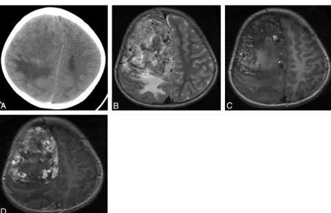
Intracranial Meningeal Hemangiopericytomas in Children and Adolescents: CT and MR Imaging Findings
Full text
Figure


Related documents
Cryptococcal meningitis Patient No. Cryptococcal meningitis compared to viral meningitis. The peaks found in the cryptococcal menin- gitis CSF are numbered to correspond to
production and development of theory from data through identification of concepts and categories, production of theoretical elements to theoretical fragments (Chapter 5, section
case study at Somers Park was performed fo r the purposes of theory
Data collection: CAD- 4 Software (Enraf±Nonius, 1989); cell re®nement: CAD- 4 Software ; data reduction: HELENA (Spek, 2002); program(s) used to solve structure: SHELXS 97
The Rumagesic capsule a marketed siddha formulation showed potent anti-arthritic activity. Evaluation of the anti-arthritic activity of this formulation was the initial step
related to the distorted structure of the complex and the different rigidity of the 2-(2-pyridyl)-4-methoxycarbonyl- quinoline ligand and the bipyridine.. Similar distortions have
No sig- nificant difference was observed among lung squamous cell carcinoma (SCC) patients ( P = 0.42, Figure 2B ), whereas we observed L1CAM- positive expression
While there is no sub- stantial change of retinal structure after expo- sure to hypergravity (+10 Gz/3 min), the expres- sion levels of the Müller cell marker GFAP and the
