ISSN Online: 2160-5629 ISSN Print: 2160-5440
DOI: 10.4236/oju.2018.86019 Jun. 20, 2018 177 Open Journal of Urology
Adolescent Varicocele
Serhan Çimen
Malatya Education and Research Hospital Urology Clinic, Malatya, Turkey
Abstract
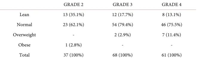
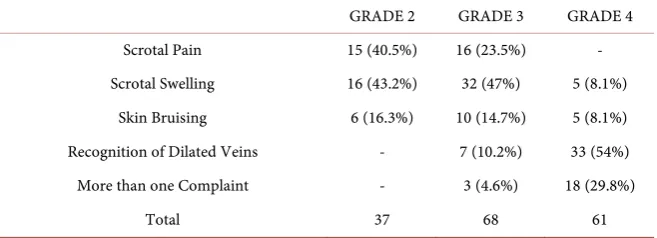
Introduction: Adolescent varicocele is still a controversial issue in pediatric urology. In this study, we aimed to evaluate adolescent patients with varico-cele, who were operated in our clinic, in the light of literature. Materials and Methods: The data of adolescent patients who underwent varicocelectomy between January 2012 and January 2018 in the Urology Clinic of Malatya Training and Research Hospital were examined retrospectively. The age and admission complaint of the patients, the localization of varicocele, the varico-cele grade determined by color Doppler ultrasound (US), the presence of tes-ticular atrophy, the height, weight, body mass index of the patients as well as their relationship with each other were evaluated. Results: The mean age of the patients is 14.74 years (10 - 17 years). Of the patients, 153 (92.2%) had a left-sided varicocele and 13 (7.8%) had a right-sided varicocele. When admis-sion complaints of the patients were examined, 31 (18.6%) had scrotal pain, 53 (32.2%) had scrotal swelling, 21 (12.6%) had skin bruising, 40 (24%) had dilated veins recognized by the family, and 21 (12.6%) had more than one complaint. When the relationship between varicocele grade and BMI was examined, 13 (35.1%) of the 37 patients with a grade 2 varicocele were lean, 23 (62.1%) were normal weight, and 1 (2.8%) was obese. 12 (17.7%) of the 68 pa-tients with a grade 3 varicocele were lean, 54 (79.4%) were normal weight, and 2 (2.9%) were overweight. 8 (13.1%) of the 61 (36.7%) with a grade 4 varico-cele were lean, 46 (75.5%) were normal weight, and 7 (11.4%) were over-weight. Conclusion: Adolescent varicocele is still one of the controversial is-sues today; the diagnosis should be made with the help of physical examina-tion and radiological tests besides the family’s applicaexamina-tion in boys in this age group in order to prevent possible problems.
Keywords
Adolescent, Varicocele, Body Mass Index
1. Introduction
Varicocele is defined as dilatation of the spermatic veins and the plexus pampi-How to cite this paper: Çimen, S. (2018)
Adolescent Varicocele. Open Journal of Urology, 8, 177-183.
https://doi.org/10.4236/oju.2018.86019 Received: April 20, 2018
Accepted: June 16, 2018 Published: June 20, 2018
Copyright © 2018 by author and Scientific Research Publishing Inc. This work is licensed under the Creative Commons Attribution International License (CC BY 4.0).
http://creativecommons.org/licenses/by/4.0/
DOI: 10.4236/oju.2018.86019 178 Open Journal of Urology niformis which is a scrotal extension of these veins [1]. Although it is rarely seen in the prepubertal period, it increases during puberty. It peaks around the age of thirteen. This rate increases up to 15% between ten and nineteen years of age. This rate is similar to the rates in adults [2] [3]. Several rates on the prevalence of adolescent varicocele have been reported in the literature. These rates have ranged from 9% to 42.8% [4] [5]. Adolescent varicocele is still a controversial issue in pediatric urology [6]. Children and adolescents with varicocele may ex-perience fertility problems in the future. Therefore, it is still being discussed that they need to undergo surgical treatment and can benefit from this treatment [7]. In this study, we aimed to evaluate adolescent patients with varicocele, who were operated in our clinic, in the light of literature.
2. Materials and Methods
The data of adolescent patients who underwent varicocelectomy between Janu-ary 2012 and JanuJanu-ary 2018 in the Urology Clinic of Malatya Training and Re-search Hospital were examined retrospectively. The age and admission com-plaint of the patients, the localization of varicocele, the varicocele grade deter-mined by color Doppler ultrasound (US), the presence of testicular atrophy, the height, weight, body mass index of the patients as well as their relationship with each other were evaluated.
3. Results
DOI: 10.4236/oju.2018.86019 179 Open Journal of Urology Table 1. Admission complaint.
Scrotal Pain 31 (18.7%)
Scrotal Swelling 53 (31.9%)
Skin Bruising 21 (12.7%)
Recognition of Dilated Veins 40 (24%)
More than one Complaint 21 (12.7%)
Total 166
Table 2. Relationship between varicocele grade and BMI.
GRADE 2 GRADE 3 GRADE 4
Lean 13 (35.1%) 12 (17.7%) 8 (13.1%)
Normal 23 (62.1%) 54 (79.4%) 46 (75.5%)
Overweight - 2 (2.9%) 7 (11.4%)
Obese 1 (2.8%) - -
Total 37 (100%) 68 (100%) 61 (100%)
varicocele had scrotal pain, 16 (43.2%) had scrotal swelling, and 6 (16.3%) had skin bruising. 16 (23.5%) of the 68 patients with a grade 3 varicocele had scrotal pain, 32 (47%) had scrotal swelling, 10 (14.7%) had had skin bruising, 7 (10.2%) had dilated veins recognized by the family, and 3 (4.6%) had more than one complaint. 5 (8.1%) of the 61 (36.7%) with a grade 4 varicocele had scrotal swel-ling, 5 (8.1%) had skin bruising, 33 (54%) had dilated veins recognized by the family, and 18 (29.8%) had more than one complaint (Table 3).
4. Discussion
Varicocele is defined as dilatation of the spermatic veins and the plexus pampi-niformis which is a scrotal extension of these veins. The right internal spermatic vein is approximately 10 cm shorter than the left internal spermatic vein and drains directly into the vena cava inferior. Therefore, it creates a hydrostatic pressure difference and sets the ground for the formation of varicoceles. Varico-cele is frequently observed on the left side when the literature is examined. It is rarely seen on the right side or both sides [8] [9]. In our study, the majority of patients had a left-sided varicocele in accordance with the literature.
[image:3.595.210.540.225.324.2]DOI: 10.4236/oju.2018.86019 180 Open Journal of Urology Table 3. Relationship between varicocele grade and admission complaint.
GRADE 2 GRADE 3 GRADE 4
Scrotal Pain 15 (40.5%) 16 (23.5%) -
Scrotal Swelling 16 (43.2%) 32 (47%) 5 (8.1%)
Skin Bruising 6 (16.3%) 10 (14.7%) 5 (8.1%)
Recognition of Dilated Veins - 7 (10.2%) 33 (54%)
More than one Complaint - 3 (4.6%) 18 (29.8%)
Total 37 68 61
more accurate results when compared to other volume measurements [12]. Tes-ticular volume differential (TVD) is calculated by the following formula: volume of unaffected testis – volume of affected testis/total testicular volume × 100(%). Testicular atrophy index (TAI) is calculated by the following formula: volume of unaffected testis – volume of affected testis/volume of unaffected testis × 100(%) [13] [14]. The presence or absence of testicular atrophy is calculated in this way. Testicular hypotrophy is defined as testicular volume below 2 ml by ultrasound and more than 20% difference in testicular volume relative to the contralateral testis [15]. Testicular hypotrophy is among the indications for surgery [16]. In our study, approximately 1/10 of the adolescent patients who underwent surgery due to varicocele had testicular atrophy.
Although varicocele in childhood and adolescence is typically asymptomatic, few patients have symptoms and complaints. They seldom present with pain and are sometimes detected due to dilated veins and skin bruising recognized by the family. Sometimes, they are detected when parents recognize the difference be-tween the two testes and thereby bring their children to the clinical examination
[10] [17] [18] [19]. In our study, the most common admission complaint of the
patients was scrotal swelling. This was followed by dilated veins recognized by the family and local skin bruising.
The incidence of varicocele in the patients whose first-degree relatives (such as a father or brother) had varicocele was found to be 3 - 4 times higher than the normal population [20] [21]. In our study, 24 (14.4%) of the patients had a posi-tive family history of varicocele.
The relationship between adolescent varicocele, weight and BMI is still un-clear [22]. Although some studies have revealed a positive correlation between the incidence of adolescent varicoceles and weight gain, it has been shown to be inversely proportional to reduced body mass index [23]-[28]. When the litera-ture is examined, various studies have shown that the incidence of varicocele is decreased in overweight and obese men [29] [30] [31] [32]. In a large-scale study by Liu et al., it was shown that varicocele grade was decreased with reduced body mass index [22] [28]. In our study, while the patients with a low BMI had a low-grade varicocele, the patients with a high BMI had a high-grade varicocele.
DOI: 10.4236/oju.2018.86019 181 Open Journal of Urology conducted on adults. Fazeli et al. found that the patients with a low BMI had a high-grade varicocele [33]. In our study, it was seen that varicocele grade in-creased as BMI inin-creased.
Varicocele causes progressive damage to the testis through various mechan-isms and can lead to infertility. Although adolescent varicocele is still one of the controversial issues today, the diagnosis should be made with the help of physi-cal examination and radiologiphysi-cal tests besides the family’s application in boys in this age group in order to prevent possible problems. We think that a large number of studies are needed in order to clarify the relationship of adolescent varicocele with demographic characteristics.
References
[1] Robinson, S.P., Hampton, L.J. and Koo, H.P. (2010) Treatment Strategy for the Adolescent Varicocele. Urologic Clinics of North America, 37, 269-278.
https://doi.org/10.1016/j.ucl.2010.03.011
[2] Oster, J. (1971) Varicocele in Children and Adolescents. An Investigation of the In-cidence among Danish School Children. Scandinavian Journal of Urology and Ne-phrology, 5, 27-32. https://doi.org/10.3109/00365597109133569
[3] Akbay, E., Cayan, S., Doruk, E., Duce, M.N. and Bozlu, M. (2000) The Prevalence of Varicocele and Varicocele-Related Testicular Atrophy in Turkish Children and Adolescents.BJU International, 86, 490-493.
https://doi.org/10.1046/j.1464-410X.2000.00735.x
[4] Berger, O.G. (1980) Varicocele in Adolescence. Clinical Pediatrics (Phila), 19, 810-811. https://doi.org/10.1177/000992288001901205
[5] Pfeiffer, D., Berger, J., Schoop, C. and Tauber, R. (2006) A Doppler-Based Study on the Prevalence of Varicocele in German Children and Adolescents. Andrologia, 38, 13-19. https://doi.org/10.1111/j.1439-0272.2006.00680.x
[6] Haddad, N.G., Houk, C.P. and Lee, P.A. (2014) Varicocele: A Dilemma in Adoles-cent Males. Pediatric Endocrinology Reviews, 11, 274-283.
[7] Kass, E.J. and Belman, A.B. (1987) Reversal of Testicular Growth Failure by Varicocele Ligation. Journal of Urology, 137, 475-476.
https://doi.org/10.1016/S0022-5347(17)44072-9
[8] Höllwarth, M.E. (2006) Varicocele. In: Puri, P. and Höllwarth, M., Eds., Pediatric Surgery, Springer-Verlag Berlin Heidelberg, 569.
https://doi.org/10.1007/3-540-30258-1_54
[9] Hutson, J.M. (2006) Undescended Testis, Torsion and Varicocele. In: O’Neill, J.A., Grosfeld, J.L., James, A., Fonkalsrud, E.W. and Coran, A.G., Eds., Pediatric Surgery, Mosby, Philadelphia, 1193.
[10] Uğuz, S. and Irkılataa, H.C. (2015) Adolescent Varicocele. Turkiye Klinikleri Jour-nal of Urology Special Topics, 8, 86-92.
[11] Lambert, B. (1951) The Frequency of Mumps and of Mumps Orchitis and the Con-sequences for Sexuality and Fertility. Acta Genetica Et Statistica Medica, 2, 1-166. [12] Hsieh, M.L., Huang, S.T., Huang, H.C., Chen, Y. and Hsu, Y.C. (2009) The
DOI: 10.4236/oju.2018.86019 182 Open Journal of Urology [13] Niedzielski, J., Paduch, D. and Raczynski, P. (1997) Assessment of Adolescent
Varicocele. Pediatric Surgery International, 12, 410-413. https://doi.org/10.1007/BF01076952
[14] Christman, M.S., Zderic, S.A. and Kolon, T.F. (2014) Comparison of Testicular Volume Differential Calculations in Adolescents with Varicoceles. Journal of Pedi-atric Urology, 10, 396-398.https://doi.org/10.1016/j.jpurol.2013.12.007
[15] Diamond, D.A., Zurakowski, D., Bauer, S.B., et al. (2007) Relationship of Varicocele Grade and Testicular Hypotrophy to Semen Parameters in Adolescents. Journal of Urology, 178, 1584-1588.https://doi.org/10.1016/j.juro.2007.03.169
[16] Ünal, D., Erbağcı, A., Güneş, A., Ersay, A., Semerciöz, A., Satar, N. and Sarıca, K. (2002) Peripubertal Variıations of Testicular Size in Adolescent Varicocele: Gapug Series. Türk Üroloji Dergisi, 28, 161-165.
[17] Özkan, S., Özbek, E., Gürpınar, T., Sarıyüce, O., Güneş, A. and Özsan, Ö. (1996) Prevalence of Varicocele in Adolescence and Use of Doppler USG. Turgut Özal Tıp Merkezi Dergisi, 3, 210-212.
[18] Öztürk, E. and Soygür, T. (2013) Adölesan varikosel. Androloji Bülteni, 53, 103-105.
[19] Aksoy, Y., Ziypak, T. and Adanur, Ş. (2012) Turkiye Klinikleri. Journal of Urology, 5, 61-66.
[20] Raman, J.D., Walmsley, K. and Goldstein, M. (2005) Inheritance of Varicoceles. Urology, 65, 1186-1189.https://doi.org/10.1016/j.urology.2004.12.057
[21] Gökçe, A., Davarci, M., Yalçinkaya, F.R., Güven, E.O., Kaya, Y.S., Helvaci, M.R., et al. (2010) Hereditary Behavior of Varicocele. Journal of Andrology, 31, 288-290. https://doi.org/10.2164/jandrol.109.008698
[22] Liu, J., Zhang, S., Liu, M., Wang, Q., Shen, H., Zhang, Y. and Yan, D. (2017) Preva-lence of Varicocoele and Its Association with Body Mass Index among 39,559 Rural Men in Eastern China: A Population-Based Crosssectional Study. Andrology, 5, 562-567.https://doi.org/10.1111/andr.12345
[23] Delaney, D.P., Carr, M.C., Kolon, T.F., Snyder, H.M. and Zderic, S.A. (2004) The Physical Characteristics of Young Males with Varicocele. BJU International, 94, 624-626.https://doi.org/10.1111/j.1464-410X.2004.05013.x
[24] Shin, J.I. and Lee, J.S. (2007) Changes in Body Mass Index and Prevalence of Varicoceles during Adolescence. Urologia Internationalis, 78, 178.
https://doi.org/10.1159/000098079
[25] Prabakaran, S., Kumanov, P., Tomova, A., Hubaveshki, S. and Agarwal, A. (2006) Adolescent Varicocele: Association with Somatometric Parameters. Urologia Inter-nationalis, 77, 114-117.https://doi.org/10.1159/000093902
[26] Nielsen, M.E., Zderic, S., Freedland, S.J. and Jarow, J.P. (2006) Insight on Patho-genesis of Varicoceles: Relationship of Varicocele and Body Mass Index. Urology, 68, 392-396.https://doi.org/10.1016/j.urology.2006.02.005
[27] Kumanov, P., Robeva, R.N. and Tomova, A. (2008) Adolescent Varicocele: Who Is at Risk? Pediatrics, 121, e53-e57.https://doi.org/10.1542/peds.2007-0340
[28] May, M., Taymoorian, K., Beutner, S., Helke, C., Braun, K.P., Lein, M., et al. (2006) Body Size and Weight as Predisposing Factors in Varicocele. Scandinavian Journal of Urology and Nephrology, 40, 45-48.https://doi.org/10.1080/00365590500407795
DOI: 10.4236/oju.2018.86019 183 Open Journal of Urology 663-669.https://doi.org/10.1111/j.2047-2927.2013.00113.x
[30] Gokce, A., Demirtas, A., Ozturk, A., Sahin, N. and Ekmekcioglu, O. (2013) Associa-tion of Left Varicocoele with Height, Body Mass Index and Sperm Counts in Infer-tile Men. Andrology, 1, 116-119.https://doi.org/10.1111/j.2047-2927.2012.00014.x
[31] Doğantekin, E., Görgel, S.N., Şahin, E. and Girgin, C. (2014) Relationship between Varicocele and Anthropometric Indices in Infertile Population. Dicle Medical Journal, 41, 59-63.https://doi.org/10.5798/diclemedj.0921.2014.01.0373
[32] Hassanzadeh, K., Yavari-Kia, P., Soleymanpour, H., Ebrahimpour, N. and Alikhan, H. (2011) Effect of Body Mass Index and Prevalence of Varicocele. Pakistan Journal of Biological Sciences, 14, 806-875.https://doi.org/10.3923/pjbs.2011.869.875