Pulsed and Tissue Doppler Echocardiographic
Abnormalities in Patients with Diastolic Heart Failure
with and without Atrial Fibrillation
Taysir Said Garadah1,2, Najat Hassan Mahdi1,2, Mohamed Ahmed Al Alwai1, Ahmed Abdulkareem Jaradat, Zuheir Ahmed Hasan2
1Salmaniya Medical Complex, Ministry of Health, Manama, Bahrain; 2College of Medicine and Medical Sciences, Arabian Gulf
University, Manama, Bahrain. Email: garadaht@hotmail.com
Received September 11th, 2012; revised October 20th, 2012; accepted November 9th, 2012
ABSTRACT
Background: Symptoms of heart failure (HF) are identical in both systolic (SHF) and diastolic hear failure (DHF). The prevalence of atrial fibrillation (AF) in heart failure varies in different studies depending on the criteria of enrollment and the use of echocardiographic parameters in the definition of HF. Aim: To assess the clinical characteristic of pa- tients with DHF complicated by AF and compare with those with SHF in regard of echocardiographic abnormalities and causative agents. Furthermore, evaluate the clinical and biochemical markers for the prediction of AF in HF. Method:
Over the duration of 12 months, each patient diagnosed as HF based on admission code was enrolled in the study. Patients were classified into two groups: group 1: DHF, with preserved LVPEF% > 50%, n = 204 (60%), and group 2, with SHF, with LVREF% ≤ 50%, n = 140 (40%). The presence or absence of AF on ECG was recorded. The predictive value of different clinical and biochemical variables for the development of AF was evaluated using logistic multiple regression analysis. Results: Three hundred and forty four eligible patients were admitted to hospital with heart failure out of 7650 who had other medical problems. The prevalence of HF in this population was 4.5%, those with DHF were 2.7% and SHF of 1.8%. The incidence of AF on ECG was 35% in the whole study population and 65% were in sinus rhythm (SR). The occurrence of AF was twice higher in DHF patients of 22% compared with 11% in SHF. Echo pulsed Doppler in DHF and AF compared with those in SR showed a severe restrictive pattern with significantly thick septum wall, higher LV mass index, shorter DT and higher E/e− ratio of 12.4 vs. 9.73, P < 0.05. The predictive risk (odd ratio) of different clinical variables for development of AF in HF was positive for LV hypertrophy on ECG of 2.4, history of hypertension of 1.6, history of DM of 1.4, BMI > 28 of 1.7. Conclusions: The prevalence of HF was 4.5% in the study population, with SHF of 1.8% and DHF of 2.7%. Patients with DHF and AF were older with a higher female ratio with severe restrictive pattern compared with those of SHF. The incidence of AF in the whole study was 35%. The best predictor of AF in HF was left ventricle hypertrophy followed by history of hypertension and DM.
Keywords: Diastolic Heart Failure; Atrial Fibrillation; Tissue Doppler; Bahrain
1. Introduction
The symptoms of heart failure (HF) may be identical whether failure is secondary to systolic dysfunction (SHF) with reduction of left ventricle (LV) systolic function or diastolic heart failure (DHF) with normal LV function. In patients with SHF the left ventricle (LV) is dilated and LV ejection fraction on echocardiogram of ≤50%.
DHF had normal LV cavity dimensions with no valve disease and LVEF > 50%, [1] but abnormal echo indices of left ventricle diastolic filling such as abnormal relaxa- tion and or dispensability (restriction) of the LV [2]. DHF accounts for 30% - 50% of patients with heart fail-
ure and is an independent predictor of atrial fibrillation in the elderly [3]. Heart failure is a recognized risk factor for atrial fibrillation (AF) as a coexistent complication [4].
and hospitalization [8,9].
Randomized pathophysiologic studies showed impor- tant differences between patients with systolic heart fail- ure (SHF) and diastolic heart failure (DHF) as the two conditions were suggested to be distinct syndromes rather than a continuous spectrum of one disorder [10, 11]. The difference between both type of heart failure in clinical and histopathological levels indicates that they are distinct phenotypes [12,13].
Brain natriuretic peptide (BNP), a cardiac neurohor- mone, had been recognized as a marker of HF usually released by the left ventricle in response to volume ex- pansion and pressure overload [14]. BNP had been shown earlier to be sensitive biomarkers for the detection of a symptomatic LV dysfunction and that they had im-portant diagnostic and prognostic implications in patients with LV dysfunction [15].
To date, studies on the relationship between HF with DHF and the development of AF in nonwhite population are limited.
In this study we aim to first: to assess the incidence of AF in patients with DHF and compare with those with SHF in regard of clinical characteristics, causative agents and echo abnormalities. Second: to evaluate the clinical and biochemical markers of for the prediction of AF in HF.
2. Material and Methods
Patients admitted to the Salmaniya hospital with diagno- sis of acute heart failure, based on hospital admission code, were included and clinical data was extracted from patient’s files. The study duration was of 12 months from 1.1.2010 to 31.12.2010.
Salmaniya hospital is the main governmental hospital with a catchment area of 900,000 populations. There were 344 patients that had heart failure based on admis- sion code. The Total number of patients who were ad- mitted with acute medical problems for the same dura- tion was 7650 patients.
Patient’s data regarding clinical history and physical findings on examination, biochemical laboratory results were obtained. The results of echocardiography and elec- trocardiographic data were available for all patients on admission or during hospital stay.
A constitutional ethical committee approval was ob- tained prior to the data extraction and analysis.
2.1. Inclusion Criteria
Subjects were included if they were presented with acute dyspnea and clinical diagnosis of HF by code of admis- sion. The history of dyspnea class was obtained based on New York Heart Association (NYHA) functional classi- fication [16].
2.2. Clinical Assessment
The clinical findings on examination of S3 gallop and bibasal crackle and raised jugular venous pressure, heap- tomegaly and ankle edema were all recorded. Patients were labeled as biventricular failure if both left and right HF signs are present clinically.
Clinical demographic data and past medical history of heart failure, history of hypertension and diabetes melli- tus (DM) prior to admission, history of smoking and pre- vious documentation of AF were extracted from the pa- tients file.
Twelve leads ECG was evaluated for the presence of voltage criteria of left ventricle hypertrophy using Pe- rugia score [17].
The presence of Q wave and conduction abnormality with ST segment elevation or depression or T wave changes ware all recorded. Atrial fibrillation was defined as absence of P wave on 12 leads electrocardiogram (ECG), with irregular ventricular rhythm and last for >30 seconds [18].
Echocardiographic data of M mode, 2 D. Pulsed and tissue doppler were tabulated for the dimension of LV cavity dimensions, wall thickness on M mode and calcu- lated LV mass index, the calculated left ventricle ejection fraction (LVEF%), valve abnormalities and indices of left ventricle diastolic fillings were tabulated [19].
Patients were subdivided into those with preserved LVPFE% > 50% and reduced LVREF% of ≤50%. Those with LVREF% ≤ 50% patients were grouped based on calculation of EF % using M mode, Teichholz formula as very mild (LVREF% <43% - ≥50%), mild (LVREF of <33% - ≥43%), moderate (<23% - ≥33%) and severe (<22%) [20,21].
The diastolic indices and the severity of diastolic dys- function based on E/A ratio, deceleration time (DT) of E wave and E/e− was assessed in all patient as mild, mod-
erate or severe if the E/A ratio is E/A <1, 1 - 2 and ≥2 respectively [22]. Estimated LV mass index on echo of >131 g/m2) for men and >113 g/m2 for women was de-
fined as cut off points for severe LVH [23].
Heart failure was documented as a clinical diagnosis in 344 patients. The total number of patients who were ad- mitted to emergency room with acute medical disease was 7650. There were 120 (35%) patients who had AF on 12 leads ECG and 224 (60%) were in sinus rhythm (SR). The study population was subdivided based on LVEF% into group 1, patients with DHF with LVPEF > 50%, n = 204 (60%) and group 2, with SHF and LVREF
≤ 50%, n = 140.
2.3. Clinical History
hood, history hypertension and alcohol intake were all recoded. The clinical presentation on admission such as fast palpitation, angina pectoris pain, dyspnea, dizziness, and focal neurological deficit were recorded. The NYHA class of I to IV on admission was also recorded.
The height and weight, the presence or absence of goiter, cardiac murmur suggestive of aortic or mitral or pulmonary valve disease was all recorded. The signs of pulmonary edema such as bibasal crackles and gallop, elevated JVP, ankle odema were recorded.
The results of available serum level of brain natriuretic peptide (BNP), uric acid, estimated Glomerular Filtration Rate (eGFR) and serum potassium were tabulated.
2.4. Statistical Analysis
All data were entered and analyzed using the Statistical Package of Social Sciences (SPSS) version 17.1. Data are presented as mean ± SD. Unpaired student-test was
used to analyze the differences between the mean vari- ables of M mode for septal wall thickness, LV cavity and LV mass index in the two groups.
Student’s t-test was applied for continuous variables
and Chi-square analysis for frequency non-continuous
data such as Doppler ratio for pulsed derived indices of E wave to A wave velocity (E/A) ratio, deceleration time (DT) of E wave and pulsed to tissue derived Doppler ratio of E wave velocity (E/e−).
Multiple logistic regression analysis was used to cal- culate the odds ratio for different clinical and biochemi- cal variables for the development of AF in the study group. Clinical variables are the history of hypertension, DM, smoking, body mass index, LV hypertrophy on ECG and the biochemical variables are serum level of uric acid, potassium level, BNP and eGFR.
All reported P-values are two tailed and P-value was
regarded as significant at level of <0.05.
3. Results
Three hundred and forty four patients with clinical diag- nosis of heart failure were enrolled and divided into two groups based on LVEF% on echo: group 1, diastolic heart failure with preserved systolic LV-PEF, n = 204 (60%) and group 2, with SHF and reduced systolic func- tion LV-REF, ≤50%, n = 140 (40%).
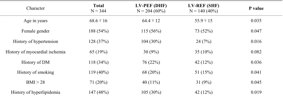
Patients with DHF were older than SHF, mean age of 64.4 ± 12 and 55.9 ± 15 respectively. In the whole study there were 195 females and 150 males. Sixty five percent of patients were female gender DHF patients and there were 52% in those with SHF. Table 1 shows the clinical characteristic of patients in both groups.
Patients with DHF compared with SHF had signify- cantly a higher number of patients with history of hyper- tension of 34% vs. 8, DM of 25% vs. 14%, history of smoking of 23% vs. 17% and BMI > 28 of 14% vs. 10% and history of hyperlipidemia of 104 (30%) vs. 42 (12%). Those with history of myocardial ischemia were of no significant difference in both groups.
3.1. ECG
The total number of patients who had atrial fibrillation on admission were 120 (35%) and 223 (65%) were in sinus rhythm. In the whole study out of those who had AF (35%), the new onset AF with no previous history of AF was detected in 72 (21%) patients and 48 (14%) patients had history of chronic AF.
[image:3.595.60.538.549.714.2]Patients with left ventricle hypertrophy based on ECG criteria were 160 (46%). Among those with LVH there were 89 (26%) patients with AF and 71 (21%) were in sinus rhythm. The overall sensitivity and specificity of
Table 1. Demographic data of all patients who were admitted with heart failure (n = 344).
Character N = 344 Total LV-PEF (DHF) N = 204 (60%) LV-REF (SHF) N = 140 (40%) P value
Age in years 68.6 ∓ 16 64.4 ∓ 12 55.9 ∓ 15 0.035
Female gender 188 (54%) 115 (56%) 73 (52%) 0.047
History of hypertension 128 (37%) 104 (30%) 24 (7%) 0.016
History of myocardial ischemia 65 (19%) 30 (9%) 35 (10%) 0.082
History of DM 118 (34%) 76 (22%) 42 (12%) 0.036
History of smoking 119 (40%) 68 (20%) 51 (15%) 0.041
BMI > 28 71 (20%) 40 (11%) 31 (9%) 0.045
History of hyperlipidemia 147 (48%) 105 (30%) 42 (12%) 0.019
Abbreviation: LV-PEF: left ventricle with preserved ejection fraction; LV-REF: left ventricle with reduced ejection fraction; DHF: diastolic heart failure; SHF: ystolic heart failure; BMI: body mass index; DM: diabetes mellitus.
ECG for detection of LVH compared with BMI derived by echo as gold standard 75% and 60% respectively.
3.2. Clinical Findings
All patients had heart failure on admission, 154 (45%) patients had left ventricle failure manifested with pul- monary odema and 120 (35%) had both left ventricular and right ventricular failure (biventricular). There were 20 patients in NYHA functional class IV, 114 were in class III, 210 patients were in functional class II and none was in class I. History of medication on prior to admis- sion showed 270 (78%) patients were taking angiotensin receptor antagonist (ACE) tablet, 198 (57%) were on diuretics, 125 (36%) on beta blocker, and 58 (17%) were on digoxin medication.
[image:4.595.309.537.95.192.2]3.3 Heart Failure and Patient’s Age
Figure 1 show patients age distribution for both groups. In the age category of 20 - 35 years, patients with DHF had the ratio of 1.9 higher than SHF, for category of (35 - 50) the ratio was 1.6, for age category of (50 - 65) years it was 2, and in age category of >65 years the ratio was nearly equal of 0.96 for both types of HF.
3.4. Echocardiographic and Biochemical Variables
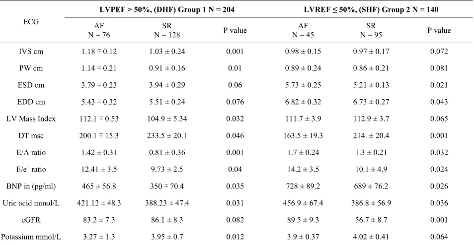
There were 204 (60%) patients who had DHF and 140 (40%) were with SHF. Table 2 shows biochemical, Pulsed and tissue Doppler results in both groups. In those with DHF and AF on ECG compared with those with SR, M mode echocardiogram showed significantly thicker septal and posterior wall of LV. The LV mass index was significantly higher of 112.1 vs. 104.9 gm/m2. Further,
Pulsed Doppler showed significantly higher E/A ratio of 1.42 ± 0.31 vs. 0.81 ± 0.2.5 lower DT 200 ∓ 15.3 m sec vs. 233.5 ± 20.1 m sec suggesting restrictive pattern. The tissue Doppler showed higher E/e− ratio of 12.41 ∓ 3.5 vs.
9.73 ± 2.5.
In SHF and AF on ECG compared with S R, M mode Echo showed no significant difference in wall thickness but of LV septal and posterior wall but LV cavity showed significant dilation of LV cavity in both systole and diastole. Pulse and Tissue Doppler showed signifi- cantly higher E/A ratio, shorter DT and higher E/e−. The
LV mass index was lower but the difference was of no significant.
3.5. Biochemical Markers
In DHF Blood chemistry in patients with AF showed significantly higher level of uric acid, BNP and lower potassium level, however difference between eGFR was
Figure 1. Distribution of patients of both groups in HF ac-cording to patients age, patients with DHF (LV-PEF), n = 204) and those with SHF (LV-REF), n = 140.
of no significance. In SHF patients and AF compared with those of SR, there significantly higher level of uric acid, higher eGFR and BNP but difference of potassium level was not significant.
Segmental wall motion abnormality was detected in DHF and AF in 32 (9%) patients versus 14 (4%) in those with SR. In SHF and AF those with segmental wall mo- tion abnormality were detected in 4 (12%) patients ver- sus 24 (7%) in those with SR. Figure 2 shows the distri- bution of patients with AF and SR in both groups. In DHF the ratio between those with AF to SR was 0.51 and in SHF it was 0.6.
Medication prior to admission showed 270 (78%) pa- tients using angiotensin receptor antagonist (ACE) tablet, 198 (57%) were on diuretics, 125 (36%) on Beta blocker, and 58 (17%) were on digoxin tablet.
3.6. Etiological Diagnosis
The etiology in DHF, n = 204, showed 74 (36%) patients in hypertensive emergency with systolic BP > 180 mmHg complicated with pulmonary edema. Seventy (34%) pa- tients had ischemic heart disease, among them 39 had acute myocardial infarction and 31 had unstable angina. Ten (5%) patients had thyrotoxicosis, 10 (5%) had his- tory suggestive of Dilated Myopathy, 10 patients had valve disease (5%), seven had severe mitral regurgitation and three with moderate to severe aortic regurgitation. There were thirty (15%) patients with no definite etio- logical diagnosis.
In SHF, n = 140. There were 45 (32%) patients with dilated cardiomyopathy, nine (9%) had hypertension heart failure, 30 (21%) had ischemic heart disease), 27 (19%) had significant valve disease with 16 of mitral valve regurgitation and 11 had severe aortic regurgitation, 13 (9%) patients had thyroid disease and 15 (10%) were of unknown etiology.
3.7. The Odds Ratio for Risk of AF in HF Patients
Table 2. The mean value ∓ standard deviation (SD) of biochemical markers and echocardiographic indices in all patients presented with HF, n = 344.
LVPEF > 50%, (DHF) Group 1 N = 204 LVREF ≤ 50%, (SHF) Group 2 N = 140
ECG AF
N = 76
SR
N = 128 P value
AF N = 45
SR
N = 95 P value
IVS cm 1.18 ∓ 0.12 1.03 ± 0.24 0.001 0.98 ± 0.15 0.97 ± 0.17 0.072
PW cm 1.14 ∓ 0.21 0.91 ± 0.16 0.01 0.89 ± 0.24 0.86 ± 0.21 0.081
ESD cm 3.79 ∓ 0.23 3.94 ± 0.29 0.06 5.73 ± 0.25 5.21 ± 0.13 0.021
EDD cm 5.43 ∓ 0.32 5.51 ± 0.24 0.076 6.82 ± 0.32 6.73 ± 0.27 0.043
LV Mass Index 112.1 ∓ 0.53 104.9 ± 5.34 0.032 111.7 ± 3.9 112.9 ± 3.7 0.065
DT msc 200.1 ∓ 15.3 233.5 ± 20.1 0.046 163.5 ± 19.3 214. ± 20.4 0.001
E/A ratio 1.42 ± 0.31 0.81 ± 0.36 0.001 1.7 ± 0.24 1.3 ± 0.21 0.032
E/e− ratio 12.41 ± 3.5 9.73 ± 2.5 0.04 14.2 ± 3.5 10.1 ± 4.9 0.024
BNP in (pg/ml) 465 ± 56.8 350 ∓ 70.4 0.035 728 ± 89.2 689 ± 76.2 0.026
Uric acid mmol/L 421.12 ± 48.3 388.23 ± 47.4 0.031 456.9 ± 67.4 386.8 ± 56.9 0.036
eGFR 83.2 ± 7.3 86.1 ± 8.3 0.082 89.5 ± 9.3 56.7 ± 8.7 0.001
Potassium mmol/L 3.27 ± 1.3 3.95 ± 0.7 0.012 3.9 ± 0.37 4.02 ± 0.41 0.064
[image:5.595.61.538.124.371.2]Abbreviations: diastolic heart failure (DHF); systolic heart failure (SHF); interventricular septum (IVS); posterior wall (PW); end systolic dimension (ESD); end diastolic dimension (EDD); deceleration time (DT); estimated glomerular filtration rate (eGFR); atrial fibrillation (AF); sinus rhythm (SR) (ratio of E ve-locity to tissue doppler veve-locity of septum (E/e−).
Table 3. The hazard ratio for the development for AF in patients presented with heart failure using logistic multiple regression analysis in patients presented with heart failure, n = 344.
Variable Odds ratio Confidence interval P value
History of
hypertension 1.6 (CI 95%: 1.1 - 2.1) 0.036
History of DM 1.4 (CI 95%: 0.7 - 2.25) 0.037
History of smoking 0.8 (CI 95%: 0.5 - 1.1) 0.073
BMI > 28 1.7 (CI 95%: 1.1 - 2.3) 0.028
LV hypertrophy
on ECG 2.4 (CI 95%: 1.8 - 3.0) 0.021
K < 3.4 mmol/L 1.4 (CI 95%: 1.1 - 1.7) 0.043
Uric acid > 420
mmol/L 2.5 (CI 95%: 2.1 - 2.9) 0.021
eGFR < 50 2.1 (CI 95%: 1.6 - 2.6) 0.013
BNP pg/L 3.8 (CI 95%: 2.7 - 5.7) 0.001
Abbreviation: BNP: brain natriuretic peptide; eGFR: estimated glomerular filtration rate; K: potassium; BMS: body mass index.
the study for the developing of AF in patients with HF. After adjusting for age and sex multiple regression analysis showed positive predictive value for: history of hypertension of 1.6, history of DM of 1.4, for BMI > 28
Figure 2. Distribution arial fibrillation AF, n = 76 and sinus rhythm SR, n = 148 in group 1, with diastolic heart failure and group 2, with systolic heart failure: AF n = 45 and SR, n = 75.
of 1.7, LVH on ECG of 2.
Biomarker positive predictors were hyperuricemia > 420 mmol/l of 2.5, higher BNP > 500 pg/ml of 3.8, and low estimated glomerular filtration rate (eGFR) < 5 of 2.1, hypokalemia of < 3.4 mmol/l of 1.4. Smoking was of negative predictive value.
4. Discussion
[image:5.595.308.539.415.535.2]who had DHF with LVEF > 50% and both were evalu- ated regarding the development of AF on ECG.
Patients with DHF compared with SHF had high fe- male distribution and were older. This finding is similar to previous report, where 50% - 70% of patients above the age of 70 had diastolic heart failure and 49% had systolic heart failure [9].
In this hospital based study with total of 7650 patients, the overall prevalence of heart failure over one year was 4.4%. This was higher than other studies with prevalence of 4.1% and 4.2 respectively [24,25].
The prevalence in of SHF was 1.8% and for DHF it was 2.3%, which differ from one study by Mureddu GF
et al., where the prevalence was 1.8% and 4.9% respec-
tively with no difference by gender in patients with DHF [26].
The rate of occurrence of AF patients with HF in the study was of 35% which was higher than previous re- ports of 31% and 25% respectively [4,27]. The rate of occurrence of AF in both subgroups was nearly equal of 38% for SHF and of 37% in DHF.
In different clinical trials, the use of different diagnos- tic criteria and different age groups leads to variation of prevalence of HF. In one study by J. Grewal, et al., the
prevalence of HF was 1.9% [28] and another study by S. J. Phillips et al.,[29] the prevalence of HF was of 1.7%.
In both studies the prevalence was lower than this study although the prevalence was increasing as the patient’s age increased.
Sixty eight percent of patients with DHF and AF were in severe diastolic dysfunction of restrictive pattern while 23% had mild dysfunction of abnormal relaxation pattern. This finding was in agreement with previous reports where severe diastolic dysfunction was associated with worse outcome including AF and mortality [30,31].
In previous studies women were less likely than men to have SHF accounting to 25% - 35% of systolic heart failure cases [32]. In one report men have double the risk of developing blood-pumping (systolic) problems com- pared with women [33]. In this study percentage of fe- male gender was higher in both DHF and SHF than male.
The age distribution for both groups showed increment in the rate of HF in old age patient, the percentage of DHF was two folds higher compared with SHF in young age patients but was equal in patients of >65 years. Pa- tients of >65 years in SHF were twice higher than those of 50 - 65 years. In one study the estimated prevalence of diastolic dysfunction among patients with HF was 15, 33, and 50 percent at ages less than 50, 50 - 70, and >70 years, respectively [34].
In one trial, a higher incidence of DHF occur in old age <65 years mainly due to ischemia and hypertension [35].
The level of BNP was significantly lower in those with DHF compared with SHF. This finding had been shown previously, where BNP was significantly high in SHF with no identifiable threshold to distinguish between the two forms of HF [28]. In one report higher level of BNP > 100 pg/ml was an independent predictor of adverse cardiovascular outcome in patients with DHF [36].
There was a significantly lower serum level of potas- sium in AF patients in patients with DHF but not in SHF. Previous studies showed AF is positively associated with hypokalemia [37].
In this study a higher rate of segmental wall motion abnormality on echo was detected in patients with AF both groups of HF.
The causative agents in DHF was mainly hypertension, followed by coronary artery disease while in SHF it was dilated myopathy followed valvular heart disease. In one reports by Topol et al., hypertension and ischemia were
the main etiology of DHF [38].
In this study the best clinical predictors for develop- ment of AF in heart failure was the history of hyperten- sion, followed by DM and obesity.
In patients with a SHF and dilated LV the heart failure, tachycardia-mediated cardiomyopathy was the presumed the causative agent in 29% of patients with HF however in this subset of patients improvement of function was seen with rhythm or rate control [39,40].
Limitation of the Study
The echocardiographic findings were observed on ad- mission and were not repeated after improvement of pa- tient’s clinical condition as tachycardia mediated HF cannot be ruled out as an etiological diagnosis on admis- sion.
REFERENCES
[1] A. Mosterd, A. W. Hoes, M. C. de Bruyne, et al., “Preva- lence of Heart Failure and Left Ventricular Dysfunction in the General Population; The Rotterdam Study,” Euro-
pean Heart Journal, Vol. 20, No. 6, 1999, pp. 447-455.
doi:10.1053/euhj.1998.1239
[2] J. P. Bounhoure, P. Massabuau, M. Galinier, et al., “[Heart Failure with Preserved Left Ventricular Function: Clinical, Echocardiographic, and Clinical Course Features. Prognostic Factors],” Bulletin de l’Academie Nationale de
Medecine, Vol. 186, No. 6, 2002, pp. 1003-1014.
[3] R. Nagarakanti and M. Ezekowitz, “Diastolic Dysfunc- tion and Atrial Fibrillation,” Journal of Interventional
Cardiac Electrophysiology, Vol. 22, No. 2, 2008, pp.
111-118.doi:10.1007/s10840-008-9203-8
[4] E. J. Benjamin, D. Levy, S. M. Vaziri, et al., “Independ-
American Medical Association, Vol. 271, No. 11, 1994, pp. 840-844.doi:10.1001/jama.1994.03510350050036 [5] R. Nieuwlaat, L. W. Eurlings, J. G. Cleland, et al., “Atrial
Fibrillation and Heart Failure in Cardiology Practice: Re-ciprocal Impact and Combined Management from the Perspective of Atrial Fibrillation: Results of the Euro Heart Survey on Atrial Fibrillation,” Journal of the
American College of Cardiology, Vol. 53, No. 18, 2009,
pp. 1690-1698.doi:10.1016/j.jacc.2009.01.055
[6] J. G. Cleland, K. Swedberg, F. Follath, et al., “The
Eu-roHeart Failure Survey Programme—A Survey on the Quality of Care among Patients with Heart Failure in Europe. Part 1: Patient Characteristics and Diagnosis,”
European Heart Journal, Vol. 24, No. 5, 2003, pp. 442-
463.doi:10.1016/S0195-668X(02)00823-0
[7] A. J. Camm and I. Savelieva, “Atrial Fibrillation: Ad-vances and Perspectives,” Dialogues in Cardiovascular
Medicine, Vol. 8, 2003, pp. 183-202.
[8] M. Klapholz, M. Maurer, A. M. Lowe, et al., “Hospitali-zation for Heart Failure in the Presence of a Normal Left Ventricular Ejection Fraction: Results of the New York Heart Failure Registry,” Journal of the American College
of Cardiology, Vol. 43, No. 8, 2004, pp. 1432-1438.
doi:10.1016/j.jacc.2003.11.040
[9] S. Yusuf, M. A. Pfeffer, K. Swedberg, et al., “Effects of
Candesartan in Patients with Chronic Heart Failure and Preserved Left-Ventricular Ejection Fraction: The CHARM- Preserved Trial,” Lancet, Vol. 362, No. 9386, 2003, pp. 777-781.doi:10.1016/S0140-6736(03)14285-7
[10] M. M. Redfield, G. N. Kay, L. S. Jenkins, et al., “Tachy-
cardia-Related Cardiomyopathy: A Common Cause of Ventricular Dysfunction in Patients with Atrial Fibrilla- tion Referred for Atrioventricular Ablation,” Mayo Clinic
Proceedings, Vol. 75, No. 8, 2000, pp. 790-795.
doi:10.4065/75.8.790
[11] T. S. Tsang, B. J. Gersh, C. P. Appleton, et al., “Left Ventricular Diastolic Dysfunction as a Predictor of the First Diagnosed Nonvalvular Atrial Fibrillation in 840 Elderly Men and Women,” Journal of American College
of Cardiology, Vol. 40, No. 9, 2002, pp. 1636-1644.
doi:10.1016/S0735-1097(02)02373-2
[12] R. S. Bhatia, J. V. Tu, D. S. Lee, et al., “Outcome of
Heart Failure with Preserved Ejection Fraction in a Popu- lation-Based Study,” New England Journal of Medicine,
Vol. 355, No. 3, 2006, pp. 260-269.
doi:10.1056/NEJMoa051530
[13] T. Fujino, T. Yamashita, S. Suzuki, et al., “Characteris-tics of Congestive Heart Failure Accompanied by Atrial Fibrillation with Special Reference to Tachycardia-In- duced Cardiomyopathy,” Circulation Journal, Vol. 71, No. 6, 2007, pp. 936-940.doi:10.1253/circj.71.936 [14] K. Dickstein, “Natriuretic Peptides in Detection of Heart
Failure,” Lancet, Vol. 351, No. 9095, 1998, pp. 9-13.
doi:10.1016/S0140-6736(05)78100-9
[15] A. S. Maisel, P. Krishnaswamy, R. M. Nowak, et al., “Rapid Measurement of B-Type Natriuretic Peptide in the Emergency Diagnosis of Heart Failure,” New England
Journal of Medicine, Vol. 347, No. 3, 2002, pp. 161-167.
doi:10.1056/NEJMoa020233
[16] K. Dickstein, A. Cohen-Solal, G. Filippatos, et al., “ESC
Guidelines for the Diagnosis and Treatment of Acute and Chronic Heart Failure 2008: The Task Force for the Di-agnosis and Treatment of Acute and Chronic Heart Fail-ure 2008 of the European Society of Cardiology. Devel-oped in Collaboration with the Heart Failure Association of the ESC (HFA) and Endorsed by the European Society of Intensive Care Medicine (ESICM),” European Journal
of Heart Failure, Vol. 10, No. 10, 2008, pp. 933-989.
doi:10.1016/j.ejheart.2008.08.005
[17] P. Verdecchia, G. Schillaci, C. Borgioni, et al.,
“Prognos-tic Value of a New Electrocardiographic Method for Di-agnosis of Left Ventricular Hypertrophy in Essential Hy-pertension,” Journal of the American College of
Cardi-ology, Vol. 31, No. 2, 1998, pp. 383-390.
doi:10.1016/S0735-1097(97)00493-2
[18] A. S. Go, E. M. Hylek, K. A. Phillips, et al., “Prevalence of Diagnosed Atrial Fibrillation in Adults: National Im-plications for Rhythm Management and Stroke Preven-tion: The AnTicoagulation and Risk Factors in Atrial Fib-rillation (ATRIA) Study,” Journal of the American Medi-
cal Association, Vol. 285, No. 18, 2001, pp. 2370-2375.
doi:10.1001/jama.285.18.2370
[19] D. J. Sahn, A. DeMaria, J. Kisslo, et al., “Recommenda- tions Regarding Quantitation in M-Mode Echocardiogra- phy: Results of a Survey of Echocardiographic Measure- ments,” Circulation, Vol. 58, No. 6, 1978, pp. 1072-1083.
doi:10.1161/01.CIR.58.6.1072
[20] J. G. F. Cleland, E. Erdmann, R. Ferrari, et al., “Guide-
lines for the Diagnosis of Heart Failure,” European Heart
Journal, Vol. 16, 1995, p. 741.
[21] R. M. Lang, M. Bierig, R. B. Devereux, et al., “Recom- mendations for Chamber Quantification: A Report from the American Society of Echocardiography’s Guidelines and Standards Committee and the Chamber Quantifica-tion Writing Group, Developed in ConjuncQuantifica-tion with the European Association of Echocardiography, a Branch of the European Society of Cardiology,” Journal of the
American Society of Echocardiography, Vol. 18, No. 12,
2005, pp. 1440-1463.doi:10.1016/j.echo.2005.10.005 [22] S. F. Nagueh, C. P. Appleton, T. C. Gillebert, et al.,
“Recommendations for the Evaluation of Left Ventricular Diastolic Function by Echocardiography,” European Jour-
nal of Echocardiography, Vol. 10, No. 2, 2009, pp. 165-
193.doi:10.1093/ejechocard/jep007
[23] M. J. Koren, R. B. Devereux, P. N. Casale, et al., “Rela-tion of Left Ventricular Mass and Geometry to Morbidity and Mortality in Uncomplicated Essential Hypertension,”
Annals of Internal Medicine, Vol. 114, No. 5, 1991, pp.
345-352.
[24] F. Ceia, C. Fonseca, T. Mota, et al., “Prevalence of
Chronic Heart Failure in Southwestern Europe: The EPICA Study,” European Journal of Heart Failure, Vol.
4, No. 4, 2002, pp. 531-539.
doi:10.1016/S1388-9842(02)00034-X
Preventive Strategies and Comprehensive Disease Man- agement,” American Heart Journal, Vol. 133, No. 6,
1997, pp. 703-712.doi:10.1016/S0002-8703(97)70173-X [26] G. F. Mureddu, N. Agabiti, V. Rizzello, et al., “Preva-
lence of Preclinical and Clinical Heart Failure in the Eld- erly. A Population-Based Study in Central Italy,” Euro-
pean Journal of Heart Fail, Vol. 14, No. 7, 2012, pp.
718-729.doi:10.1093/eurjhf/hfs052
[27] J. G. Cleland, A. Khand and A. Clark, “The Heart Failure Epidemic: Exactly How Big Is It?” European Heart Jour- nal, Vol. 22, No. 8, 2001, pp. 623-626.
doi:10.1053/euhj.2000.2493
[28] J. Grewal, R. S. McKelvie, H. Persson, et al., “Usefulness
of N-Terminal Pro-Brain Natriuretic Peptide and Brain Natriuretic Peptide to Predict Cardiovascular Outcomes in Patients with Heart Failure and Preserved Left Ventricu- lar Ejection Fraction,” American Journal of Cardiology,
Vol. 102, No. 6, 2008, pp. 733-737.
doi:10.1016/j.amjcard.2008.04.048
[29] S. J. Phillips, J. P. Whisnant, W. M. O’Fallon, et al.,
“Prevalence of Cardiovascular Disease and Diabetes Mel-litus in Residents of Rochester, Minnesota,” Mayo Clinic
Proceedings, Vol. 65, No. 3, 1990, pp. 344-359.
doi:10.1016/S0025-6196(12)62535-X
[30] T. A. McDonagh, C. E. Morrison, A. Lawrence, et al.,
“Symptomatic and Asymptomatic Left-Ventricular Sys- tolic Dysfunction in an Urban Population,” Lancet, Vol.
350, No. 9081, 1997, pp. 829-833.
doi:10.1016/S0140-6736(97)03033-X
[31] J. E. Moller, E. Sondergaard, S. H. Poulsen, et al., “Pseu-
donormal and Restrictive Filling Patterns Predict Left Ventricular Dilation and Cardiac Death after a First Myo- cardial Infarction: A Serial Color M-Mode Doppler Echo- cardiographic Study,” Journal of the American College of
Cardiology, Vol. 36, No. 6, 2000, pp. 1841-1846.
doi:10.1016/S0735-1097(00)00965-7
[32] F. Bursi, S. A. Weston, M. M. Redfield, et al., “Systolic
and Diastolic Heart Failure in the Community,” Journal
of American Medical Association, Vol. 296, No. 18, 2006,
pp. 2209-2216.doi:10.1001/jama.296.18.2209
[33] N. Ilksoy, M. Hoffman, R. H. Moore, et al., “Comparison
of African-American Patients with Systolic Heart Failure versus Preserved Ejection Fraction,” American Journal of
Cardiology, Vol. 98, No. 6, 2006, pp. 806-808.
doi:10.1016/j.amjcard.2006.03.066
[34] E. P. Havranek, F. A. Masoudi, K. A. Westfall, et al.,
“Spectrum of Heart Failure in Older Patients: Results from the National Heart Failure Project,” American Heart
Journal, Vol. 143, No. 3, 2002, pp. 412-417.
doi:10.1067/mhj.2002.120773
[35] J. I. Haft and L. E. Teichholz, “Echocardiographic and Clinical Risk Factors for Atrial Fibrillation in Hyperten-sive Patients with Ischemic Stroke,” American Journal of
Cardiology, Vol. 102, No. 10, 2008, pp. 1348-1351.
doi:10.1016/j.amjcard.2008.07.009
[36] F. L. Dini, U. Conti, P. Fontanive, et al., “Prognostic Value of N-Terminal Pro-Type-B Natriuretic Peptide and Doppler Left Ventricular Diastolic Variables in Patients with Chronic Systolic Heart Failure Stabilized by Ther- apy,” American Journal of Cardiology, Vol. 102, No. 4, 2008, pp. 463-468.doi:10.1016/j.amjcard.2008.03.083 [37] B. Al-Aloul, J. M. Li, D. Benditt, et al., “Atrial
Fibrilla-tion Associated with Hypokalemia Due to Primary Hy-peraldosteronism (Conn’s Syndrome),” Pacing and Clini-
cal Electrophysiology, Vol. 29, No. 11, 2006, pp. 1303-
1305.doi:10.1111/j.1540-8159.2006.00536.x
[38] E. J. Topol, T. A. Traill and N. J. Fortuin, “Hypertensive Hypertrophic Cardiomyopathy of the Elderly,” New
Eng-land Journal of Medicine, Vol. 312, No. 5, 1985, pp.
277-283.doi:10.1056/NEJM198501313120504
[39] M. A. Allessie, P. A. Boyden, A. J. Camm, et al., “Patho-physiology and Prevention of Atrial Fibrillation,”
Circu-lation, Vol. 103, No. 5, 2001, pp. 769-777.
doi:10.1161/01.CIR.103.5.769
[40] J. S. Shinbane, M. A. Wood, D. N. Jensen, et al., “Tachy-
cardia-Induced Cardiomyopathy: A Review of Animal Models and Clinical Studies,” Journal of the American
College of Cardiology, Vol. 29, No. 4, 1997, pp. 709-715.