The early stage of polyphosphate accumulation in
Saccharomyces cerevisiae
: comparative study by extraction
and DAPI staining
Lubov Ryazanova1, Nadeschda Andreeva1, Tatiana Kulakovskaya1, Airat Valiakhmetov1, Valerii Yashin2, Vladimir Vagabov1, Igor Kulaev1
1Skryabin Institute of Biochemistry and Physiology of Microorganisms, Russian Academy of Sciences, Moscow, Russia; 2Institute of Cell Biophysics, Russian Academy of Sciences, Moscow, Russia.
Email: [email protected]
Received 16 June 2011; revised 13 July 2011; accepted 20 July 2011.
ABSTRACT
Inorganic polyphosphate (PolyP), a bioactive polymer with multiple functions, plays a key role in biominer-alizaion and phosphorus homeostasis in yeasts. After phosphate starvation, the cells of Saccharomyces cere-visiae restored their pool of PolyP during the first 30 min of incubation in the media containing phosphate and carbon sources. The cells of parent strain accu-mulated PolyP both in glucose and ethanol-containing media. In the medium with glucose, the strain with inactivated PPX1 and PPN1 genes (encoding two ma-jor yeast polyphosphatases) accumulated 2-fold more PolyP than the parent strain. The PolyP in the mutant cells had a greater average chain length compared to the parent strain. The strain with inactivated exopo- lyphosphatase genes РРХ1 and PPN1 was incapable of PolyP synthesis in the medium with ethanol. The in vivo staining of cells with DAPI show that in the cells of parent strain PolyP appeared first in cytoplasm and mitochondria under cultivation in glucose-con- taining medium and in cytoplasm and vacuoles in the medium with ethanol. In the ∆PPX1∆PPN1 double mutant PolyP accumulated in cytoplasm and vacuoles under cultivation in glucose-contained medium. Keywords:Saccharomyces Cerevisiae; Inorganic
Polyphosphate; Localization; Cytosol; Mitochondria; Vacuole; Exopolyphosphatase; Glucose; Ethanol; DAPI
1. INTRODUCTION
Inorganic polyphosphates (PolyP) are the linear poly-mers containing a few to several hundred orthophosphate (Pi) residues, which are linked by energy-rich phospho-anhydride bonds and perform in living cells a set of
func-tions including participation in stress response, mem-brane transport, and gene expression regulation [1-5]. PolyP plays the key role in the processes of biominerali-zation and phosphorus homeostasis [6,7]. The cells of
Saccharomyces cerevisiae under certain growth condi-tions are able to accumulate PolyP in great amounts strongly depending on the growth stage, culture condi-tions, and a carbon source [8,9].
One of convenient method of PolyP detection in situ
is fluorescence microscopy using a fluorochrome 4’,6’- diamino-2-phenylindole 2HCl (DAPI), which is com-mon for DNA detection at concentration of 0.1 to 1 µg/mL. At a high concentration (3 - 50 µg/mL), it stains also PolyP granules and lipid inclusions [10-12]. The emission maximum of DAPI is 456 nm. DNA induces a strong increase in the fluorescence intensity depending on the concentration. PolyP shows a shift of the fluores-cence maximum to 525 nm. Pure DAPI-DNA complexes fluorescence is blue-white, while pure DAPI-PolyP com-plexes fluorescence is yellow [1,12]. DAPI-lipid fluo-rescence is white-yellow, week and fades in a few sec-onds [12]. In living cells mixed fluorescence spectra may be observed [13,14]. The green fluorescence of DAPI appears if PolyP are co-localyzed with DNA, or if a mixed spectrum of DAPI and DAPI-PolyP appears.
DAPI staining was used for identification of the vacuolar PolyP [10,13,14] and cell surface PolyP [11] in yeast. However, these studies were carried out with sta-tionary growth stage cells without a comparison of DAPI staining results and chemical extraction of PolyP.
differ in their properties and localization in cells [17,18]. So, it is possible to observe the accumulation of PolyP under suppression of its degradation. The effect of car-bon source and inactivation of the PPN1 and PPX1 genes on PolyP accumulation and DAPI staining at early stage of biosynthesis of these biopolymers in S. cerevisiae
cells is also taken into account in this study.
2. MATERIALS AND METHODS
[image:2.595.309.539.633.723.2]2.1. Research Objects and Culture Conditions Saccharomyces cerevisiae strains CRY (parent strain) and the strain CNX with inactivated PPX1 and PPN1 genes CNX (∆PPX1∆PPN1) were kindly provided by Prof. N. Rao and Prof. A. Kornberg (Stanford University) [15,16]. The yeast was cultivated in the YPD medium supplemented with 20 mM potassium phosphate (+P medium) or the YPD phosphate-deficient medium con-taining 0.02 mM of Pi (−P medium). The (−P) medium was prepared according to [19]. For studying the early stage of PolyP accumulation, the cells grown in the (−P) medium to the stationary growth stage were re-inocu- lated into the (+P) medium with 2% glucose or 1% ethanol as a carbon source. The initial cell concentration in these experiments was 5 g·L–1 of wet biomass. The cells were cultivates for 30 min and harvested by cen-trifugation at 3000 g for 10 min, twice washed with dis-tilled water.
2.2. Extraction and Assay of Phosphorus Compounds
Five PolyP fractions were obtained accordingly [8]: PolyP1 was extracted with 0.5 N HClO4; PolyP2 was extracted with NaClO4 (2 g) and HClO4 (0.5 mL) per 1 g of wet biomass; PolyP3 was extracted with weak solution of NaOH, pH 9 - 10; and PolyP4 was extracted with 0.05 N NaOH, pH 12. All fractions were extracted at 0˚C. PolyP5 was assayed by the appearance of Piduring hydrolysis of residual biomass in 0.5 N HClO4 at 90˚C for 20 min [8]. After each extraction procedure, the residual biomass was separated by centrifugation and the supernatants were analyzed. PolyP and Pi were assayed as described previously [8,20].
2.3. Electrophoresis
PolyP was precipitated from extracts with saturated Ba(NO3)2 solution, pH 8.2 [8], and subjected to electro-phoresis in 30% polyacrylamide gel with 7 M urea. The gels were stained with 0.05% toluidine blue, 25% metha-nol and 1% glycerol, and then washed with distilled water.
2.4. Fluorescence Microscopy
The cell suspension in PBS, pH 7.4, was incubated in the
presence of 10 µg·mL–1 DAPI (Sigma, USA) at 30˚ for 15 min. Then the cells were centrifuged, re-suspended in PBS with 1% of agarose, placed onto specimen slides, covered with a cover glass, and sealed with nail polish at the periphery of the cover glass to prevent evaporation. The preparations were studied in a Leica TSC SP5 laser scanning microscope (Leica, Germany) with a PL APO 63x/1.40 Oil λbl (lambda blue) objective. Fluorescence was excited by a laser with λ = 405 nm and registered in two spectral ranges: the first channel of 416 - 500 nm and the second channel of 500 - 635 nm. The images were recorded in transmitted light (as a contrasting method) by the DIC method.
3. RESULTS AND DISCUSSION
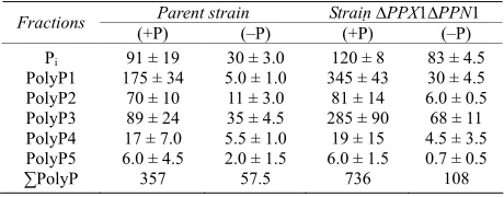
The amount of PolyP decreased in the cells of parent strain and mutant strain ∆PPX1∆PPN1without exopoly- phosphatases during cultivation in the (−P) medium ( Ta-ble 1). However, the content of PolyP in ∆PPX1∆PPN1
cells still higher than that in the parent cells. DAPI- stained cells of the parent strain under Pi-limitation had insignificant fluorescence (Figure 1(a)). It is in
agree-ment with the low level of PolyP. The cells of the mutant strain showed blue fluorescence of the nuclei and green fluorescence of the cytoplasm and the vacuole (Figure 2(a)). Green fluorescence may be a sum of DAPI self-
fluorescence (blue) and DAPI-PolyP-fluorescence (yel-low) in the case of cytoplasm and vacuoles and a sum of DAPI-DNA and DAPI-PolyP complexes fluorescence in the case of nuclei and mitochondria. The fluorescence of cytoplasm and vacuoles seems to result from the fact the
∆PPX1∆PPN1 cells grown in Pi-deficient medium con-tains 2-fold more PolyP than the cells of parent strain (Table 1).
After 30 min of cultivation in the (+P) medium (Table 2) the PolyP pools restored to the levels observed at the
stationary stage of growth in the (+P) medium (Tables 1
and 2). The cells of parent strain restored the PolyP pool
both under use of glucose and ethanol as energy sources. Ethanol did not provide PolyP accumulation in mutant
Table 1. The content of Pi and PolyP (µmol/g of dry biomass)
in the cells of S. cerevisiae at stationary growth stage in the YPD medium (+Р) (20 mM Рi) and YPD medium (–P) (0.05
mM Рi) with glucose. The average results of four experiments
are presented.
Parent strain Strain ∆PPX1∆PPN1 Fractions (+Р) (–P) (+Р) (–P)
Pi PolyP1 PolyP2 PolyP3 PolyP4 PolyP5
∑PolyP
91 ± 19 175 ± 34
70 ± 10 89 ± 24 17 ± 7.0 6.0 ± 4.5 357
30 ± 3.0 5.0 ± 1.0 11 ± 3.0 35 ± 4.5 5.5 ± 1.0 2.0 ± 1.5 57.5
120 ± 8 345 ± 43
81 ± 14 285 ± 90
19 ± 15 6.0 ± 1.5
736
83 ± 4.5 30 ± 4.5 6.0 ± 0.5 68 ± 11 4.5 ± 3.5 0.7 ± 0.5 108 *The work was supported by the Russian Foundation for Basic
Figure 1. The cells of S. cerevisiae (parent strain) stained by DAPI: left—transmission mode, right—fluorescent mode, (a) the cells grown in (−P) medium with glucose, (b) the cells grown for 30 min in (+P) medium with glucose, (c) the cells grown for 30 min in (+P) medium with ethanol. 1—nucleus, 2—vacuole, 3—cytoplasm, 4—mitochondrial network.
Table 2. The content of Pi and PolyP (µmol/g of dry biomass)
in the cells of S. cerevisiae after 30 min cultivation in (+P) medium (20 mM Рi) with glucose or ethanol. The average results
of four experiments are presented.
Parent strain Strain ∆PPX1∆PPN1 Fractions Glucose Ethanol Glucose Ethanol
Pi PolyP1 PolyP2 PolyP3 PolyP4 PolyP5
∑PolyP
59 ± 10 95 ± 11 69 ± 6.5 130 ± 12 13 ± 4 12 ± 3.0
318
46 ± 2.0 41 ± 3.0 72 ± 3.5 121 ± 14 35 ± 1.0 28 ± 13 343
87 ± 17 208 ± 9.5
89 ± 19 375 ± 40
48 ± 14 86 ± 4.5 806
115 ± 8 40 ± 0.8 11 ± 1.5 71 ± 7.5 4.6 ± 0.5 7.0 ± 0.5 133
cells (Table 2). In the parent strain, with ethanol used as
a carbon source, the content of acid-soluble fractions was lesser than that on glucose, while the content of PolyP3 and PolyP5 was higher. Such effect was observed earlier in the wild type strain [9].
The results of electrophoresis of PolyP accumulated
Figure 2. The cells of S. cerevisiae (strain with inactivated genes PPX1 and PPN1) stained by DAPI: left—transmission mode, right—fluorescent mode, (a) the cells grown in (–P) medium with glucose, (b) the cells grown for 30 min in (+P) medium with glucose, (c) the cells grown for 30 min in (+P) medium with ethanol. 1—nucleus, 2—vacuole, 3—cytoplasm.
during 30 min in the (+P) medium with glucose are shown in Figure 3. The average chain length of PolyP in
the mutant was greater compared to the parent strain, except for fraction PolyP4. This fraction is believed to be associated with the cell wall, and its metabolism is different from other fractions under study [1].
As whole, the ∆PPX1∆PPN1 cells accumulated more PolyP in the first 30 min than the cells of parent strain in the medium with glucose. It suggested that exopolyphos- phatases participate in PolyP metabolism at the first time of its biosynthesis, and biosynthesis and degradation of these polymers occur in parallel.
[image:3.595.58.286.546.635.2]Figure 3. Electrophoregram of PolyP in 20% polyacrylamide gel in the presence of 7 M urea. PolyP were extracted from the cells after 30 min cultivation in (+P) medium with glucose.
(Figure 1(b)). These granules, according to the literature
data, are fragments of the mitochondrial network [13]. Previously we have shown that under PolyP accumula-tion in glucose-containing medium the mitochondrial fraction accumulates PolyP [21]. It seems that the green fluorescence in this case is associated with formation of DAPI-mitochondrial DNA and DAPI-PolyP complexes. The green fluorescence of cytoplasm may be associated with accumulation of PolyP in this compartment.
A different picture was observed with ethanol used as a carbon source (Figure 2(c)). Mitochondria devoid of
PolyP [22] under these conditions fluoresced in the blue spectral region typical of DAPI-DNA complexes, along with the nuclei. The cytoplasm and the vacuole fluoresced to approximately the same extent in the green region of the spectrum (Figure 2(c)).
In the mutant strain, under PolyP accumulation in the glucose-containing medium, the green fluorescence of the cytoplasm and vacuoles increased and single areas of yellow fluorescence appeared (Figure 2(b)). The green
granules (parts of the mitochondrial network) were not observed. Probably, it was due to defectiveness of mito-chondria in this strain [23]. It should also be noted that mitochondria of the mutant strain contain relatively little PolyP at the early growth stage; PolyP in these organ-elles of mutant strain is accumulated later [23]. With ethanol, cell fluorescence was not different in intensity or character from that of the cells grown under
phos-phate deficiency (Figures 2(a) and (c)). It is in
agree-ment with the chemical extraction data showing that mutant cells cannot accumulate PolyP in the medium with ethanol as a carbon source. It would be interesting to note that blue fluorescence of the nuclei typical of DAPI-DNA complexes in the mutant strain is much more pronounced than in the parent strain. The nature of this phenomenon cannot be explained as yet; probably, the nuclear envelope permeability for the dye is different for different strains.
Earlier, we demonstrated that cytoplasm, mitochon-dria, vacuoles and nuclei of S. cerevisiae cells possesses PolyP by subcellular fractionation [24]. However the methods for obtaining of pure fractions of the organelles need long time. The in vivo DAPI staining is a simple method for vital observing of PolyP. The data obtained suggest, that PolyP localization at the initial stage of its synthesis in S. cerevisiae cells depends on the carbon source. PolyP appears first of all in the cytoplasm and mitochondria under cultivation in glucose-containing medium and in the cytoplasm and vacuoles in the me-dium with ethanol. In the mutant with inactivated genes encoding two exopolyphosphatases (PPX1 and PPN1) PolyP accumulates in the cytoplasm and vacuoles. The role of mitochondria in PolyP synthesis needs further investigation. Comparison of chemical extraction data (Table 2) and DAPI staining (Figures 1 and 2) shows
that staining character actually depends on PolyP content. However this dependence is not simple. Nevertheless, DAPI staining is useful for visualization of PolyP local-ization in living cells. The method of confocal micros-copy in vivo should be accompanied with chemical ex-traction of PolyP. The results show that at the early stage of PolyP biosynthesis after Pi starvation the polymer is immediately localized in different cell compartments, first of all in the cytoplasm. The vacuolar localization of PolyP is more typical of depleted (including Pi-depleted) media [10,25], while formation of DAPI-staining PolyP- containing granules in S. cerevisiae is observed at the stationary growth stage [14]. Commercial PolyP with average chain length of 25, 45, 75 phosphate residues from Sigma (USA) and with average chain length of 188 phosphate residues from Monsanto (USA) were used as markers.
The comparison of PolyP content and chain length at the early stage of accumulation after Pi deprivation sug-gest participation of exopolyphosphatases in PolyP me-tabolism at the first stage of its accumulation. The con-tent and chain lengths of the PolyP1 and PolyP3 frac-tions increased to the maximum under inactivation of the
exopoly-phosphatases, this degradation did not occur and the PolyP content and chain length generally increased.
4. ACKNOWLEDGEMENTS
We thank E. Makeeva for help in preparation of the manuscript.
REFERENCES
[1] Kulaev, I.S., Vagabov, V.M. and Kulakovskaya, T.V. (2004) The biochemistry of inorganic polyphosphates. John Wiley & Sons Ltd., Chichester.
doi:10.1002/0470858192
[2] Rao, N.N., Gomez-Garcia, M.R. and Kornberg, A. (2009) Inorganic polyphosphate: Essential for growth and sur-vival. Annual Review of Biochemistry, 78, 605-647. doi:10.1146/annurev.biochem.77.083007.093039 [3] Docampo, R., Ulrich, P. and Moreno, S.N. (2010)
Evolu-tion of acidocalcisomes and their role in polyphosphate storage and osmoregulation in eukaryotic microbes. Phi-losophical Transactions of the Royal Society B: Biologi-cal Sciences, 365, 775-784. doi:10.1098/rstb.2009.0179 [4] Chaves, F.P., Lunsdorf, H.L. and Jerez, C.A. (2004)
Growth of polychlorinated-biphenyl-degrading bacteria in the presence of biphenyl and chlorobiphenyls gener-ates oxydative stress and massive accumulation of inor-ganic polyphosphate. Applied and Environmental Micro-biology, 70, 3064-3072.
doi:10.1128/AEM.70.5.3064-3072.2004
[5] Campos, E., Façanha, A., Moraes, J. et al. (2007) A mi-tochondrial exopolyphosphatase activity modulated by phosphate demand in Rhipicephalus (Boophilus) micro-plus embryo. Insect Biochemistry and Molecular Biology, 37, 1103-1107. doi:10.1016/j.ibmb.2007.06.003
[6] Gebremariam, S.Y., Beutel, M.W., Christian, D. and Hess, T.F. (2011) Research advances and challenges in the mi-crobiology of enhanced biological phosphorus removal-a critical review. Water Environment Research, 83, 195- 219. doi:10.2175/106143010X12780288628534
[7] Omelon, S.J. and Grynpas, M.D. (2008) Relationships between polyphosphate chemistry, biochemistry and apa-tite biomineralization. Chemical Reviews, 108, 4694- 4715. doi:10.1021/cr0782527
[8] Vagabov, V.M., Trilisenko, L.V. and Kulaev, I.S. (2000) Dependence of inorganic polyphosphate chain length on the orthophosphate content in the culture medium of the yeast Saccharomyces cerevisiae. Biochemistry, 65, 349- 355.
[9] Vagabov, V.M., Trilisenko, L.V., Kulakovskaya, T.V., et al. (2008) Еffect of carbon source on polyphosphate ac-cumulation in Saccharomyces cerevisiae. FEMS Yeast Research, 8, 877-882.
doi:10.1111/j.1567-1364.2008.00420.x
[10] Allan, R.A. and Miller, J.J. (1980) Influence of S-ade- nosylmethionine on DAPI-induced fluorescence of poly- phosphate in the yeast vacuole. Canadian Journal of Mi-crobiology, 26, 912-920. doi:10.1139/m80-158
[11] Tijssen, J.P.F., Beekes, H.W. and Van Steveninck, J. (1982) Localization of polyphosphate in Saccharomyces fragilis, as revealed by 4’6’-diamidino-2-phenylindole fluorescence. Biochimica et Biophysica Acta, 721, 394-
398. doi:10.1016/0167-4889(82)90094-5
[12] Serafim, L.S., Lemos, O.C., Levantesi, C., et al. (2002) Methods for detection and visualization of intracellular polymers stored by polyphosphate-accumulating micro-organisms. Journal of Microbiological Methods, 51, 1- 18. doi:10.1016/S0167-7012(02)00056-8
[13] Puchkov, E.O. (2006) The viability assessment of etha-nol-producing yeast by computer-aided fluorescence mi-croscopy. Mikrobiologiia, 75, 193-200.
[14] Puchkov, E.O. (2010) Brownian motion of polyphos-phate complexes in yeast vacuoles:characterization by fluorescence microscopy with image analysis. Yeast, 27, 309-315.
[15] Wurst, H., Shiba, T. and Kornberg, A. (1995) The gene for a major exopolyphosphatase of Saccharomyces cere-visiae. Journal of Bacteriology, 177, 898-906.
[16] Sethuraman, A., Rao, N.N. and Kornberg, A. (2001) The endopolyphosphatase gene: Essential in Saccharomyces cerevisiae. Proceedings of the National Academy of Sci-ences, 98, 8542-8547. doi:10.1073/pnas.151269398 [17] Lichko, L.P., Andreeva, N.A., Kulakovskaya, T.V., et al.
(2003) Exopolyphosphatases of the yeast Saccharomyces cerevisiae. FEMS Yeast Research, 3, 233-238.
doi:10.1016/S1567-1356(02)00205-2
[18] Andreeva, N.A., Kulakovskaya, T.V. and Kulaev, I.S. (2006) High molecular mass exopolyphosphatase from the cytosol of the yeast Saccharomyces cerevisiae is en-coded by the PPN1 gene. Biochemistry, 71, 975-977. [19] Rubin, G.M. (1973) The nucleotide sequence of
Sac-charomyces cerevisiae 5.8 S ribosomal ribonucleic acid. Journal of Biological Chemistry, 11, 3860-3875.
[20] Kulakovskaya, T.V., Andreeva, N.A., Karpov, A.V., et al. (1999) Hydrolysis of tripolyphosphate by purified exopo- lyphosphatase of Saccharomyces cerevisiae cytosol: Ki-netic model. Biochemistry, 64, 990-993.
[21] Pestov, N.A., Kulakovskaya, T.V. and Kulaev, I.S. (2004) Inorganic polyphosphate in mitochondria of Saccharo-myces cerevisiae at phosphate limitation and phosphate excess. FEMS Yeast Research, 4, 643-648.
doi:10.1016/j.femsyr.2003.12.008
[22] Andreeva, N.A., Kulakovskaya, T.V., Kulakovskaya, E.V., et al. (2008) Polyphosphates and exopolyphosphatases in cytosol and mitochondria of Saccharomyces cerevisiae during growth on glucose or ethanol under phosphate surplus. Biochemistry, 73, 65-69.
[23] Pestov, N.A., Kulakovskaya, T.V. and Kulaev I.S. (2005) Effects of inactivation of the PPN1 gene on exopoly-phosphatases, inorganic polyphosphates and function of mitochondria in the yeast Saccharomyces cerevisiae. FEMS Yeast Research, 5, 823-828.
doi:10.1016/j.femsyr.2005.03.002
[24] Lichko, L., Kulakovskaya, T., Pestov, N. and Kulaev, I. (2006) Inorganic polyphosphates and exopolyphosphata-ses in cell compartments of the yeast Saccharomyces cerevisiae under inactivation of PPX1 and PPN1 genes. Bioscience Reports, 26, 45-54.
doi:10.1007/s10540-006-9003-2