https://www.scirp.org/journal/ojpathology ISSN Online: 2164-6783
ISSN Print: 2164-6775
DOI: 10.4236/ojpathology.2019.94010 Sep. 30, 2019 86 Open Journal of Pathology
Metastatic Breast Cancer Survival
in Pointe Noire: Analysis of 30 Cases
C. F. S. Ngatali
1*, A. F. Bolenga Liboko
2, M. L. Eouani
3, L. M. A. Boumba
4, P. E. G. Sounga Bandzouzi
5,
E. Ndounga
2, Y. Mabila
2, D. Moukassa
6, J. B. Nkoua-Mbon
21Department of Oncology and Internal Medicine, Loandjili General Hospital, Pointe Noire, Congo 2Medical Oncology Department, CHU-Brazzaville, Brazzaville, Congo
3Department of Gynecology, Loandjili General Hospital, Pointe Noire, Congo 4Laboratoire d’Anatomie Pathologie, Loandjili General Hospital, Pointe Noire, Congo 5Departement of Neurology, Loandjili General Hospital, Pointe Noire, Congo 6Hospital General Edith Lucie Bongo, Oyo, Congo
Abstract
Introduction: Breast cancer is the leading cancer and the leading cause of cancer death in women worldwide. About 5% to 10% of breast cancer pa-tients present with metastases. While the 5-year survival of papa-tients with local breast cancer varies around 98.8%, this survival rate drops to around 26.3% for metastatic patients. The objective of this study was to determine the sur-vival of patients with metastatic breast cancer in resource-limited settings. Patients and Methods: This was a cross-sectional descriptive study that took place in the Cancer Department of the General Hospital of Loandjili in Pointe Noire during the period from January 1, 2013 to December 31, 2018, for du-ration of 6 years. 30 records of patients over 18 years of age and with histo-logical evidence who received at least 3 courses of chemotherapy were col-lected. The variables studied were: age, level of education, socio-economic level, menopausal status, history, WHO status, menopausal status, tumor size, histological type, tumor location, the type of treatment and survival. Survival was calculated by Kaplan Meier method. Fisher’s exact test was used to search for correlation between variables. Results: The average age was 52.62 ± 10.96 years old. The extremes were 33 years and 75 years old. The most represented level of education was the primary level in 67% of cases. The majority of pa-tients had low socioeconomic status in 50% of cases. The papa-tients were me-nopausal in 57% of cases. The antecedents of cancer were present in 13% of cases. 50% of patients had a WHO status performance at 2. The tumor size was greater than 2 cm in 77% of cases. The most represented histological type was invasive ductal carcinoma in 93% of cases. The most represented histo-logical grade was Scharff grade III Richardson bloom in 80% of cases. The How to cite this paper: Ngatali, C.F.S.,
Liboko, A.F.B., Eouani, M.L., Boumba, L.M.A., Bandzouzi, P.E.G.S., Ndounga, E., Mabila, Y., Moukassa, D. and Nkoua-Mbon, J.B. (2019) Metastatic Breast Cancer Survival in Pointe Noire: Analysis of 30 Cases. Open Journal of Pathology, 9, 86-99.
https://doi.org/10.4236/ojpathology.2019.94 010
Received: July 5, 2019 Accepted: September 27, 2019 Published: September 30, 2019
Copyright © 2019 by author(s) and Scientific Research Publishing Inc. This work is licensed under the Creative Commons Attribution-NonCommercial International License (CC BY-NC 4.0). http://creativecommons.org/licenses/by-nc/4.0/
DOI: 10.4236/ojpathology.2019.94010 87 Open Journal of Pathology
most represented metastatic localization was pulmonary in 33% of cases. The metastatic localizations were unique in 47% of cases and multiple in 53% of cases. Anthracycline-based chemotherapy was more used in 53% of cases. Bi-variate analysis revealed a correlation between tumor size and number of metastases, p < 0.05. The mean patient follow-up time was 22 ± 15.45 months. The median overall survival was 35.35 months. Brain metastases (18.2 months) had a poor prognosis compared to liver metastases (25.4 months). The median survival of pulmonary metastases was 36.5 months, p > 0.05. Patients treated with anthracyclines were greater than that of patients treated with taxanes in combination was 26.48 months, p > 0.05. Conclusion: Metastatic breast cancer remains an incurable disease, its survival remains low despite diagnostic and therapeutic advances that remain difficult to access for our resource-poor developing countries. Patients are treated with conven-tional chemotherapy (anthracyclines and taxanes). The most common me-tastases are respectively pulmonary, hepatic and cerebral in our context.
Keywords
Breast Cancer, Metastatic, Survival, Pointe Noire, Congo
1. Introduction
DOI: 10.4236/ojpathology.2019.94010 88 Open Journal of Pathology
2. Patients and Methods
This was a cross-sectional descriptive study that took place in the Cancer De-partment of the General Hospital of Loandjili in Pointe Noire during the period from January 1, 2013 to December 31, 2018, for duration of 6 years. Have been included in our study: all patients over 18 years old; all patients with a complete file that is to say with a histological diagnosis and an extension assessment per-formed through an abdominal chest CT scan and/or an ultrasound or chest x-ray; all patients diagnosed with primary or de novo breast cancer (synchronous) and secondary or metachronous breast cancer; all patients who received at least 3 courses of chemotherapy [chemotherapy was based on anthracyclines (FAC protocol = 5 fluorouracil, dose 500 mg/m2, doxorubicin 50 mg/m2
cyclophospha-mide 500 mg/m2) and taxanes (docetaxel protocol dose 100 mg/m2)]; all patients
with no histological diagnosis were excluded from our study; all men with breast cancer. The variables studied were: Sociodemographic parameters: age, level of education, socio-economic level; Clinical parameters: WHO status performance, menopausal status, tumor size, number of lymph node involvement, metastatic localization of metastasis, number of metastases; Histological type and histolog-ical grade; The type of treatment; The survival. The data collection was done from a survey sheet, comprising the different variables studied.
Bivariate analysis was done between size and number of metastasis. The data entry was made from the Excel version 2016 software. Qualitative variables were represented in numbers and percentages. Quantitative variables were represented in numbers and on average. Statistical analysis and data processing were per-formed by the Excel 2016 software and graphpad Prism version 7 software. The statistical test used was Fisher’s exact test for finding correlation between va-riables. Survival was calculated by Kaplan Meier method. The initial date was the date of diagnosis of metastasis; the point date was the end date of the study. The final event was the occurrence of death. Patients were followed throughout the study period, from January 1, 2013 to December 31, 2018, for a period of 6 years. The comparison of the curves was made by the logrank test. The results were statistically significant for p < 5%.
3. Results
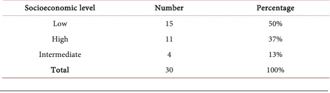
At the end of our study, 30 files of metastatic breast cancer patients fulfilling the criteria of inclusions were collected. The average age was 52.62 ± 10.96 years old. The extremes were 33 years and 75 years old. The most represented age group was the age group from 53 to 62 in 33%, followed by the age group from 43 to 52 (Table 1). The highest level of education was the primary level in 67% of cases followed by the higher level of education in 20% and the secondary level in 13% of cases (Table 2). The majority of patients had a low socio-economic level in 50% of cases; the intermediate socio-economic level and higher were represented in 37% and 17% of cases respectively (Table 3). Patients were menopausal status in 57% of cases (Table 4). We found a history of cancer in 13% of cases (Table 5).
DOI: 10.4236/ojpathology.2019.94010 89 Open Journal of Pathology
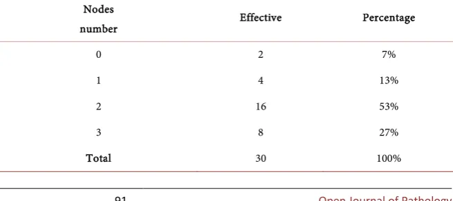
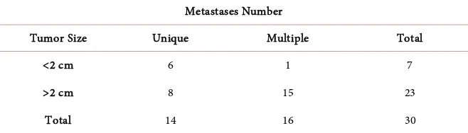
performance at 1, 7% of patients had at 0 and 3% of patients had at 1 (Table 6). Tumor size was greater than 2 cm in 77% of cases and was less than 2 cm in 23% of cases (Table 7). The patients had respectively two nodes invaded in 53% of cases and 3 nodes in 27% of cases (Table 8). The most represented histological type was invasive ductal carcinoma in 87% of cases, lobular carcinoma in 10% of cases and a 3% breast sarcoma (Figure 1). The most represented histological grade was grade III of Scharff bloom Richardson in 80% of cases followed re-spectively by grade II and grade I with respective percentages of 10% and 5%. Metastatic localization the most represented was that of the lungs in 33% of cas-es (Table 9). The metastatic localization was unique in 47% of cases and mul-tiple in 53% of cases (Table 10). The most widely used chemotherapies were chemotherapy-based anthracyclines in 53% and taxan-based chemotherapy alone or in combination with anthracyclines in 34% of cases. Only one patient had received targeted therapy (Table 11). Bivariate analysis found a correlation between tumor size and number of metastases, the result was statistically signif-icant (Table 12). The mean patient follow-up time was 22 ± 15.45 months. The median overall survival was 35.35 months (Figure 2). The median survival of patients with lung, liver and brain metastases, with the lungs and liver (lungs + liver) associated with the lungs and lymph nodes (lungs + lymph nodes), was respectively 36.5 months, 25.4 months, 18.26 months, 24.3 months, 37.5 months. There was no statistically significant difference (Figure 3). The survival of pa-tients treated with anthracyclines was 36.35 months that of Papa-tients treated with Texan was 26.48 months, the result was not statistically significant (Figure 4).
Figure 1. Distribution of patients by histological type.
Figure 2. Representation of the survival curve of patients with metastatic breast cancer.
87%
10% 3%
DOI: 10.4236/ojpathology.2019.94010 90 Open Journal of Pathology
[image:5.595.245.458.224.332.2]Figure 3. Comparison of survival curves versus metastatic localization. P > 0.05 statistically insignificant.
[image:5.595.209.540.397.506.2]Figure 4. Comparison of survival curves by type of treatment. P > 0.05 non significant results.
Table 1. Distribution of patients according to group age.
Group age Number Percentage
33 - 42 5 17%
43 - 52 9 30%
53 - 62 10 33%
63 - 72 4 13%
73 - 82 2 7%
Total 30 100%
Table 2. Distribition of patients according to study level.
Study level Number Percentage
Primairy 20 67%
Secondary 4 13%
Superior 6 20%
Total 30 100%
Table 3. Distribution of patients according to socioeconomic level.
Socioeconomic level Number Percentage
Low 15 50%
High 11 37%
Intermediate 4 13%
[image:5.595.210.539.536.616.2] [image:5.595.207.539.646.740.2]DOI: 10.4236/ojpathology.2019.94010 91 Open Journal of Pathology
Table 4. Distribution of patients according to menopausal status.
Menopausal
status Number Percentage
No 13 43%
yes 17 57%
[image:6.595.209.539.209.293.2]Total 30 100%
Table 5. Distribution of patients according to cancer histoty. Cancer
history Number Percentage
No 26 87%
yes 4 13%
[image:6.595.210.538.327.449.2]Total 30 100%
Table 6. Distribution of patients according to performance status of WHO.
Performance
status Number Percentage
0 2 7%
1 12 40%
2 15 50%
3 1 3%
Total 30 100%
Table 7. Distribution of patients according to tumor size.
Tumor
size Number Percentage
<2 cm 7 23%
>2 cm 23 77%
Total 30 100%
Table 8. Distribution of patients according to the number of invade nodes.
Nodes
number Effective Percentage
0 2 7%
1 4 13%
2 16 53%
3 8 27%
[image:6.595.210.540.483.568.2] [image:6.595.210.538.601.747.2]DOI: 10.4236/ojpathology.2019.94010 92 Open Journal of Pathology
Table 9. Distribution of patients according to metastatic localization.
Metastatic localization Number Percentage
bone + foie 2 7%
brain 2 7%
liver 2 7%
lung 10 33%
lung + node + rachis 3 10%
lung + liver 5 17%
lung + nodes 2 7%
Lung + bone + liver 4 13%
[image:7.595.209.539.302.370.2]Total 30 100%
Table 10. Distribution of patients according to number of metastases.
Metastases number Number Percentage
Multiple 16 53%
Unique 14 47%
Total 30 100%
Table 11. Distribution of patients according to treatment.
Chemotherapy type Number Percentage
Fac 17 53%
fac + taxotere 11 34%
taxotere 2 6%
taxotere + avastin 1 3%
capecitabine 1 3%
Total 32 100%
Table 12. Distribution of patients according to tumor size and number of metastases
Metastases Number
Tumor Size Unique Multiple Total
<2 cm 6 1 7
>2 cm 8 15 23
Total 14 16 30
P < 0.05 results statistically significant.
[image:7.595.208.539.402.528.2] [image:7.595.209.538.559.653.2]DOI: 10.4236/ojpathology.2019.94010 93 Open Journal of Pathology
4. Discussion
DOI: 10.4236/ojpathology.2019.94010 94 Open Journal of Pathology
in 87% of cases. This result corroborates to the literature [5] [18] [24] [25]. The risk of developing metastases according to the histological type is practically the same, which explains the same treatment between invasive ductal carcinoma and invasive lobular carcinoma [12]. In our study, visceral metastases were most represented by pulmonary metastases in 30% of cases and hepatic metastases in 10% of cases. Bone metastases were represented in association with other metas-tases in two cases. These results do not corroborate with those of some authors of the literature. Indeed several authors have described the predominance of bone metastases in their series [5] [7] [20], this could probably be explained by an underestimation of bone metastases due to the lack of other balance sheet examinations, such as bone scans and pet scan that are not available in our li-mited resource context. On the other hand, one study reported the same pul-monary prevalence as in our study [25]. The multiple metastases were the most represented in our study contrary to the literature that reported predominance. Metastatic breast cancer is incurable, under these conditions the goal of treat-ment is the improvetreat-ment of the quality of life and the prolongation of survival. Nowadays the advent of targeted therapies and immunotherapy combined with conventional chemotherapy has helped to achieve the goal of these treatments that of improving the quality of life and prolonging survival. Thus, in our study, patients received anthracycline chemotherapy in 53% of cases, continuously or sequentially associated with taxanes in 34% of cases. These results corroborate those of the literature [5] [20] [25]. The absence of health insurance, the high cost of targeted therapies was a limiting factor in the use of these, so only one patient was able to benefit from targeted therapies. The absence of immunohis-tochemistry for the detection of hormone receptors made it impossible to use hormonal treatment. In our study we observed a positive correlation between tumor size and number of metastases. The number of metastases increased with tumor size. Size is a prognostic factor for breast cancer.
DOI: 10.4236/ojpathology.2019.94010 95 Open Journal of Pathology
36.5 months, 18.2 months.
Brain metastases were those with the lowest median survival. Indeed, brain metastases are difficult to access to cytotoxic treatments because of the blood- brain barrier that prevents the passage of anticancer drugs. Moreover, no patient has been able to benefit from a specific local treatment (surgery and radiothera-py). The liver is a common site of metastasis of breast cancer, with bone and lung [30] [31] [32]. Liver metastases in breast cancer patients are a prognostic factor independent of other risk factors [15] [33], since the median survival of patients with breast cancer with hepatic metastasis varies from 4.8 to 15 months [34] [35] [36] [37] [38]. In contrast, breast cancer patients with lung or bone metastases have median survival rates of 9 to 27.4 months [37] [38] and 16.3 to 56 months [37] [39] [40], respectively. The survival of breast cancer patients with cerebral metastases was estimated in the literature between 4 and 16 months [41] [42] [43] [44]. This rate is close to that of our study which was 18.6 months. The median overall survival of anthracycline-treated patients was 36.35 months, while that of taxane and anthracycline-treated patients was 26.48 months. These results were statistically insignificant. In the literature, associa-tions with texanes are superior to those without taxanes [45].
5. Conclusion
Metastatic breast cancer remains an incurable disease, the goal of its treatment is to prolong survival and improve quality of life. Despite the limitations of our study, the survival of patients with metastatic breast cancer remains low. The various advances in diagnosis and therapeutics of recent years are currently dif-ficult to access in developing countries with limited resources. Anthracyc-line-based chemotherapy remains the basis of treatment. The most frequent me-tastatic sites are pulmonary, hepatic and cerebral. However, studies with a larger sample are needed to support its observations in our context with limited re-sources.
Conflicts of Interest
The authors declare no conflicts of interest regarding the publication of this pa-per.
References
[1] Jemal, A., Bray, F., Center, M.M., et al. (2011) Global Cancer Statistics. CA: A Cancer Journal for Clinicians, 61, 69-90.https://doi.org/10.3322/caac.20107
[2] Bray, F., Ferlay, J., Soerjomataram, I., Siegel, R.L., Torre, L.A. and Jemal, A. (2018) Global Cancer Statistics: GLOBOCAN Estimates of Incidence and Mortality Worldwide for 36 Cancers in 185 Countries. CA: A Cancer Journal for Clinicians, 68, 394-424. https://doi.org/10.3322/caac.21492
DOI: 10.4236/ojpathology.2019.94010 96 Open Journal of Pathology
https://doi.org/10.1056/NEJMsr1504363
[4] Dafni, U., Grimani, I., Xyrafas, A., Eleftheraki, A.G. and Fountzilas, G. (2010) Fif-teen-Year Trends in Metastatic Breast Cancer Survival in Greece. Breast Cancer Re-search and Treatment, 119, 621-631. https://doi.org/10.1007/s10549-009-0630-8
[5] Barinoff, J., Heitz, F., Kuemmel, S., Dittmer, C., Hils, R., Lorenz-Salehi, F., Traut, A. and Bois, A. (2013) Improvement of Survival in Patient with Primary Metastatic Breast Cancer over a 10-Year Periode: Prospective Analyses Based on Individual Pa-tient Date from a Multicenter Data Bank. Journal of Cancer Therapy, 4, 1306-1312.
https://doi.org/10.4236/jct.2013.48154
[6] Kotsakis, A., Ardavanis, A., Koumakis, G., Samanthas, E., Psyrri, A. and Papadimi-triou, C. (2019) Epidemiological Characteristics, Clinical Outcomes and Manage-ment Patterns of Metastatic Breast Cancer Patients in Routine Clinical Care Settings of Greece: Results from the EMERGE Multicenter Retrospective Chart Review Study.
BMC Cancer, 19, 88.https://doi.org/10.1186/s12885-019-5301-5
[7] Dia, J., Saki, C., Mouhideen, O., Bohoussou, E., Touré, M., Okon, G., Guié, P. and Anongba, S. (2017) Epidemiological Aspects of Metastatic Relapse of Breast Cancer in an African Context. Open Journal of Obstetrics and Gynecology, 7, 552-561.
https://doi.org/10.4236/ojog.2017.75058
[8] Smigal, C., Jemal, A., Ward, E., et al. (2006) Trends in Breast Cancer by Race and Ethnicity: Update 2006. CA: A Cancer Journal for Clinicians, 56, 168-183.
https://doi.org/10.3322/canjclin.56.3.168
[9] Brewster, A.M., Hortobagi, G.N., Brogilo, K.R., et al. (2008) Residual Risk of Breast Cancer Recurrence 5 Years after Adjuvant Therapy. Journal of the National Cancer Institute, 100, 1179-1183. https://doi.org/10.1093/jnci/djn233
[10] Schon, D., Bertz, J., Gorsch, B., Haberland, J. and Kurth, B.-M. (2004) Die Dachdo- kumentation Krebs. Gesundheitsbl-Gesundheitsforsch-Gesundheitsschutz, 47, 429-436.
https://doi.org/10.1007/s00103-004-0830-7
[11] Surveillance Epidemiology and End Results Program (2005) 1973–2002. Division of Cancer Control and Population Science, National Cancer Institute.
https://seer.cancer.gov
[12] Rugo, H.S., Majure, M., Dragun, A., Buxton, M. and Esserman, L. (2017) Neop-lasms of Breast. In: Bast Jr., R.C., et al., Eds., Holland-Frei Cancer Medicine, 9th Edition,Wiley-Blackwell, Hoboken, NJ, 1368-1438.
[13] Gnerlich, J., Jeffe, D.B., Deshpande, A.D., Beers, C., Zander, C. and Margenthaler, J.A. (2007) Surgical Removal of the Primary Tumor Increases Overal Survival in pa-tients with Metastatic Breast Cancer: Analysis of the 1998-2003 SEER Data. Annals of Surgical Oncology, 14, 2187-2194.
https://doi.org/10.1245/s10434-007-9438-0
[14] Lu, J., Steeg, P.S., Price, J.E., Krishnamurthy, S., Mani, S.A., Reuben, J., et al. (2009) Breast Cancer Metastasis: Challenges and Opportunities. Cancer Research, 69, 4951- 4953. https://doi.org/10.1158/0008-5472.CAN-09-0099
[15] Bonotto, M., Gerratana, L., Poletto, E., Driol, P., Giangreco, M., Russo, S., et al. (2014) Measures of Outcome in Metastatic Breast Cancer: Insights from a Real-World Scenario. Oncologist, 19, 608-615. https://doi.org/10.1634/theoncologist.2014-0002
[16] Chevallier, B., Heintzman, F. and Mosseri, V. (1989) Quels sont les facteurs pronostics du cancer du sein opérable sans envahissement ganglionnaire axillaire histologique? Résultats d’une analyse uni et multifactorielle. Bulletin du Cancer, 76, 51-60. [17] De La Rochefordière, A., Asslain, B. and Campana, F. (1993) Age Prognostic Factor
DOI: 10.4236/ojpathology.2019.94010 97 Open Journal of Pathology
https://doi.org/10.1016/0140-6736(93)92407-K
[18] Djanhan, L.E., Dia, J.M., Mian, B., Oyelade, M., Diallo, A., Guié, P. and Anongba, S. (2017) Occurrence Delays of Metastatic Relapses of Breast Cancers Treated at Uni-versity Hospital of Treichville (Abidjan-Cote d’Ivoire). Journal of Cancer Therapy, 8, 924-932. https://doi.org/10.4236/jct.2017.811081
[19] Toure, M., Nguessan, E., Bambara, A.T., Kouassi, Y.K.K., Dia, J.M.L. and Adoubi, I. (2013) Factors Linked to Late Diagnosis in Breast Cancer in Sub-Saharan Africa: Case of Cote D’Ivoire. Gynécologie Obstétrique et Fertilité, 41, 696-700.
https://doi.org/10.1016/j.gyobfe.2013.08.019
[20] Korpela, J., Mali, P., Kaljonen, A. and Salminen, E. (2011) Quality of Life of Patients with Metastatic Breast Cancer Treated with Epirubicin and Docetaxel. International Journal of Clinical Medicine, 2, 346-351. https://doi.org/10.4236/ijcm.2011.23060
[21] Rosen, P.P., Groshen, S., Kinne, D.W. and Norton, L. (1993)Factors Influencing Prognosis in Node Negative Breast Carcinoma: Analysis of 767 T1N0M0/T2N0M0 Patients with Long Term Follow-Up. Journal of Clinical Oncology, 11, 2090-2100.
https://doi.org/10.1200/JCO.1993.11.11.2090
[22] Hanrahan, E.O., Gonzalez-Angulo, A.M., Giordano, S.H., et al. (2007) Overall Sur-vival and Cause-Specific Mortality of Patients with Stage T1a,bN0M0 Breast Carci-noma. Journal of Clinical Oncology, 25, 4952-4960.
https://doi.org/10.1200/JCO.2006.08.0499
[23] Nemoto, T., Vana, J., Bedwani, R.N., etal. (1980) Management and Survival of Fe-male Breast Cancer: Results of a National Survey by the American College of Surge-ons. Cancer, 45, 2917-2924.
https://doi.org/10.1002/1097-0142(19800615)45:12<2917::AID-CNCR2820451203> 3.0.CO;2-M
[24] Hammoud, H., Saleh, J., Bachour, M. and Salamoon, M. (2014) Serum Caspase-3 and Caspase-7 as Predictive Factors of Response in Locally Advanced and Metastat-ic Breast Carcinoma. Journal of Cancer Therapy, 5, 584-590.
https://doi.org/10.4236/jct.2014.56067
[25] Gogia, A., Deo, S.V.S., Sharma, D., Thulkar, S., Kumar, R., Malik, P.S. and Mathur, S. (2019) Clinicopathologic Characteristics and Treatment Outcomes of Patients with up-Front Metastatic Breast Cancer: Single-Center Experience in India. Journal of Global Oncology, 5,1-9. https://doi.org/10.1200/JGO.18.00265
[26] National Comprehensive Cancer Network (2016) NCCN Clinical Practice Guide-lines in Oncology: Breast Cancer, Version 2. http://www.nccn.com
[27] Cardoso, F., Costa, A., Senkus, E., Aapro, M., André, F., Barrios, C.H., et al. (2017) 3rd ESO-ESMO International Consensus Guidelines for Advanced Breast Cancer (ABC 3). Annals of Oncology, 28, 16-33.
[28] Colombo, P.E., Milanezi, F., Weigelt, B. and Reis-Filho, J.S. (2011) Microarrays in the 2010s: The Contribution of Microarray-Based Gene Expression Profiling to Breast Cancer Classification, Prognostication and Prediction. Breast Cancer Re-search, 13, 212. https://doi.org/10.1186/bcr2890
[29] Parise, C.A. and Caggiano, V. (2014) Breast Cancer Survival Defined by the ER/PR/HER2 Subtypes and a Surrogate Classification According to Tumor Grade and Immunohistochemical Biomarkers. Journal of Cancer Epidemiology, 2014, Ar-ticle ID: 469251. https://doi.org/10.1155/2014/469251
DOI: 10.4236/ojpathology.2019.94010 98 Open Journal of Pathology [31] Berman, A.T., Thukral, A.D., Hwang, W.T., Solin, L.J. and Vapiwala, N. (2013) In-cidence and Patterns of Distant Metastases for Patients with Early-Stage Breast Cancer after Breast Conservation Treatment. Clinical Breast Cancer, 13, 88-94.
https://doi.org/10.1016/j.clbc.2012.11.001
[32] Savci-Heijink, C.D., et al. (2015) Retrospective Analysis of Metastatic Behaviour of Breast Cancer Subtypes. Breast Cancer Research and Treatment, 150, 547-557.
https://doi.org/10.1007/s10549-015-3352-0
[33] Gerratana, L., et al. (2015) Pattern of Metastasis and Outcome in Patients with Breast Cancer. Clinical & Experimental Metastasis, 32, 125-133.
https://doi.org/10.1007/s10585-015-9697-2
[34] Wyld, L., et al. (2003) Prognostic Factors for Patients with Hepatic Metastases from Breast Cancer.British Journal of Cancer, 89, 284-290.
https://doi.org/10.1038/sj.bjc.6601038
[35] Tarhan, M.O., et al. (2013) The Clinicopathological Evaluation of the Breast Cancer Patients with Brain Metastases: Predictors of Survival. Clinical & Experimental Metastasis, 30, 201-213.https://doi.org/10.1007/s10585-012-9528-7
[36] Tseng, L.M., et al. (2013) Distant Metastasis in Triple-Negative Breast Cancer. Neo- plasma, 60, 290-294. https://doi.org/10.4149/neo_2013_038
[37] Liu, X.H., Man, Y.N., Cao, R., Liu, C. and Wu, X.Z. (2014) Individualized Chemo-therapy Based on Organ Selectivity: A Retrospective Study of Vinorelbine and Cape-citabine for Patients with Metastatic Breast Cancer. Current Medical Research and Opinion, 30, 1017-1024. https://doi.org/10.1185/03007995.2014.895310
[38] Ahn, S.G., et al. (2013) Prognostic Factors for Patients with Bone-Only Metastasis in Breast Cancer. Yonsei Medical Journal, 54, 1168-1177.
https://doi.org/10.3349/ymj.2013.54.5.1168
[39] Purushotham, A., et al. (2014) Age at Diagnosis and Distant Metastasis in Breast Cancer—A Surprising Inverse Relationship. European Journal of Cancer, 50, 1697-1705.
https://doi.org/10.1016/j.ejca.2014.04.002
[40] Karimi, A., Delpisheh, A., Sayehmiri, K., Saboori, H. and Rahimi, E. (2014) Predic-tive Factors of Survival Time of Breast Cancer in Kurdistan Province of Iran be-tween 2006-2014: A Cox Regression Approach. Asian Pacific Journal of Cancer Prevention, 15, 8483-8488. https://doi.org/10.7314/APJCP.2014.15.19.8483
[41] Bourdeanu, L., Chen, L. and Luu, T. (2017) RetrospectiveReview for Prevalence and Survival in MetastaticBreast Cancer with Brain Metastasis in Two Patient Cohorts: One Collected 2000-2005 and the Second Collected 2006-2011. Journal of Cancer Therapy, 8, 490-505. https://doi.org/10.4236/jct.2017.85042
[42] Niikura, N., Hayashi, N., Masuda, N., Takashima, S., Nakamura, R., Watanabe, K.,
et al. (2014) Treatment Outcomes and Prognostic Factors for Patients with Brain Metastases from Breast Cancer of Each Subtype: A Multicenter Retrospective Anal-ysis. Breast Cancer Research and Treatment, 147, 103-112.
https://doi.org/10.1007/s10549-014-3090-8
[43] Wadasadawala, T., Gupta, S., Bagul, V. and Patil, N. (2007) Brain Metastases from Breast Cancer: Management Approach. Journal of Cancer Research and Therapeutics, 3, 157-165. https://doi.org/10.4103/0973-1482.37409
[44] Ushio, A., Sawaki, M., Fujia, T., Hattori, M., Kondo, N., Horio, A., Gondou, N., Iwata, H., et al. (2012) Clinicopathological Analysis of Breast Cancer Patients with Brain Metastases. CancerResearch, 72, P3-12-06-P3-12-06.