Patients’ specific simulations of coronary fluxes in case of
three-vessel disease
Mahmoud Maasrani1, Issam Abouliatim2,3, Majid Harmouche2,3, Jean-Philippe Verhoye2,3, Hervé Corbineau2,3, Agnès Drochon4
1Faculty of Sciences, Lebanese University, Tripoli, Lebanon;
2Department of Thoracic and Cardiovascular Surgery, Rennes Hospital Center, Rennes, France; 3Research Unit INSERM U642, Rennes, France;
4University of Technology of Compiègne, Compiègne, France. Email: agnes.drochon@utc.fr
Received 2 November 2010; revised 4 November 2010; accepted 8 November 2010.
ABSTRACT
In this work, we propose a model of the coronary circulation based on hydraulic/electric analogy. This model aims to provide quantitative estimations of the distribution of flows and pressures across the coro-nary network for patients with stenoses of the left main coronary artery (LMCA), left anterior de-scending artery (LAD) and left circumflex branch (LCx), and chronic occlusion of the right coronary artery (RCA), undergoing off-pump coronary sur-gery. The results of the simulations are presented for 10 patients with various stenoses grades and collat-eral supply. For each patient, the four revasculariza-tion situarevasculariza-tions (no graft operating, pathological situa-tion (0G); right graft only (1G), left grafts only (2G), complete revascularization (3G)) are considered. It is shown that: 1) the complete revascularization is fully justified for these patients because neither the right graft alone, nor the left grafts alone can ensure a suf-ficient perfusion improvement for the heart; 2) the capillary and collateral resistances (and the propor-tion between them) have a major impact on the flows and pressures everywhere in the network; 3) in the presence of the left grafts, the flows in the native stenosed arteries become low and this could promote the development of the native disease in these branches.
Keywords: Coronary Disease; Off-Pump Surgery; Patients’ Specific Simulations; Lumped Parameter Model
1. INTRODUCTION
Bypass grafting is commonly performed to obtain myo-cardial reperfusion distal to critical coronary stenoses or thromboses. An accurate model of the coronary
circula-tion would be helpful for predicting the effects of pathophysiological and pharmacological interventions, especially bypass grafts. Developing such a model is a complicated task due to the complexity of coronary hemodynamics, especially in pathological situations. Coronary artery diseases may induce the development of a coronary collateral circulation, which is generally es-timated from a patient’s angiogram. However, well de-veloped collaterals are a risk factor for restenosis after bypass grafting due to competing hemodynamic forces. In this work, we study the case of severe coronary dis-eases: patients have stenoses of the left main coronary artery (LMCA), left anterior descending artery (LAD) and left circumflex branch (LCx), and chronic occlusion of the right coronary artery (RCA). In this clinical situa-tion, the collateral circulation to the occluded artery is difficult to ascertain from preoperative measurements and it is debatable whether it is necessary to revascular-ize the right artery.
In a previously published paper [1], we proposed a model based on hydraulic/electric analogy that describes the coronary artery system mathematically. This model has been modified in order to include possible stenosis of the LMCA and to take into account variable stenosis coefficients on all left branches. In the present paper, we provide the detailed hemodynamic results for 10 patients with different stenoses severities. The simulations allow to know the pressures and flow rates in the stenosed na-tive arteries, the collateral branches, the capillary areas, depending on the revascularization status (no grafts, right graft only, left grafts only, complete revasculariza-tion).
2. MATERIALS AND METHODS
2.1. Clinical Measurements for Each Patient
Informed, signed consent was obtained from the patients before participating in the study.
The reductions in diameter and area of the stenosed arteries were estimated from angiographic observations, before surgery.
The off-pump coronary surgical procedure has been described previously [2]. The RCA is first revascularized via a saphenous vein graft. Two series of measurements are performed: Pao (aortic pressure), Pv (central venous pressure), Pw (pressure distal to the RCA occlusion), with the right graft clamped (0G); and Pao, Pv, QRCAg (flow rate in the RCA graft) with the right graft opened (1G). The left coronary arteries are then revascularized via the internal thoracic arteries. Two additional series of measurements are performed: Pao, Pv, Pw, QLADg and QLCxg (flow rates in the LAD and LCx grafts) with the right graft clamped (2G); and Pao, Pv, QLADg, QLCxg and QRCAg with the right graft opened (3G). Flow rates are measured with an ultrasonic transit time flowmeter (Butterfly Flowmeter 2001; Medi-Stim, Oslo, Norway), after hemodynamic stabilization.
2.2. Biomechanical Model
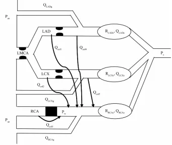
A lumped biomechanical model of this coronary network is proposed in Figure 1. In this model, the capillaries are represented by their hydraulic resistances (RLADc, RLCXc, RRCAc are the resistances of the capillaries vascularized by the LAD, LCx and RCA arteries, respectively). The blood flow rates across the LAD, LCx and RCA capil-laries are denoted by QLADc, QLCXc, QRCAc respectively. Qcol1 and Qcol4 are the collateral flow rates from LAD towards RCA (before and after LAD stenoses, respec-tively), Qcol2 and Qcol5 are the collateral flow rates from LCx (before and after LCx stenoses, respectively) and Qcol3 is the collateral flow rate from the aorta towards the RCA.
Full resolution of the fluid mechanics equations in such a network is complicated. As suggested by several authors, prediction of flows and pressures can be facili-tated by the use of an analog electrical model [3].
2.3. The Electrical Model
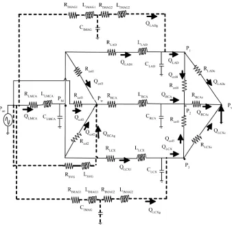
[image:2.595.129.470.387.675.2]The electrical analog model corresponding to the coro-nary network of Figure 1 is shown in Figure 2. Each segment of the coronary artery can be simulated by an
Figure 2.Analog electrical model for the network shown in Figure 1. The grafts are repre-sented with dotted lines.
equivalent analog circuit model with resistance R (hy-draulic resistance of the vessel), capacitance C (compli-ance of the vessel) and induct(compli-ance L (inertia of the flowing blood). The LMCA is represented by RLMCA, CLMCA and LLMCA. The LAD is represented by RLAD, CLAD and LLAD. The LCx is represented by RLCX, CLCX and LLCX. The RCA is represented by RRCA, CRCA and LRCA. Our coronary artery system was modeled in the presence of bypasses. Pietrabissa et al. suggested some
representation of internal mammary artery grafts (IMAG, used for left coronary artery bypasses) and saphenous vein grafts (SVG, used for the RCA) that are supported by physiological observations [4]. To take into account tapering, the IMAG is artificially divided into two seg-ments of equal length (70 mm) but of different diameters (2.8 mm and 2 mm, respectively); consequently, it is modeled by five elements: two resistances RIMAG1, R I-MAG2; two coils LIMAG1, LIMAG2; and a capacitor CIMAG. SVG is modeled by two elements: a resistance RSVG and a coil LSVG. The SVG model does not include an electric capacitance as experimental data confirm that when a vein is exposed to arterial pressure it loses its high com-pliance characteristics. The myocardial capillaries fed by
the left and right coronary arteries are represented only by their resistances RLADc, RLCXc and RRCAc. This ap-proximation is convenient since the resistive effects are preponderant for small diameter vessels like capillaries [3].For the same reason, the collateral vessels are also represented only by their resistances Rcoli, i = 1-5.
In this hydraulic/electric analogy, pressure and flow rate correspond to electrical voltage and current, respec-tively.
According to the network presented in Figure 2, the flow rate in the LAD artery, QLAD, is defined as the sum of the flow rate in the LAD graft, QLADg, (if it exists), and of the flow rate in the stenosed native artery, QLAD1. The same notations are used for the LCx branch.
The sum of the right and left coronary flows is de-noted Qt:
t LAD LCx RC
Q Q Q Q A (1)
2.4. Parameter Determination
2.4.1. Vessel Resistance, Inductance and Compliance As suggested by Wang et al. [5] and Pietrabissa et al. [4],
follows: 4 128 l R D
(2a)
2 4 l L D
(2b)
3 4 D l C Eh
(2c)
[image:4.595.146.286.92.184.2]where = 4 10-3 kg.m-1.s-1 is the blood viscosity; = 103 kg.m-3 is the blood density; E = 2 105 Pa is the Young modulus of the vessel; l (m) is the vessel length; D (m) is the vessel diameter and h (m) is the vessel wall thickness (estimated as: h = 0.08D).
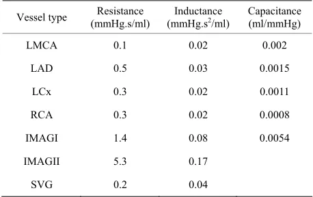
Table 1 shows the values of R, L and C for the left and right coronary arteries and grafts. These values were provided by Pietrabissa et al. [4] except for the data for
the RCA. In our study, the useful length of the RCA corresponds to the part distal to the thrombosis. This has been estimated as half of the LAD length and R, C and L for the RCA were deduced from those provided by Pietrabissa et al. for the LAD [4].
Left coronary stenoses were considered by varying the parameters of specific segments of the net as follows [5]:
2 0
RR (3a)
3 2 0
CC (3b)
1 0
LL (3c)
where = 1 – p, p is the percentage of area reduction of the stenosed vessel. R0, C0, and L0 are the values when p = 0.
2.4.2. Capillary Resistances
These resistances (RLADc, RLCXc and RRCAc) are patient– specific. Their determination is performed using the ex-perimental data of the case (3G) for each patient. The detailed calculations are given in [6].
Table 1.Values of resistance R, inductance L and capacitance C for the vessels represented in the model.
Vessel type (mmHg.s/ml) Resistance (mmHg.sInductance 2/ml) Capacitance (ml/mmHg)
LMCA 0.1 0.02 0.002
LAD 0.5 0.03 0.0015
LCx 0.3 0.02 0.0011
RCA 0.3 0.02 0.0008
IMAGI 1.4 0.08 0.0054
IMAGII 5.3 0.17
SVG 0.2 0.04
2.4.3. Collateral Resistances
Due to the difficulty of determining the exact character-istics of the collateral pathways, it was assumed that all the collateral resistances are the same [1]:
1 2 3 4 5
col col col col col col
R R R R R R (4)
This resistance is also specific to each patient.
In the case of RCA occlusion and three vessel disease, the value of Rcol is strongly related to the value of pres-sure Pw. Thus, the Pw value measured in case (2G) is used as a convergence criterion to numerically determine the convenient value of Rcol for the patient. The numeri-cal simulations are performed using the Matlab Simulink program. The value of Rcol is changed until the calcu-lated Pw value converges towards the clinically measured one.
2.5. Flow Rate and Pressure Simulations
2.5.1. Aortic Pressure
The input of the model is the aortic pressure wave, Pao(t), measured for each patient and each situation (0G, 1G, 2G, 3G).
2.5.2. Matlab Simulations
Once the model parameters are determined, flow and pressure predictions can be performed in any branch of the model and for all surgical cases. The calculated flows and pressures are time-dependent, but we focus on average cardiac cycle values. This is consistent with the fact that the collected clinical data are also average car-diac cycle values.
The influence of ventricular contraction upon coro-nary vascular bed resistance and compliance is not taken into account in our simulations. The impact of this as-sumption is probably less important in the case of our study than it would be for healthy patients. Wang et al.
[5] have indeed shown that when severe stenoses exist, the significance of the collapse effect of intramural ar-teries due to myocardial contraction is reduced.
3. RESULTS
3.1. Stenoses Severity
The percentages of area reduction of the stenosed vessels for each patient are given in Table 2. For all patients, the RCA is totally occluded. Patient 7 has no stenosis on LAD; Patient 9 and 10 have no stenosis on LCx. Patients 1, 4 and 5 have moderate lesions on LMCA. Patient 1 has a very severe lesion on LAD.
3.2. Capillary and Collateral Resistances
[image:4.595.56.285.574.718.2]Table 2.Percentage of area reduction of the stenosed vessels for each patient.
Patient % area LMCA % area LAD % area LCx
1 26 99 90
2 46 89 95
3 91.6 84.8 95.6
4 19 86 97
5 20 88 92
6 85 94 82
7 80 0 85
8 87 70 90
9 83 78 0
[image:5.595.56.284.345.533.2]10 75 93 0
Table 3. Values of the capillary resistances, RLADc, RLCxc, RRCAc, and collateral resistances, Rcol, for the patients consid-ered in the study. All these values are given in mmHg.s/ml.
Patient RLADc RLCxc RRCAc Rcol
1 83.3 207.9 54.1 160
2 174.6 210.9 96.9 430
3 213 94.2 62.8 350
4 47.5 119.1 147.2 565
5 175.3 68.7 56.1 205
6 240.4 135.5 117.6 1055
7 50.2 118.4 76 650
8 77.6 196 347.6 970
9 374.8 33.7 80.7 420
10 155.9 62.1 213.8 405
Mean ± σ 159.3 ± 96.6 124.6 ± 59.9 125.3 ± 87.6 521 ± 282.3
this paper is more precise than that presented in the pre-vious paper [1], the mean values obtained for these 10 patients are in the same range than those previously pub-lished. As discussed in [1], these values are in good agreement with literature data. Important values of Rcol indicate that the collateral network is not very developed or not functional. The values of resistances presented in Table 3 are affected by a great variability. This finding supports the motivation of our work: the necessity to provide patients’ specific simulations.
3.3. Flows in LAD Branch and in LCx Branch
In the following, all the quantities are calculated values,
except when indicated with the notation “Cli” (used to denote measured clinical values).
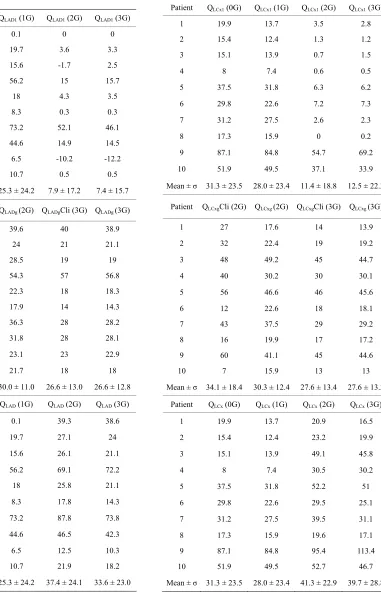
The values of the flow rates in the native stenosed LAD (QLAD1), in the LAD graft (QLADg) and the total flow rate in the LAD branch (QLAD) are given in Table 4.
The same type of values, for the LCx branch, are given in Table 5.
One can notice that the flow rates in the native stenosed artery, QLAD1 or QLCx1, is only slightly affected by the presence of the right graft (mean value of QLAD1 in the case (1G) = 25.3 ± 24.2 and in the case (0G) = 28.4 ± 24.1; mean value of QLCx1 in the case (1G) = 28.0 ± 23.4 and in the case (0G) = 31.3 ±2 3.5). This slight de-crease is due to the fact that, in the presence of the right graft, the pressure drop across the LAD or LCx stenosis is slightly modified (because the pressure gradients across the collaterals are modified).
On the contrary, there is an important decrease of QLAD1 and QLCx1 in the presence of the left grafts (mean value of QLAD1 in the case (2G) = 7.9 ± 17.2, in the case (3G) = 7.4 ± 15.7 and in the case (0G) = 28.4 ± 24.1; mean value of QLCx1 in the case (2G) = 11.4 ± 18.8, in the case (3G) = 12.5 ± 22.3 and in the case (0G) = 31.3 ± 23.5). This decrease is less important for Patient 7 (no stenosis on LAD) and for Patient 9 and 10 (no stenosis on LCx). When the LAD graft or LCx graft are present, the pressure drops across the LAD or LCx stenosis are reduced and since the hydraulic resistances of these stenosed arteries remain the same, the flow rate drops. Some sort of flow compensation appears between the graft and the native artery, especially if the native artery is not too severely obstructed (for example, this remark does not apply for the Patient 1, whose LAD branch is almost totally obstructed). Overall, the perfusion of the LAD territory (QLAD) or of the LCx territory (QLCx) is improved in the presence of the left grafts, but this im-provement remains moderate (lower than 10ml/min). This finding is important because it means that the graft could thus promote progression of native disease. It is hoped that these results will bring some new arguments to the controversy that exists regarding the competitive flows between the native stenosed artery and the graft [7-11].
For Patient 9, a negative QLAD1 flow is obtained in the presence of the left grafts (cases (2G) and (3G)).
Table 4. Values of the flow rates (ml/min) in the native ste-nosed LAD (QLAD1), in the LAD graft (QLADg), and total flow rate in the LAD branch (QLAD). These values are given for each patient, in the four different revascularization situations (0G, 1G, 2G, 3G).
Patient QLAD1 (0G) QLAD1 (1G) QLAD1 (2G) QLAD1 (3G)
1 0.1 0.1 0 0
2 23.6 19.7 3.6 3.3
3 20.7 15.6 -1.7 2.5
4 56.2 56.2 15 15.7
5 25.9 18 4.3 3.5
6 11.6 8.3 0.3 0.3
7 77.5 73.2 52.1 46.1
8 45.7 44.6 14.9 14.5
9 10.4 6.5 -10.2 -12.2
10 12.1 10.7 0.5 0.5
Mean ± σ 28.4 ± 24.1 25.3 ± 24.2 7.9 ± 17.2 7.4 ± 15.7
Patient QLADgCli (2G) QLADg (2G) QLADgCli (3G) QLADg (3G)
1 34 39.6 40 38.9
2 23 24 21 21.1
3 22 28.5 19 19
4 59 54.3 57 56.8
5 24 22.3 18 18.3
6 11 17.9 14 14.3
7 28 36.3 28 28.2
8 38 31.8 28 28.1
9 24 23.1 23 22.9
10 20 21.7 18 18
Mean ± σ 28.3 ± 13.1 30.0 ± 11.0 26.6 ± 13.0 26.6 ± 12.8
Patient QLAD (0G) QLAD (1G) QLAD (2G) QLAD (3G)
1 0.1 0.1 39.3 38.6
2 23.6 19.7 27.1 24
3 20.7 15.6 26.1 21.1
4 56.2 56.2 69.1 72.2
5 25.9 18 25.8 21.1
6 11.6 8.3 17.8 14.3
7 77.5 73.2 87.8 73.8
8 45.7 44.6 46.5 42.3
9 10.4 6.5 12.5 10.3
10 12.1 10.7 21.9 18.2
Mean ± σ 28.4 ± 24.1 25.3 ± 24.2 37.4 ± 24.1 33.6 ± 23.0
Table 5. Values of the flow rates (ml/min) in the native stenosed LCx (QLCx1), in the LCx graft (QLCxg), and total flow rate in the LCx branch (QLCx). These values are given for each patient, in the four different revascularization situations.
Patient QLCx1 (0G) QLCx1 (1G) QLCx1 (2G) QLCx1 (3G)
1 19.9 13.7 3.5 2.8
2 15.4 12.4 1.3 1.2
3 15.1 13.9 0.7 1.5
4 8 7.4 0.6 0.5
5 37.5 31.8 6.3 6.2
6 29.8 22.6 7.2 7.3
7 31.2 27.5 2.6 2.3
8 17.3 15.9 0 0.2
9 87.1 84.8 54.7 69.2
10 51.9 49.5 37.1 33.9
Mean ± σ 31.3 ± 23.5 28.0 ± 23.4 11.4 ± 18.8 12.5 ± 22.3
Patient QLCxgCli (2G) QLCxg (2G) QLCxgCli (3G) QLCxg (3G)
1 27 17.6 14 13.9
2 32 22.4 19 19.2
3 48 49.2 45 44.7
4 40 30.2 30 30.1
5 56 46.6 46 45.6
6 12 22.6 18 18.1
7 43 37.5 29 29.2
8 16 19.9 17 17.2
9 60 41.1 45 44.6
10 7 15.9 13 13
Mean ± σ 34.1 ± 18.4 30.3 ± 12.4 27.6 ± 13.4 27.6 ± 13.3
Patient QLCx (0G) QLCx (1G) QLCx (2G) QLCx (3G)
1 19.9 13.7 20.9 16.5
2 15.4 12.4 23.2 19.9
3 15.1 13.9 49.1 45.8
4 8 7.4 30.5 30.2
5 37.5 31.8 52.2 51
6 29.8 22.6 29.5 25.1
7 31.2 27.5 39.5 31.1
8 17.3 15.9 19.6 17.1
9 87.1 84.8 95.4 113.4
10 51.9 49.5 52.7 46.7
[image:6.595.57.290.142.713.2]LAD and LCx, so that retrograde flow is lower. Some situations of retrograde flow have been reported previ-ously in the literature: for example, in arterial conduits grafted to coronary arteries with lower grade stenosis [9, 11,12]. However, the situation studied in this paper is somewhat different because of the LMCA stenosis.
This observation about Patient 9 is consistent with the high flow rates predicted in its LCx branch (this patient has no stenosis on LCx and a low RLCxc value). Patient 10 has also no stenosis on LCx, but he does not present such a disproportion between RLCxc and RLADc.
This demonstrates that the values of the capillary and collateral resistances have a major impact on all the pressures and flow rates, including the flow rates in the grafts. For example, an elevated value of QLADg is ob-tained for Patient 4 because RLADc is specially low for this patient (on the contrary, Patient 4 has severe stenosis on LCx; consequently, low QLCx flow rates are predicted for this patient in the case 0G and 1G). Patients who have rather high values of RLCxc have rather low flow rates in the LCx graft (for example, Patients 1, 2, 6, 8). Conversely, patients who have rather low values of RLCxc have rather large flow rates in the LCx graft (for exam-ple, Patients 3, 5, 9). Such a correlation between graft flow and grafted perfusion area has been demonstrated previously by Hirotani et al. [13].
As we previously found in [14], a small decrease (a few ml/min) of the flow rates in the left grafts can be noticed in the case (3G) compared to the case (2G).
3.4. Flows and Pressures in the RCA Branch
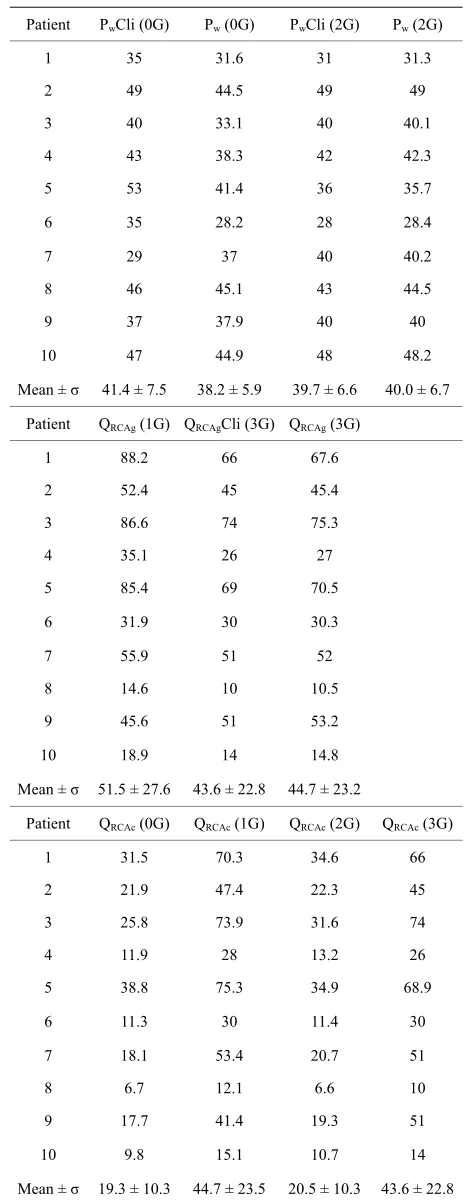
The values of the pressures distal to the thrombosis on RCA (Pw), of the flow rates in the RCA graft (QRCAg), and of the flow rates in the right capillary area (QRCAc) are presented in Table 6.
As we previously found in [2], no significant change in the mean value of Pw in the presence of the left grafts (case (2G) compared to case (0G)) can be demonstrated. Moreover, no evident correlation appears between the values of Pw and the corresponding flow rates in the right capillary area, QRCAc.
The flow rates QRCAg (1G) are slightly higher than QRCAg (3G) (mean value of QRCAg in the case (1G) = 51.5 ± 27.6 and in the case (3G) = 44.7 ± 23.2) because of the negative collateral flows that exist in the case (1G) (For example, for Patient1, because of the severe obstruction of the LAD artery, Qcol4 is important (= -15ml/min, see Table 7)).
[image:7.595.306.538.114.715.2]For all patients, the perfusion of the right territory, QRCAc, is improved in the presence of the right graft (mean value of QRCAc in the case (1G) = 44.7 ± 23.5, and in the case (3G) = 43.6 ± 22.8; mean value of QRCAc in the case (0G) = 19.3 ± 10.3, and in the case (2G) = 20.5 ±
Table 6. Values of the pressures distal to the thrombosis on RCA (Pw, in mmHg), of the flow rates (ml/min) in the RCA graft (QRCAg), and in the right capillary area (QRCAc).
Patient PwCli (0G) Pw (0G) PwCli (2G) Pw (2G)
1 35 31.6 31 31.3
2 49 44.5 49 49
3 40 33.1 40 40.1
4 43 38.3 42 42.3
5 53 41.4 36 35.7
6 35 28.2 28 28.4
7 29 37 40 40.2
8 46 45.1 43 44.5
9 37 37.9 40 40
10 47 44.9 48 48.2
Mean ± σ 41.4 ± 7.5 38.2 ± 5.9 39.7 ± 6.6 40.0 ± 6.7
Patient QRCAg (1G) QRCAgCli (3G) QRCAg (3G)
1 88.2 66 67.6
2 52.4 45 45.4
3 86.6 74 75.3
4 35.1 26 27
5 85.4 69 70.5
6 31.9 30 30.3
7 55.9 51 52
8 14.6 10 10.5
9 45.6 51 53.2
10 18.9 14 14.8
Mean ± σ 51.5 ± 27.6 43.6 ± 22.8 44.7 ± 23.2
Patient QRCAc (0G) QRCAc (1G) QRCAc (2G) QRCAc (3G)
1 31.5 70.3 34.6 66
2 21.9 47.4 22.3 45
3 25.8 73.9 31.6 74
4 11.9 28 13.2 26
5 38.8 75.3 34.9 68.9
6 11.3 30 11.4 30
7 18.1 53.4 20.7 51
8 6.7 12.1 6.6 10
9 17.7 41.4 19.3 51
10 9.8 15.1 10.7 14
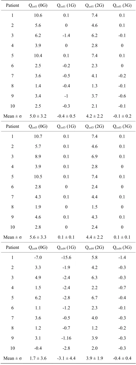
Table 7. Values of the collateral flow rates (ml/min), for each patient, in the four different revascularization situations.
Patient Qcol1 (0G) Qcol1 (1G) Qcol1 (2G) Qcol1 (3G)
1 10.6 0.1 7.4 0.1
2 5.6 0 4.6 0.1
3 6.2 -1.4 6.2 -0.1
4 3.9 0 2.8 0
5 10.4 0.1 7.4 0.1
6 2.5 -0.2 2.3 0
7 3.6 -0.5 4.1 -0.2
8 1.4 -0.4 1.3 -0.1
9 3.4 -1 3.7 -0.6
10 2.5 -0.3 2.1 -0.1
Mean ± σ 5.0 ± 3.2 -0.4 ± 0.5 4.2 ± 2.2 -0.1 ± 0.2
Patient Qcol3 (0G) Qcol3 (1G) Qcol3 (2G) Qcol3 (3G)
1 10.7 0.1 7.4 0.1
2 5.7 0.1 4.6 0.1
3 8.9 0.1 6.9 0.1
4 3.9 0.1 2.8 0
5 10.5 0.1 7.4 0.1
6 2.8 0 2.4 0
7 4.3 0.1 4.4 0.1
8 1.9 0 1.5 0
9 4.6 0.1 4.3 0.1
10 2.8 0 2.4 0
Mean ± σ 5.6 ± 3.3 0.1 ± 0.1 4.4 ± 2.2 0.1 ± 0.1
Patient Qcol4 (0G) Qcol4 (1G) Qcol4 (2G) Qcol4 (3G)
1 -7.0 -15.6 5.8 -1.4
2 3.3 -1.9 4.2 -0.3
3 4.9 -2.4 6.3 -0.3
4 1.5 -2.4 2.2 -0.7
5 6.2 -2.8 6.7 -0.4
6 1.1 -1.2 2.3 -0.1
7 3.6 -0.5 4.0 -0.3
8 1.2 -0.7 1.2 -0.2
9 3.1 -1.16 3.9 -0.3
10 -0.4 -2.8 2.0 -0.3
Mean ± σ 1.7 ± 3.6 -3.1 ± 4.4 3.9 ± 1.9 -0.4 ± 0.4
Patient Qcol5 (0G) Qcol5 (1G) Qcol5 (2G) Qcol5 (3G)
1 6.6 -2.6 6.7 -0.4
2 1.8 -3.1 4.3 -0.2
3 -0.4 -7.5 5.9 -0.8
4 -1.2 -4.7 2.5 -0.3
5 1.5 -7.4 5.9 -1.3
6 2.3 -0.4 2.2 -0.1
7 3.0 -1.0 4.0 -0.3
8 0.9 -0.9 1.3 -0.1
9 3.3 -1.0 3.7 -0.7
10 2.4 -0.3 2.1 -0.2
Mean ± σ 2.0 ± 2.1 -2.9 ± 2.8 3.9 ± 1.8 -0.4 ± 0.4
10.3). However, for patients 8 and 10, this perfusion appears to be critically low. This seems to be related to their high values of the right capillary resistance RRCAc. For patients 4 and 6, QRCAc is low in the case (0G) and (2G), and this seems to be related to high values of the collateral resistance Rcol.
For patients 7, 9 and 10, the important QLAD1 or QLCx1 flow rates due to the absence of stenoses on the LAD or LCx branch are not associated with any particular im-provement of the right territory perfusion.
More generally, it appears from the results of Table 6, that, in the presence of the left grafts, the QRCAc flow rates are not significantly modified (mean value of QRCAc in the case (2G) = 20.5 ± 10.3, and mean value of QRCAc in the case (0G) = 19.3 ± 10.3). Most of the blood brought by the left grafts is used to perfuse the distal left territories (QLADc and QLCxc). This observation is consis-tent with our previous results [1,15].
3.5. Collateral Flows
The results of the collateral flows are presented in Table 7, for each patient and each revascularization situation. Because of the assumption that the collateral resistances are the same (Eq.4), we have PM – Pw= Rcol. Qcol1 = Rcol. Qcol2 (see Figure 2); consequently, Qcol1 = Qcol2.
[image:8.595.56.285.107.722.2]negative because severe stenoses are present on the LCx artery.
Such reverse collateral flow also exists when the right graft is present, especially in the case (1G): due to the presence of the right graft, the right territory is better perfused and the pressures in the right area become higher than those of the left territories. This has been also demonstrated by other authors: Miyamoto et al. [16]
have shown that revascularization of the receiving artery can reverse the pressure gradient across the collateral network, establishing collateral flow in the opposite di-rection.
Qcol1 and Qcol2, in the case 0G, are slightly higher than Qcol1 and Qcol2, in the case 2G (mean value of Qcol1 in the case (0G) = 5.0 ± 3.2, and in the case (2G) = 4.2 ± 2.2). This is consistent with the fact that, in the presence of the left grafts, the flow in the native artery decreases, and consequently, Qcol1 and Qcol2 decrease.
For patients with moderate lesions on LMCA and se-vere stenoses on LAD and LCx (for example, patient 1 and 5), the blood is forced to flow through Rcol1 and Rcol2, yielding values of Qcol1 and Qcol2 in the case (0G) higher than those obtained for other patients. For these patients, Qcol3 is high also, because Rcol is low. For the same rea-son, the collateral flows of the case (2G) for these pa-tients are somewhat higher than those obtained for other patients.
We also notice that, in the case (0G) and (2G), Qcol3 directly varies as the pressure drop (Pao-Pw).
In the case (3G), the collateral flows become negligi-ble. Loss of collateral flow after revascularization agrees with the findings of previous studies: Wang et al. found that collateral flow disappeared in 12/14 patients after grafting of the RCA [17]. Werner et al. also demon-strated regression of collateral function after recanaliza-tion of chronic total coronary occlusions [18].
3.6. Total Flow Qt
[image:9.595.308.539.107.309.2]The results obtained for Qt are shown in Table 8. It ap-pears that the better perfusion will be obtained with the three grafts. In the case (2G), there is an improvement of Qt, compared to the pathological case (0G) (mean value of Qt in the (2G) case = 91.4 ± 26.4, and in the (0G) case = 75.3 ± 24.7), but this improvement is related to an in-crease of QLAD and QLCx, and the presence of the left grafts does not really improve QRCAc. Conversely, in the case (1G), QRCAc is improved by the presence of the right graft, but QLAD and QLCx remain rather the same as in the pathological case. This demonstrates that, for the pa-tients considered in this study, complete revasculariza-tion (with the 3 grafts) is fully justified. However, for Patient 8 and 10 (and in a lesser extent, 6), the revascu-larization yields a very small improvement of Qt.
Table 8. Values of the total flow rates Qt (ml/min), for each patient, in the four different revascularization situations.
Patient Qt (0G) Qt (1G) Qt (2G) Qt (3G)
1 51.9 102.2 82.3 122.9
2 55.9 84.5 64.1 89.4
3 57.1 113.3 94.5 141.9
4 75.8 98.7 108.1 129.4
5 94.5 135.3 100.2 142.8
6 49.3 62.5 54.2 69.6
7 120.2 155.6 139.9 156.4
8 67.7 74.2 70.2 69.7
9 108.8 134.9 119.6 175.7
10 71.7 78.5 81.2 79.4
Mean ± σ 75.3 ± 24.7 104 ± 30.5 91.4 ± 26.4 117.7 ± 38.2
4. DISCUSSION
To the best of our knowledge, the current study is unique because of the specific three vessel disease situation, and because our simulations take into account simultane-ously the effect of revascularization, the grade of native arteries stenoses and the collaterality.
The values presented in this paper for the flow rates, pressures, capillary and collateral resistances, are in complete agreement with the results previously obtained with a less sophisticated version of the model [1]. The range of these values and the main assumptions and limitations of the model have already been discussed in [1].
In spite of these limitations, the clinical relevance of our results seem good; the results confirm the surgeons professional experience and agree with the data and analysis that can be found in the literature.
The most important features shown by the calcula-tions can be summarized as follows:
Important variability between individuals for their capillary resistances and collateral resistances (RLADc, RLCxc, RRCAc, Rcol). This supports the moti-vation of our work to develop patient’s specific simulations.
When the three grafts are functional, the collateral channels play no more role and a severe reduction of the flow rates in the native left arteries is dem-onstrated (depending on their degree of occlusion). The left grafts could thus promote progression of the native disease.
The values of the capillary resistances and collat-eral resistances have a major impact on the flows and pressures everywhere in the network and it appears that each variable depends on all the oth-ers. For example, it is difficult to study separately the influence of the LAD stenosis or LCx stenosis, because the flow in the corresponding branch also depends on the LMCA stenosis, on the capillary resistance RLADc or RLCxc, on the collateral resis-tance Rcol, on the pressure difference between the left and right branches, on the aortic and venous pressures, …
However, further work is necessary to improve the physiological relevance of the present model:
Improve the representation of collateral vessels (the assumption that all the collateral resistances are the same is probably not very realistic)
Take into account the capillary resistance variation during the cardiac cycle, due to myocardial con-traction
In a further step, the model could then be transformed in order to work with pre-operative clinical measure-ments and thus become a full predictive model.
REFERENCES
[1] Maasrani, M., Verhoye, J.P., Corbineau, H. and Drochon, A. (2008) Analog electrical model of the coronary circu-lation in case of multiple revascularizations. Annals
Biomedical Engineering, 36, 1163-1174.
doi:10.1007/s10439-008-9500-5
[2] Verhoye J. Ph., De Latour B., Drochon A. and Corbineau H. (2005) Collateral flow reserve and right coronary oc-clusion: evaluation during off-pump revascularization.
Interactive Cardiovascular Thoracic Surgery, 4, 23-26.
doi:10.1510/icvts.2004.093088
[3] Olufsen, M. and Nadim, A. (2004) On deriving lumped models for blood flow and pressure in the systemic ar-teries. Math Biosciences Engineering, 1, 61-80.
[4] Pietrabissa, R., Mantero, S., Marotta, T. and Menicanti, L. (1996) A lumped parameter model to evaluate the fluid dynamics of different coronary bypasses. Medical
Engi-neering & Physics, 18, 477-484.
doi:10.1016/1350-4533(96)00002-1
[5] Wang, J.Z., Tie, B., Welkowitz, W., Kostis, J. and Semmlow, J. (1989) Incremental network analogue model of the coronary artery. Medical & Biological En-gineering & Computing., 27, 416-422.
doi:10.1007/BF02441434
[6] Maasrani, M., Abouliatim, I., Ruggieri, V.G., Corbineau, H., Verhoye, J.P. and Drochon, A. (2010) Simulations of
fluxes in diseased coronary network using an electrical model. Proceedings of the XIX International Conference on Electrical Machines, IEEE –ICEM 2010, Rome. [7] Shimizu, T., Hirayama, T., Suesada, H., Ikeda, K., Ito, S.
and Ishimaru, S. (2000) Effect of flow competition on internal thoracic artery graft: postoperative velocimetric and angiographic study. Journal of Thoracic and
Car-diovascular Surgery, 120, 459-465.
doi:10.1067/mtc.2000.108166
[8] Lust, R., Zeri, R., Spence, P., Hopson, S., Sun, Y., Otaki, M., Jolly, S., Mehta, P. and Chitwood, W. (1994) Effect of chronic native flow competition on internal thoracic artery grafts. Annals of Thoracic Surgery, 57, 45-50.
doi:10.1016/0003-4975(94)90363-8
[9] Kawasuji, M., Sakakibara, N., Takemura, H., Tedoriya, T., Ushijima, T. and Watanabe, Y. (1996) Is internal thoracic artery grafting suitable for a moderately stenotic coro-nary artery? Journal of Thoracic and Cardiovascular
Surgery, 112, 253- 259.
doi:10.1016/S0022-5223(96)70246-5
[10] Berger, A., Mac Carthy, P., Siebert, U., Carlier, S., Wijns, W., Heyndrickx, G., Bartunek, J., Vanermen, H. and De Bruyne, B. (2004) Long-term patency of internal mam-mary artery bypass grafts; relationship with preoperative severity of the native coronary artery stenosis.
Circula-tion, 110, II36-II40.
doi:10.1161/01.CIR.0000141256.05740.69
[11] Berger A., Mac Carthy P., Vanermen H. and De Bruyne B. (2004) Occlusion of internal mammary grafts: a review of the potential causative factors. Acta Chirurgica
Bel-gica, 104, 630-634.
[12] Shimizu, T., Ito, S., Kikuchi, Y., Misaka, M., Hirayama, T., Ishimaru, S. and Yamashina, A. (2004) Arterial con-duit shear stress following bypass grafting for intermedi-ate coronary artery stenosis: A comparative study with saphenous vein grafts. European Journal of Cardio-
Thoracic Surgery, 25, 578-584.
doi:10.1016/j.ejcts.2003.12.039
[13] Hirotani, T., Kameda, T., Shirota, S., and Nakao, Y. (2001) An evaluation of the intraoperative transit time meas-urements of coronary bypass flow. European Journal of
Cardio-Thoracic Surgery, 19, 848-852.
doi:10.1016/S1010-7940(01)00700-X
[14] Verhoye, J. P., Abouliatim, I., Drochon, A., De Latour, B., Leclercq, C., Leguerrier, A. and Corbineau, H. (2007) Collateral blood flow between left coronary artery bypass grafts and chronically occluded right coronary circulation in patients with triple-vessel disease. European Journal
of Cardio-Thoracic Surgery, 31, 49-54.
doi:10.1016/j.ejcts.2006.09.033
[15] Maasrani, M., Drochon, A., Abouliatim, I., Corbineau, H. and Verhoye, J.P. (2010) Theoretical simulations of the influence of the stenoses severity and revascularization status on the right territory perfusion in case of severe coronary disease and occlusion of the right artery. Com-puter Methods in Biomechanics and Biomedical
Engi-neering, 13, 93-94.
doi:10.1080/10255842.2010.494037
[16] Miyamoto, S., Fujita, M., and Sasayama, S. (2000) Bidi-rectional function of coronary collateral channels in hu-mans. International Journal of Cardiology, 75, 249-252.
[17] Wang, J., Filipovic, M., Skarvan, K., Michaux, I., Schu-mann, R., Buser, P. and Seeberger, M. (2006) Transeo-sophageal Doppler echocardiographic detection of in-tramyocardial collateral flow to the right coronary artery and changes in the flow to the left inferior ventricular wall immediately after coronary bypass grafting.
Ameri-can Journal of Cardiology, 98, 1587-1592.
doi:10.1016/j.amjcard.2006.07.034
[18] Werner G., Emig U., Mutschke O., Schwarz G., Bahr-mann P., and Figulla H. (2003) Regression of collateral function after recanalization of chronic total coronary occlusions. A serial assessment by intracoronary pressure and Doppler recordings. Circulation, 108, 2877-2882.
NOMENCLATURE
LMCA: left main coronary artery LAD: left anterior descending artery LCx: left circumflex branch
RCA: right coronary artery Pao: aortic pressure Pv: central venous pressure
Pw: pressure distal to the RCA occlusion
RLADc: resistances of the capillaries vascularized by the LAD artery
RLCXc: resistances of the capillaries vascularized by the LCxartery
RRCAc: resistances of the capillaries vascularized by the RCA artery
QRCAg: flow rate in the RCA graft QLADg: flow rate in the LAD graft QLCxg: flow rate in the LCx graft
QLAD1: flow rate in the native stenosed LAD
QLCx1: flow rate in the native stenosed LCx QLADc: blood flow rate across the LAD capillaries QLCXc: blood flow rate across the LCx capillaries QRCAc: blood flow rate across the RCA capillaries Qcol1: collateral flow rate from LAD towards RCA before LAD stenosis
Qcol4: collateral flow rate from LAD towards RCA after LAD stenosis
Qcol2: collateral flow rate from LCX before LCx stenosis Qcol5: collateral flow rate from LCX after LCX stenosis Qcol3: collateral flow rate from the aorta towards the RCA R: resistance
C: capacitance L: inductance
IMAG: internal mammary artery graft SVG: saphenous vein graft