Original Article
Mir-200c inhibits HOTAIR expression resulting in the
decrease of chemoresistance in ovarian
cancer stem cells
Jing Wang1*, Dengyu Chen2,3*, Fangfang Shi2*, Junsong Chen2, Yuxia Zhang1,2, Fangfang Shi2, Di Wu1,2, Miao Li2, Meng Pan2, Jun Dou2
1Department of Gynecology & Obstetrics, Zhongda Hospital, Medical School, Southeast University, Nanjing
210009, China; 2Department of Pathogenic Biology and Immunology of Medical School, Southeast University,
Nanjing 210009,China; 3Department of Microbiology, Bengbu Medical School, Bengbu 233030, China. *Equal
contributors and co-first authors.
Received March 15, 2016; Accepted June 10, 2016; Epub July 15, 2016; Published July 30, 2016
Abstract: In this study, we investigated whether miR-200c overexpression would increase the sensitivity of epithe-lial ovarian cancer (EOC) stem cells (CSCs) to chemotherapy drugs through down-regulation of lncRNA HOTAIR. We used a magnetic-activated cell sorting system to isolate the SKOV3 CD44+CD117+CSCs from the selected human EOC SKOV3 cells that were stably transduced with lentivirus miR-200c. HOTAIR, a direct target of miR-200c, was validated by using the wild-type and the mutant region HOTAIR luciferase reporters. The results showed the over-expression of miR-200c in SKOV3 CD44+CD117+CSCs significantly decreased the drug resistant to paclitaxel and cisplatin compared with SKOV3 CD44+CD117+CSCs transduced with the lentivirus-mock or the wild-type of SKOV3 CD44+CD117+CSCs. Moreover, SKOV3 CD44+CD117+CSCs with miR-200c overexpression dramatically reduced its metastatic potential from the tumor tissues to the nude mouse lungs in contrast to SKOV3 CD44+CD117+CSCs without miR-200c overexpression. The direct down-regulation of HOTAIR was miR-200c dependent because lucifer-ase reporter and rescue assay results showed that the putative miR-200c-binding site has the inhibitory effect on HOTAIR expression. Collectively, the increased sensitivity of SKOV3 CD44+CD117+CSCs to paclitaxel or cisplatin may be modulated by overexpression of miR-200c that directly inhibits HOTAIR expression.
Keywords: Epithelial ovarian cancer, cancer stem cells, drug resistance, miR-200c, lincRNA HOTAIR
Introduction
Epithelial ovarian cancer (EOC) is the most lethal gynaecological cancer with the majority of patients succumbing to chemotherapy-resis-tant disease. Currently, there are no specific or
sensitive clinical biomarkers that maybe imple-mented to identify chemoresistance and give insight to prognosis. Thus, understanding the related mechanisms of chemotherapy resis-tance and how it can be prevented or reversed is a pivotal challenge in the treatment of EOC in the worldwide [4, 32, 33].
Accumulating studies have demonstrated that there are a subpopulation of cells that are associated with properties of stem/progenitor-like cells known as cancer stem cells (CSCs) in
drug-resistant EOC tissues, and CSCs are believed to be ‘seed cell’ in cancer-initiating potential, invasion, metastasis, and resistance to traditional chemotherapy [5, 13, 22]. The existence of EOC CSCs might explain why stan-dard chemotherapy may shrink most tumors, however, left less highly lethal malignancy re-grow and eventually cause a relapse. Therefore, it is necessary to investigate the molecular mechanisms of EOC CSC chemoresistance and
to find out the new strategies for overcoming
chemoresistance of CSCs in EOC patients [1, 2].
epigenetic states can be reversed by drug ther-apy, and thus maintenance and regulation of epigenetic change is a potential target to halt or reverse chemotherapeutic resistance [4, 5]. Indeed, there have been reports that the microRNAs (miRNAs) have emerged as poten-tial therapeutic candidates by virtue of their abi- lity to down-regulation of EOC therapeutic resis-tance. Some studies have demonstrated that epithelial-mesenchymal transition (EMT) has an established role in promoting tumor pro-gression and the acquisition of therapeutic resistance [11, 12, 23]. For example, in cisplat-in-resistant ovarian cancer tissues and cell lines, the EMT phenotype was correlated with decreased miR-186 expression, increased Twist1 expression, chemoresistance, and poor prognosis in EOC patients. While introducing miR-186 into EOC cells led to a reduction in Twist1 expression along with mesenchymal-to-epithelial transition, decrease of chemoresis-tance, and enhanced cell apoptosis [32]. In another study, miR-9 was expressed signifi-cantly higher in drug-sensitive patients than in drug-resistant ones, and inhibition of miR-9 resulted in decreased clonal formation and sensitivity to cisplatin, suggesting high expres-sion of miR-9 was associated with enhanced tumorigenesis and increased sensitivity of the tumor cells to cisplatin treatment in primary ovarian tumor cells [31]. Our previous study demonstrated that the miR-200c overexpres-sion, by modulating the EMT, specifically inhib-ited the zinc-finger E-box binding homeobox 1 expression in the SKOV3 CD117+CD44+CSCs, and reduced cell tumorigenicity in nude mouse model [5].
In addition, long intervening non-coding RNA (lincRNA) HOTAIR is well studied among lncRNA and has been shown to have an important func-tions in normal and cancer cells. Dysregulated HOTAIR correlates highly with tumor invasion and metastasis [17]. Our study showed that the downregulated HOTAIR expression in SKOV3 CD117+CD44+CSCs significantly decreased the tumor growth and lung metastasis in xenograft mice [25]. However, how to co-regulate the properties of EOC SKOV3 CD117+CD44+CSCs between the miR-200c and the HOTAIR remains a little known. To this end, we investigated the interaction between the miR-200c and the HOTAIR, and wanted to know how to modulate the resistance to chemotherapy drugs in EOC
SKOV3 CD117+CD44+CSCs by miR-200c direct down-regulation of HOTAIR.
Materials and methods
Cell line
SKOV3 cell line, a well-established ovarian can-cer model system from ovarian cancan-cer patient, was ordered from the Cellular Institute in Shanghai, China. Cells were cultured in com-plete media consisting of RPMI 1640, 2 mM L-glutamine, 100 U/ml penicillin, 100 µg/ml streptomycin, and 10% fetal bovine serum. The medium was refreshed every 3 days to main-tain adherent cells. When reached 90% conflu-ence, cells were harvested with 0.25% trypsin -1 mM EDTA (Sigma-Aldrich, St. Louis, MO, USA). Isolation of human SKOV-3 CD44+CD117+
CSCs, transduction of lentivirus miR-200c and production of stable expression colonies
CD117+CSCs were sorted from the SKOV-3 cells with stably infected lentivirus miR-200c by using above-mentioned MACS.
Cytotoxicity assay in a various CD44+CD117+CSCs
Cytotoxicity assay was used to test the ability of paclitaxel and cisplatin (Sigma-Aldrich, Mis- souri, USA) to induce cell death. 3×10⁴ CD44+ CD117+CSCs transduced with lentivirus-miR-200c were resuspended in 96-plates, and then added at concentrations of 10 μg/ml, 20 μg/ ml, 30 μg/ml, 40 μg/ml, 50 μg/ml, 60 μg/ml, and 70 μg/ml paclitaxel respectively, or 0.5 μg/ ml, 1.0 μg/ml, 1.50 μg/ml, 2,0 μg/ml, 2.5 μg/ ml, 3.0 μg/ml, 3.5 μg/ml, 4.0 μg/ml, 4.5 μg/ml, and 5.0 μg/ml cisplatin, respectively; cells were incubated for 72 h. Each concertration were done repeat three experiments. As a control, the CD44+CD117+CSCs transduced with lentivi-rus-mock and the wild type of CD44+ CD117+CSCs were used in this assay. Chemo- therapeutic sensitivity was detected by 3- (4,5-dimethylthiazol-2-yl)-2,5-diphenyltetrazoli-um bromide colorimetry (MTT) assay. The OD values were read at dual wave lengths of 450 nm and 630 nm to determine cell viability by using a microplate reader (Thermo Fisher Labsystems) [5, 30].
RNA extraction and quantitative real-time re-verse transcription-PCR (qRT-PCR)
Total RNA from the different cultured cells and mouse tumor tissues was isolated using a Qiagen RNeasy Kit (Qiagen, Valencia, CA, USA) following by manufacturer’s protocol. Single-stranded cDNA was prepared using SuperScript III reverse transcriptase (Invitrogen). qRT-PCR was carried out on an ABI step one plus real-time system (Applied Biosystems, USA). cDNAs were amplified by PCR with primers as follows: HOTAIR: sense, 5’-GGTAGAAAAAGCAACC ACG- AAGC-3’; antisense, 5’-TTGGGGAAGCATTTTCTG- AC-3’; β-actin (sense, 5’-GGACTTC GAGCAAGA- GATGG-3’; antisense, 5’-AGCACTGTGTTGGCGT- ACAG-3’). U6-RT Primer, 5’-GTCGTATCCAGTGCA- GGGTCCGAGGTATTCGCACTGGATACGACAAA- TATGGAAC-3’; sense, 5’-TGCGGGTGCTCGCTTC- GGCAGC-3’; URP (Universal Reverse Primer), 5’- CCGG CAGGGTCCGAGGT-3’; TGF-b1: sense, 5’-TGGAAACCCACAACGAAATCT-3’; antisense, 5’- GCTGAGGTATCGCCAGGAAT-3’; Bmi-1: sense, 5’- TGCTGATGCTGCCAATGG-3’; antisense, 5’-TTAC-
TTTCCGATCCAATCTGTTCTG-3’. The mRNA lev-els of the interested genes were expressed as the ratio of each gene of interest to β-actin or U6 mRNA for each sample. At least 3 biologic replicates and triplicate PCRs were used to cal-culate relative expression. The relative mRNA or miRNA levels were calculated using the com-parative cycle threshold (Ct) method (ΔΔCt). Briefly, the Ct values for the normalization gene were subtracted from Ct values of the target gene to achieve the ΔCt value. The 2−ΔC
t was calculated for each sample, and then each of the values was divided by a control sample to achieve the relative mRNA or miRNA levels (ΔΔCt) [17, 26].
Immunofluorescence detection of metastatic tumor cells in mouse lung tissues
At the end of the experiments, lung tissues were removed from the xenograft mice, fixed in 10% formalin, and then embedded in paraffin. Lung tissue sections of 4 μm thin were cut and mounted on SuperFrost Plus glass slides; the Luciferase positive cells were visualized using the immunofluorescence microscope at a mag-nification of ×200 [5, 16].
Double-luciferase reporter assay
The wild/mutated HOTAIR (NCBI: NR_0037- 16.3, site from 978 to1008 nt) was generated by PCR from human genomic DNA. These DNA fragments were cloned into the pGL3-control plasmid (Promega, Madison, WI, USA), while pGL-3.0 (firefly luciferase) was used as an inter-nal control. Briefly, HEK-293 or SKOV3 cells were seeded in 24 well cell culture clusters (Corning Incorporated; Corning, NY, USA). When reached 70% confluences, cells were cotrans-fected with the reporter constructs and hsa-miR-200c mimics for 36 h. Then the luciferase reporter assay was performed using a Double-Luciferase Assay system (Promega) per the manufacturer’s instructions [9, 21].
Statistical analysis
Statistical analysis was performed using the Student’s t-test for the difference between the experimental group and the control group. Data are expressed as mean ± standard error. Results for all analysis with a P value <0.05
indicate the statistically significant
Results
miR-200c increases chemotherapeutic sensi-tivity in SKOV3 CD44+CD117+CSCs
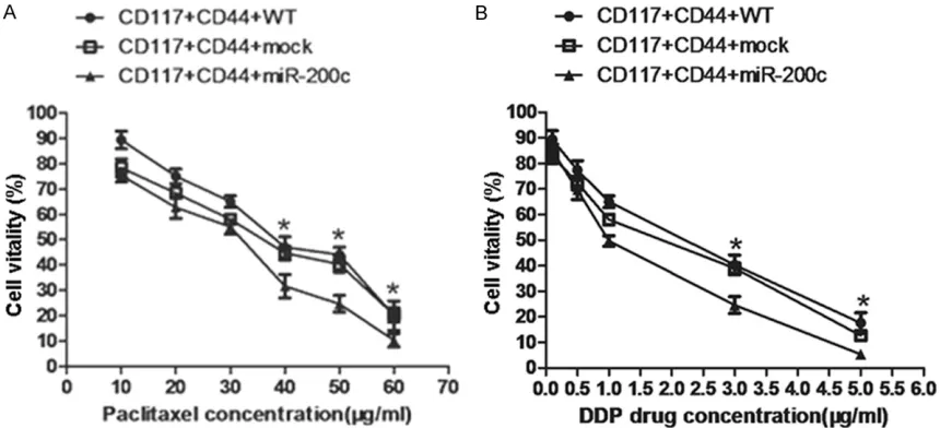
To examine whether the overexpression of miR-200c in SKOV3 CD44+CD117+CSCs would de- crease the resistance to chemotherapetic drug- s, we assessed the sensitivity changes of SKOV3 CD44+CD117+CSCs to chemotherapetic drugs paclitaxel and cisplatin respectively; these drugs are commonly used in clinic EOC patient chemotherapy. The results showed the forced overexpression of miR-200c in SKOV3 CD44+CD117+CSCs decreased the resistance to all the two drugs in vitro compared with the control cells. It was found that the cell vitality of lentivirus-miR-200c transducted SKOV3 CD44+ CD117+CSCs to paclitaxel was statistically sig-nificant decreased compared with the lentivi-rus-mock transducted SKOV3 CD44+CD117+ CSCs (31% vs 45%, *P<0.05) and the SKOV3 CD44+CD117+CSCs without miR-200c trans-duction (31% vs 49%, *P<0.05), respectively when cells were incubated with paclitaxel (40 μg/ml) for 72 hours. As this concentration was increased by 60 μg/ml, the cell vitality was simultaneously decreased in the lentivirus-miR-200c transducted SKOV3 CD44+CD117+CSCs (10%), which was statistically significant com-pared with the lentivirus-mock transducted SKOV3 CD44+CD117+CSCs (20%, *P<0.05),
and the wild type of SKOV3 CD44+CD117+CSCs (21%, *P<0.05), respectively (Figure 1A). Si- milarly, the resistance of the lentivirus-miR-200c transducted SKOV3 CD44+CD117+CSCs to cisplatin was statistically significant lower than that of the lentivirus-mock transducted SKOV3 CD44+CD117+CSCs (25% vs 41%, *P< 0.05) and the wild type of SKOV3 CD44+ CD117+CSCs (25% vs 40%, *P<0.05) when cells were incubated with cisplatin (3.0 μg/ml) for 72 hours. As predicted, an inhibitory effect of cisplatin on above various cells was signifi-cantly increased when cisplatin concentration was reached by 60 μg/ml as is shown in Figure 1B. The findings implied that the sensitivity of these various treated SKOV3 CD44+CD117+ CSCs to paclitaxel and cisplatin was mainly depended on the miR-200c overexpression or not.
miR-200c decreases the expression of TGF-β1 and Bmi-1 in SKOV3 CD44+CD117+CSCs as
well as cellular metastasis potential in mice
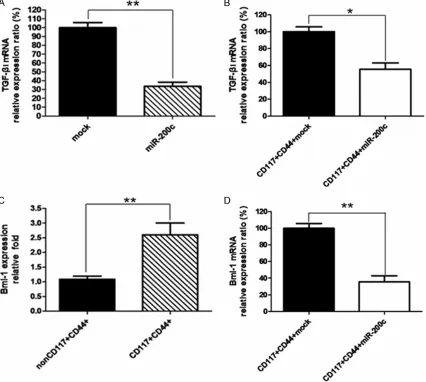
Next, we wanted to know whether decreased chemoresistance to drugs in lentivirus-miR-200c transducted SKOV3 CD44+CD117+CSCs was correlated with decreased expression of EMT promoter facror TGF-β1 and stem-like fac-tor Bmi-1. The results of qRT-PCR in Figure 2A,
[image:4.612.91.521.72.268.2]sues from the nude mice injected with lenti- virus-miR-200c transducted SKOV3 CD44+ CD117+CSCs (B) significantly reduced the TGF-β1 expression in contrast to the lentivirus-mock transducted SKOV3 CD44+CD117+CSCs, which was statistically significant (**P<0.01, SKOV3 cells) or (*P<0.05, tumor tissues). Consistently, the Bmi-1 expression was 2.5 times higher in SKOV3 CD44+CD117+CSCs than that in SKOV3 non CD44+CD117+CSCs tested by qRT-PCR, which was also statistically significant (**P< 0.01) as is shown in Figure 2C. In addition, the Bmi-1 expression was markedly reduced in the
tumor tissues from the nude mice injected with the lentivirus-miR-200c transducted SKOV3 CD44+CD117+CSCs compared to the nude mice injected with the lentivirus-mock transducted SKOV3 CD44+CD117+CSCs (Figure 2D), and the difference was statistically significant (**P< 0.01).
[image:5.612.95.521.75.457.2]capability of SKOV3 CD44+CD117+CSCs. Figure
3 gives the images of tumor metastatic lung tis-sues from the nude mice. The left side images show three kinds of different treated SKOV3 CD44+CD117+CSCs in mouse lung tissues, and no green fluorescence was found in common light microscope. The right side image (middle) shows remarkably stronger green fluorescence in lentivirus-mock transducted SKOV3 CD44+ CD117+CSCs than that in lentivirus-miR-200c transducted SKOV3 CD44+CD117+CSCs (bot-tom), suggesting a lot of lentivirus-mock trans-ducted SKOV3 CD44+CD117+CSCs migrated to the lung tissues but only a few of miR-200c overexpressed SKOV3 CD44+CD117+CSCs did migration. The top image in right side does not indicate green fluorescence this is because the wild type of SKOV3 CD44+CD117+CSCs did not
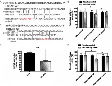
[image:6.612.90.386.69.332.2]activity of the reporter containing the wild-type of HOTAIR gene was significantly decreased fol-lowing treatment with miR-200c mimics (80% vs 100%, *P<0.05), which suggested that the direct down-regulation of HOTAIR was miR-200c-dependent because the mutation in the putative miR-200c-binding sites had rescued the inhibitory effect as is shown in Figure 4B. In this study, we meanwhile predicted the targets of common precursor miR-200c-5p since we constructed the lentivirus-miR-200c precursor that may generate two common precursors (miR-200c-3c and miR-200c-5p) when the pre-cursor was transducted to SKOV3 cells. HOTAIR mRNA contained 8 continuous base comple-mentary sites for the seed region of miR-200c-5p precursor (Figure 4C). The luciferase activity of the reporter containing the HOTAIR gene’s Figure 3. Images of tumor metastatic lung tissues from the nude mice. At the
end of the experiments, lung tissues were removed from the nude mice sub-cutaneously injected with 5×10⁴ SKOV3 CD44+CD117+CSCs with lentivirus miR-200c infection, SKOV3 CD44+CD117+CSCs with lentivirus mock infection, and SKOV3 CD44+CD117+CSCs without lentivirus infection, respectively. Lung tissue sections were made and visualized by using a light microscope or an immunofluorescence microscope at a magnification of ×200. The left and the right side images were seen under a light microscope and immunofluores -cence microscope in order. A-C represent the lung tissues from the nude mice injected with 5×10⁴ SKOV3 CD44+CD117+CSCs without lentivirus infection (A), with lentivirus mock infection (B), and with lentivirus miR-200c infection (C), respectively. These images suggested that less metastatic tumor cells were found in lung tissues that was mainly depended on the miR-200c overexpres-sion or not.
receive the lentivirus con-struct transduction. From these results, we may con-clude that the miR-200c overexpression in SKOV3 CD44+CD117+CSCs inhibit-ed the TGF-β1 and the Bmi-1 expression as well as SKO- V3 CD44+CD117+CSC lung metastatic ability in nude mice.
miR-200c binds HOTAIR and suppresses its expres-sion
wild-type was remarkably reduced after cotrasfection with miR-200c mimics (78% vs 100%, *P<0.05). Similarly, the mutation in the putative miR-200c-5p binding sites has res-cued the inhibitory effect on HOTAIR expression (*P<0.05) as is shown in Figure 4D. These results suggested the miR-200c mimics did directly suppress the HOTAIR gene expression. To further confirm whether HOTAIR is the direct target gene for miR-200c, we used qRT-PCR to test the changes of HOTAIR expression in response to forced miR-200c overexpression in SKOV3 cells. Figure 4E gives the HOTAIR expression was significant decreased in lentivi-rus-miR-200c stably transducted SKOV3 cells compared with in the lentivirus-mock stably
transducted SKOV3 cells (**P<0.01). This posi-tive data was further confirmed by HOTAIR expression in tumor tissues from the nude mice injected with SKOV3 CD117+CD44+CSCs with lentivirus-miR-200c or lentivirus-mock, indicat-ing the HOTAIR expression markedly reduced by 34% in tumor tissues from the nude mice injected with lentivirus-miR-200c transducted SKOV3 CD44+CD117+CSCs compared with the nude mice injected with lentivirus-mock trans-ducted SKOV3 non-CD44+CD117+CSCs (**P< 0.01), which was shown in Figure 5.
Discussion
[image:7.612.93.524.71.402.2]patients with advanced ovarian cancer, howev-er, a common problem faced by the EOC patients is the generated resistance to pacli-taxel and cisplatin, which results in the 5-year survival rate in patients with stage III and IV EOC less than 40% [27, 28]. One of mainly rea-sons is that paclitaxel/cisplatin-based thera-pies are able to eliminate the bulk of differenti-ated cancer cells, but are unable to eliminate CSCs. Therefore, CSCs are an attractive popu-lation of cells to target therapeutically [13]. We based on the accumulating evidence that non-coding RNAs especially miRNAs and lncRNAs represent a significant advance towards a bet-ter understanding of the mechanisms that gov-ern cancer cellular growth, and wanted to understand the possibility and the mechanisms of mir-200c inhibition of lncRNA HOTAIR in decreasing the chemotherapetic resistence of EOC CSCs.
Our current study has indicated that miR-200c is a HOTAIR-suppressive miRNA in SKOV3 CD117+CD44+CSCs, and that the inhibition function is miR-200c dependent. This is be- cause the miR-200c was found to bind HOTAIR in a sequence-specific manner, which resulted in down-regulation of the HOTAIR expression. Down-regulation of lncRNA by miRNA has only been recently observed. The suppression of HOTAIR expression by miRNA overexpression
correlated with alteration of HOTAIR function, including proliferation, invasion, metastasis,
and chemoresistance [9, 21]. Our findings indi -catd the miR-200c overexpression, which tar-gets inhibition of HOTAIR, demonstrated that the decrease of the chemoresistance of SKOV3 CD117+CD44+CSCs to paclitaxel/cisplatin treat-ment in vitro, and that suppression of expres-sion of TGF-β1 and Bmi-1 in vivo (tumor tis-sues). In addition, SKOV3 CD117+CD44+CSCs transducted with lentivirus-miR-200c signifi-cantly inhibited its metastasis in lung tissues from the SKOV3 bearing nude mice (this mice
not shown here, seen in reference 3). Our find -ing is essentially consistent with the previous
findings by others [8, 9, 29] and by us [5, 25]. It is known that Bmi-1 is an oncogene that causes neoplastic proliferation [20], and that.
TGF-β1, an EMT promoter facror, has been
shown to increase stem-like properties in
can-cer cells [3]. Both of Bmi-1 and TGF-β1 have
been found to be a target for miR-200c [18, 19]. In this study, the expression of Bmi-1 and
TGF-β1 inversely correlated with the miR-200c
expression in the tumor tissue samples at mRNA expression level (Figure 2). MiR-200c overexpression in SKOV3 CD44+CD117+CSCs
decreased the cellular metastasis potential
(Figure 3). From these consistent data, we
guess that miR-200c overexpression specifi-cally inhibited the zinc-finger E-box binding homeobox 1 expression in SKOV3 CD117+CD44+ CSCs, and reduced the expression of Bmi-1 and TGF-β1, which may inhibite the EMT, decrease the chemoresistance to paclitaxel and cisplatin as well as reduce the metastasis ability of SKOV3 CD117+CD44+CSCs by down-regulation of HOTAIR expression [5, Wang et al., 2014].
MiR-200c and HOTAIR are well studied among miRNA and lncRNA, and have been shown to have an important functions in normal and can-cer cells [21, 24], but miR-200c and HOTAIR interaction has never been studied in ovarian carcinoma cells. However, we understand that more studies are fully warranted to find out the mechanisms of how interaction between the miR-200c and the HOTAIR in SKOV3 CD117+ CD44+CSCs.
[image:8.612.91.287.71.257.2]In conclusion, our findings presented in this study demonstrated the miR-200c targets and decreases HOTAIR expression in EOC SKOV3 Figure 5. miR-200c regulates HOTAIR expression in
CD117+CD44+CSCs, and reduces cellular drug resistant to paclitaxel and cisplatin. These results suggest that interaction between the miR-200c and the HOTAIR may play a critical role to increase EOC SKOV3 CD117+CD44+CSC sensitivity to clinical therapies.
Acknowledgements
This work was supported in part by the National Natural Science Foundation of China (No. 81572887, 81202372), and in part by the Collaborative Innovation Center of Suzhou NanoScience and Technology.
Disclosure of conflict of interest
None.
Address correspondence to: Dr. Jun Dou,Depart- ment of Pathogenic Biology and Immunology, Me- dical School, Southeast University, Nanjing 210009, China. E-mail: [email protected]; Dr. Jing Wang, Department of Gynecology & Obstetrics, Zhongda Hospital, Medical School, Southeast University, Nanjing 210009, China. E-mail: [email protected]
References
[1] Baba T, Convery PA, Matsumura N, Whitaker RS, Kondoh E, Perry T, Huang Z, Bentley RC, Mori S, Fujii S, Marks JR, Berchuck A, Murphy SK. Epigenetic regulation of CD133 and tumor-igenicity of CD133+ ovarian cancer cells. Oncogene 2009; 28: 209-218.
[2] Berry NB, Bapat SA. Ovarian cancer plasticity and epigenomics in the acquisition of a stem-like phenotype. J Ovarian Res 2008; 21: 8-19. [3] Bhola NE, Balko JM, Dugger TC, Kuba MG,
Sán-chez V, Sanders M, Stanford J, Cook RS, Artea-ga CL. TGF-β inhibition enhances chemothera -py action against triple-negative breast cancer. J Clin Invest 2013; 123: 1348-58.
[4] Borley J, Brown R. Epigenetic mechanisms and therapeutic targets of chemotherapy resis-tance inepithelial ovarian cancer. Ann Med 2015; 47: 359-69.
[5] Chen D, Wang J, Zhang Y, Chen J, Yang C, Cao W, Zhang H, Liu Y, Dou J. Effect of down-regu-lated transcriptional repressor ZEB1 on the epithelial-mesenchymal transition of ovarian cancer cells. Int J Gynecol Cancer 2013; 23: 1357-1366.
[6] Chen D, Zhang Y, WangJ, Chen J, Yang C, Cai K, Wang X, Shi F, Dou J. MicroRNA-200c overex-pression inhibits tumorigenicity and metasta-sis of CD117+CD44+ ovarian cancer stem cells
by regulating epithelial-mesenchymal transi-tion. J Ovarian Res 2013; 6: 50.
[7] Chen J, Wang J, Chen D, Yang J, Yang C, Zhang Y, Zhang H, Dou J. Evaluation of characteristics of CD117+CD44+ ovarian cancer stem cells in
three dimensional basement membrane ex-tract scaffold versus two dimensional mono-cultures. BMC Cell Biol 2013; 14: 7.
[8] Chiyomaru T, Fukuhara S, Saini S, Majid S, Deng G, Shahryari V, Chang I, Tanaka Y, Enoki-da H, Nakagawa M, Dahiya R, Yamamura S. Long non-coding RNA HOTAIR is targeted and regulated by miR-141 in human cancer cells. J Biol Chem 2014; 289: 12550-12565.
[9] Chiyomaru T, Yamamura S, Fukuhara S, Yoshi-no H, KiYoshi-noshita T, Majid S, Saini S, Chang I, Tanaka Y, Enokida H, Seki N, Nakagawa M, Da-hiya R. Genistein inhibits prostate cancer cell growth by targeting miR-34a and oncogenic HOTAIR. PLoS One 2013; 8: e70372.
[10] Choi YP, Shim HS, Gao MQ, Kang S, Cho NH. Molecular portraits of intratumoral heteroge-neity in human ovarian cancer. Cancer Lett 2011; 307: 62-71.
[11] Cittelly DM, Dimitrova I, Howe EN, Cochrane DR, Jean A, Spoelstra NS, Post MD, Lu X, Bro-addus RR, Spillman MA, Richer JK. Restoration of miR-200c to Ovarian Cancer Reduces Tumor Burden and Increases Sensitivity to Paclitaxel. Mol Cancer Ther 2012; 11: 2556-2565. [12] Cochrane DR, Spoelstra NS, Howe EN,
Nor-deen SK, Richer JK. MicroRNA- 200c mitigates invasiveness and restores sensitivity to micro-tubule- targeting chemotherapeutic agents. Mol Cancer Ther 2009; 8: 1055-1066.
[13] Dou J, Gu N. Emerging strategies for the
identi-fication and targeting of cancer stem cells. Tu -mor Biol 2010; 31: 243-253.
[14] Dou J, Li Y, Zhao F, Hu W, Wen P, Tang Q, Chu L,
Wang Y, Cao M, Jiang C, Gu N. Identification of
tumor stem-like cells in a mouse myeloma cell line. Cell Mol Biol (Noisy-le-grand) 2009; 55 Suppl: OL1151-1160.
[15] Dou J, Pan M, Wen P, Li Y, Tang Q, Chu L, Zhao F, Jiang C, Hu W, Hu K, Gu N. Isolation and
identification of cancer stem-like cells from
murine melanoma cell lines. Cell Mol Immunol 2007; 4: 467-472.
[16] Hu W, Wang J, Dou J, He X, Zhao F, Jiang C, Yu F, Hu K, Chu L, Li X, Gu N. Augmenting Therapy
of Ovarian Cancer Efficacy by Secreting IL-21
Human Umbilical Cord Blood Stem Cells in Nude Mice. Cell Transplant 2011; 20: 669-680.
[17] Kogo R, Shimamura T, Mimori K, Kawahara K, Imoto S, Sudo T, Tanaka F, Shibata K, Suzuki A, Komune S, Miyano S, Mori M. Long Noncoding RNA HOTAIR Regulates Polycomb- Dependent
Poor Prognosis in Colorectal Cancers. Cancer Res 2011; 71: 6320-6326.
[18] Liu L, Qiu M, Tan G, Liang Z, Qin Y, Chen L, Chen H, Liu J. miR-200c inhibits invasion, mi-gration and proliferation of bladder cancer cells through down-regulation of BMI-1 and E2F3. J Transl Med 2014; 12: 305.
[19] Liu SJ, Tetzlaff MT, Cui R, Xu X. miR-200c Inhib-its Melanoma Progression and Drug Resis-tance through Down-Regulation of Bmi-1. Am J Pathol 2012; 181: 1823-1835.
[20] Molofsky AV, Pardal R, Iwashita T, Park IK, Clarke MF, Morrison SJ. Bmi-1 dependence distinguishes neural stem cell self-renewal from progenitor proliferation. Nature 2003; 425: 962-967.
[21] Niinuma T, Suzuki H, Nojima M, Nosho K, Ya-mamoto H, Takamaru H, YaYa-mamoto E, Maruyama R, Nobuoka T, Miyazaki Y, Nishida T, Bamba T, Kanda T, Ajioka Y, Taguchi T, Okahara S, Takahashi H, Nishida Y, Hosokawa M, Hasegawa T, Tokino T, Hirata K, Imai K, Toyota M, Shinomura Y. Upregulation of miR-196a and HOTAIR drive malignant character in gas-trointestinal stromal tumors. Cancer Res 2012; 72: 1126-1136.
[22] Pan Q, Li Q, Liu S, Ning N, Zhang X, Xu Y, Chang AE, Wicha MS. Concise Reviews: Targeting Cancer Stem Cells Using Immunologic Ap-proaches. Stem Cells 2015; 33: 2085-92. [23] Prislei S, Martinelli E, Mariani M, Raspaglio G,
Sieber S, Ferrandina G, Shahabi S, Scambia G, Ferlini C. MiR-200c and HuR in ovarian cancer. BMC Cancer 2013; 13: 72.
[24] Ulrike B, Schubert J, Wellner U, Schmalhofer O, Vincan E, Spaderna S, Brabletz T. A reciprocal repression between ZEB1 and members of the miR-200 family promotes EMT and invasion in cancer cells. EMBO Rep 2008; 9: 582-589. [25] Wang J, Chen D, He X, Zhang Y, Shi F, Wu D,
Chen J, Zhang Y, Zhao F, Dou J. Downregulated lincRNA HOTAIR expression in ovarian cancer stem cells decreases its tumorgeniesis and metastasis by inhibiting epithelial-mesenchy-mal transition. Cancer Cell Int 2015; 25: 15: 24.
[26] Wang J, Zhou D, He X, Wang Y, Hu W, Jiang L, Dou J. Effect of downregulated β-catenin on
cell proliferative activity, the sensitivity to che-motherapy drugs and tumorigenicity of ovarian cancer cells. Cell Mol Biol (Noisy-le-grand) 2011; 57: Suppl: OL1606-1613.
[27] Wu X, Zhi X, Ji M, Wang Q, Li Y, Xie J, Zhao S. Midkine as a potential diagnostic marker in epithelial ovarian cancer for cisplatin/ pacli-taxel combination clinicaltherapy. Am J Cancer Res 2015; 5: 629-638.
[28] Young M, Plosker GL. Paclitaxel: a pharmaco-economic review of its use in the treatment of ovarian cancer. Pharmacoeconomics 2001; 19: 1227-1259.
[29] Zhang L, Yang F, Yuan JH, Yuan SX, Zhou WP, Huo XS, Xu D, Bi HS, Wang F, Sun SH. Epigen-etic Activation of the MiR-200 Family Contrib-utes to H19 mediated Metastasis Suppression in Hepatocellular Carcinoma. Carcinogenesis 2013; 34: 577-586.
[30] Zhan Y, Xiang F, Wu R, Xu J, Ni Z, Jiang J, Kang X. MiRNA-149 modulates chemosensitivity of ovarian cancer A2780 cells to paclitaxel by tar-geting MyD88. J Ovarian Res 2015; 8: 48. [31] Zhao HM, Wei W, Sun YH, Gao JH, Wang Q,
Zheng JH. MicroRNA-9 promotes tumorigene-sis and mediates sensitivity to cisplatin in pri-maryepithelial ovarian cancer cells. Tumour Biol 2015; 36: 6867-73.
[32] Zhu LC, Gao J, Hu ZH, Schwab CL, Zhuang HY, Tan MZ, Yan LM, Liu JJ, Zhang DY, Lin B. Mem-branous expressions of Lewis y and CAM-DR-related markers are independent factors of chemotherapy resistance and poor prognosis in epithelial ovarian cancer. Am J Cancer Res 2015; 5: 830-843.