Original Article
Design and evaluation of poly (lactic-co-glyclic
acid)/poly (vinyl alcohol)/nano-hydroxyapatite
hydrogels for cartilage tissue engineering in vitro
Min Zeng, Jie Xie, Mingqing Li, Shaoru Lin, Weiping Su, Yihe Hu
Department of Orthopedics, Xiangya Hospital of Central South University, Changsha, Hunan, China Received January 21, 2016; Accepted May 12, 2016; Epub June 15, 2016; Published June 30, 2016
Abstract: A major challenge in cartilage tissue engineering is the scaffold. Poly (vinyl alcohol) (PVA) hydrogel has great potential because of its structure and material properties. But it still has some deficiencies, such as its insuf-ficient mechanical property and poor biocompatibility. Nano-hydroxyapatite (nano-HA), as a bone repair material, could improve the mechanical property of scaffolds, while poly (lactic-co-glyclic acid) (PLGA), a degradable polymer, could improve the biocompatibility. So far, their synergic effects on PVA hydrogels had not been studied yet. In this paper, through solvent extraction/evaporation technique and freeze-thaw cycling method, the PLGA/PVA/nano-HA composite hydrogels were prepared. It was found that the morphological characterization of the composites could be changed by altering the content of raw materials. When the concentration of PVA was on a scale from 5 wt% to 15 wt%, the moisture content of the composites was in the ranged 74-84%, average pore size 80-118 μm, porosity 57-64%. Then the human mesenchymal stem cells (MSCs) were seeded in these composites to assess the biocom-patibility and practicability in vitro by comparison to PVA/nano-HA hydrogels. The results of cell culture test showed that human MSCs were able to attach, grow and proliferate well in the composites. In addition, the composites could promote the proliferation and differentiation toward chondrocytes in vitro.
Keywords: Poly (lactic-co-glyclic acid), poly (vinyl alcohol), nano-hydroxyapatite, tissue engineering, human mesen-chymal stem cells, cartilage
Introduction
Cartilage tissue is difficult to regenerate due to its poor blood supply, special biomechanics and complex structure [1, 2]. It is generally believed that the damage can be partially or completely repaired when the diameter of the damage is less than 3 mm, and that the self-repair ability would be obviously limited when the diameter of the damage is more than 4 mm [3, 4]. Although various tissue engineering approaches to repair cartilage defects have been investigated, there is still not a perfect approach [5], and the main challenge is the scaffolds [6, 7]. The three-dimensional scaf-folds are explored for the filling of cartilage defects, which including natural and synthetic origin materials [8, 9]. The former have great biocompatibility, but poor mechanical proper-ties and immunological rejection, while the lat-ter have various mechanical properties [10].
frac-tion of nano-nano-HA is 6 wt% [20, 21], and it has great biocompatibility and suitable porosity when the mass fraction of PLGA is 30 wt% [18]. We have tried to construct a novel PVA compos-ite hydrogel that has sufficient biocompatibility and appropriate porosity and pore size. Through blending PLGA and nano-HA to improve the biocompatibility and biomechanics, we have shown that we could create super-porous and biocompatible hydrogels.
Chondrocytes, as alternative seed cells in carti-lage tissue engineering, have some limitations, such as their poor sources and dedifferentia-tion, which restrict their application [22, 23]. MSCs have great potential in cartilage tissue engineering for their great chondrogenic differ-entiation potential and rich source [24-26]. And the nature of materials and physicochemical properties of scaffolds affect the ability of chondrogenic differentiation of MSCs and the secretion of extracellular matrix [27, 28]. In this study, the composite hydrogels, formed by PVA, PLGA and nano-HA, were physically cross-linked by solvent extraction/evaporation technique and freeze-thaw cycling method. Changing the quantity of raw material could alter the physicochemical properties of the hydrogels. According to the morphological char-acterization of a series of scaffolds, this study chose the superior composites for further cell experiments. Finally, our finding evaluated the efficiency of seeding human MSCs and the practicability of using PLGA and nano-HA to improve chondrogenesis in the composites. We hypothesize that these novel composite hydro-gels would be suitable for adhesion of human MSCs and the expression of cartilage in vitro. Materials and methods
Composite hydrogels preparation
The PLAG/PVA/nano-HA composite scaffolds were fabricated using solvent extraction/evap-oration technique and freeze-thaw cycling method [29, 30]. Firstly, a certain dose of PVA (99+% hydrolyzed, Mw89000-98000, sigma, USA) and nano-HA (sigma, USA) were incorpo-rated into some double distilled water. The mix-tures were heated to 90°C for 90 minutes in water bath with the thermostatic magnetic mixer stirring. A certain dose of PLGA (lactide: glycolide 50:50, ester terminated,
Mw38000-54000, sigma, USA) was dissolved through ultrasonic stirring in dichloromethane, which was served as milk-white primary emulsion. Then, the primary emulsion was added to the PVA-nano-HA mixture. The PLGA/PVA/nano-HA solution was stirred by the thermostatic mag-netic mixer so as to evaporate the dichloro-methane. And then, the solution was carefully injected into a mould. Finally, the mould was freezing for 21 hours at -20°C and let it thaw for 3 hours at room temperature, the freeze-thaw cycle was repeated for 5 times so as to increase the density of cross linking.
As mentioned above, the composites, with 30 wt% PLGA and 5 wt% nano-HA, were divided into six groups according to the mass fraction of PVA (group A: 5 wt%; group B: 10 wt%; group C: 15 wt%; group D: 20 wt%; group E: 25 wt%; group F: 30 wt%).
Morphological characterization of hydrogels
The PLAG/PVA/nano-HA composite scaffolds and each raw material were detected at room temperature through Raman spectra analysis (LabRAM HR800, Jobin-Yvon, France). The laser wavelengths of the helium neon laser was 633 nm, the power 25 mW, the grating 1800 gr/ mm, the resolution 0.3 cm-1, and the scanning
time 10 seconds.
The moisture content of the composites was measured by calculating the difference in weight between dry and swollen composites. The PLAG/PVA/nano-HA composite scaffolds were dried to constant weight, recorded as Wd. When the dry hydrogels reached swelling
equilibrium in saline, we recorded the swollen weight as Ws. So the degree of swelling was measured as follow:
Degree of Swelling (%) = (Ws-Wd)/Wd×100%
for-mula (1)
Each value was average from three indepen-dent measurements.
The scaffolds were cut into 5×5×5 mm3 cubes,
and the moist cubes were fixed on the electron-ic conductive adhesive. Under the condition of low vacuum mode (99.75 Pa), high pressure (20, kV), and beam spot, the microscopic mor-phology of each sample was observed.
With the help of the photo in microscope, the determination of pore size and PLGA particle diameter were measured by Image J software (version: 1.48 v, National Institutes of Health, USA). Three photos in each group were select-ed, and each photo was measured three times. The porosity of hydrogel scaffolds was calcu-lated by the Archimedes principle. The sample was freeze-dried by freeze dryer (FD-1D-50, Sjialab, China), the weight and volume of which was recorded as M1 and V respectively. The weight of a pycnometer filled with alcohol was recorded as M2. Then the sample was cut into pieces and immersed in alcohol so that the pores were full of alcohol with the help of a vac-uum pump. The spilled alcohol was cleaned up, and then the weight of the pycnometer filled with alcohol and sample was recorded as M3. The actual volume (Vm) of the sample was measured as Vm = (M1+M2-M3)/ρ, where ρ is the density of alcohol. The porosity was calcu-lated by the formula:
Porosity (%) = (V-Vm)/V×100% formula (2) Each value was average from three indepen-dent measurements.
Practicability of hydrogels in cartilage tissue engineering
Based on morphological characters we have detected, we selected these groups that the mass fraction of PVA was 15 wt%, 10 wt% or 5 wt%, PLGA 30 wt%, and nano-HA 5 wt%, as experimental subjects in vitro, and the scaf-folds in the control group were only composed of nano-HA 5 wt% and PVA 20 wt% [21]. Thirty subjects in each group were in experiment. One third of that was used in detecting cellular adhesion and proliferation ability, and the rest in chondrogenesis of human MSCs.
The human MSCs (NO: 130407F01, Cyagen, USA) have been confirmed by cell morpholo-gies, cell surface markers and differentiation experiments. In summary, the steps for human MSCs amplification were as follow.
1. Preparation of human MSCs growth medi- um: mixed Glutamine solution (5 ml), Penicillin-Streptomycin solution (5 ml) and Mesenchymal Stem Cell-Qualified Fetal Bovine Serum (50 ml) with Human Mesenchymal Stem Cell Basal Medium (440 ml), and pre-warmed to 37°C. 2. Thawed out human MSCs into T25 flask and incubated with the medium above at 37°C in a 5% CO2 humidified incubator.
3. Collected sub-culture cells when cells re- ached 80-90% confluence and adjusted cell densities of the cell suspension to be 1×10⁶/
ml.
Specific steps were as follow.
1. freeze-dried the hydrogel scaffolds (5×5×5 mm3) with lyophilized (VFD-1000, Biocool, Ch-
ina), next sterilized with dichloromethane, and then rinsed with PBS solution repeatedly, final-ly, transferred the scaffolds to 24 hole cell cul-ture plate.
2. A certain amount of cell suspension (100 μl) dropped on the scaffolds, and the complexes were incubated at 37°C in a 5% CO2 humidified for 2 h, and then carefully added 2-3 ml of fresh human MSCs growth medium (pre-warmed to 37°C) to each hole, incubated at 37°C in a 5% CO2 humidified incubator for 1 day.
3. Preparation of human MSCs chondrogenic differentiation medium: the incomplete chon-drogenic differentiation medium was compo- sed of Human Mesenchymal Stem Cell Chon- drogenic Differentiation Basal Medium (97 ml), Dexamethasone (10 μL), Ascorbate (300 μL), ITS+Supplement (1 ml), Sodium Pyruvate (100 μL) and Proline (100 μL). 10 μL of TGF-β3 would convert 1 mL of incomplete medium into com-plete chondrogenic medium.
4. The next day, incubated medium should be changed into human MSCs chondrogenic differ-entiation medium.
5. Changed the medium every 2-3 days thereaf-ter and observed the morphological changes of cells by inverted microscope (Leica, Germany) everyday.
by the percentage of total adhesive cell number.
Three days after cell culture, MTT assay was used to evaluate cellular proliferation. A blank control group containing only medium and cells was included in this step. According to manu-facturer’s instructions: Firstly, after the addi-tion of 20 μL MTT (5 mg/ml) for 4 h, the super-nate was discarded. Then, with oscillations, 50 μL dimethyl sulfoxide was added to each hole. Finally, at a wavelength of 490 nanometers, the relative proliferation was assessed by enzyme-linked immunometric meter (Sunrise, Switzerland).
Chondrogenesis of MSCs in hydrogels
After 3 weeks of culture, five composites includ-ing scaffolds and cells were used for hematoxy-lin-eosin (HE) staining. The samples processed for HE staining needed to go through these steps.
1. Fixed by 4% paraformaldehyde for 24 h, then embedded by paraffin.
2. Cut the complexes into 5 μm-thick sections, and put sections under the condition of 60°C for 45 min.
3. Section dewax was done twice by xylene for 10 min, next the slices were dehydrated by
100% alcohol, 95% alcohol, 85% alcohol and 75% alcohol for 2 min in turn.
4. Firstly dyed with hematoxylin semen for 5 min, and then with eosin solution for 5 min. 5. After dehydration again with alcohol, dewax was used to make sections more transparent. 6. Phosphate buffer saline (PBS) was used to wash sections between each step.
7. After fixation with neutral resins, photomicro-graphs were taken with a microscope (Leica, Germany).
Quantitative analysis of collagen type II (COL2) and glycosaminoglycan (GAG) was measured by Western blotting after 7, 14 or 21 days in cul-ture. Following manufacturer’s instructions, first extracted protein from the complexes, next performed gel electrophoresis, then trans-ferred protein to nitro cellulose, after that con-ducted immunization response and coloration, finally the gray level distribution was analyzed by Quality one software, and the relative expres-sion of blotted protein was measured by com-paring the gray level.
Statistical analysis
[image:4.612.95.529.72.283.2]All values are reported as mean ± standard deviation (SD). When obeying normal distribu-tion and homogeneity of variance, statistical
analysis of differences between groups were evaluated using one-way ANOVA, and the pair-wise comparison among the means was done by LSD method. In addition, Tamhane’s T2 test was used for the unequal variances. With the use of SPSS19.0 statistical software, statisti-cal significance was defined as P<0.05. Result and discussion
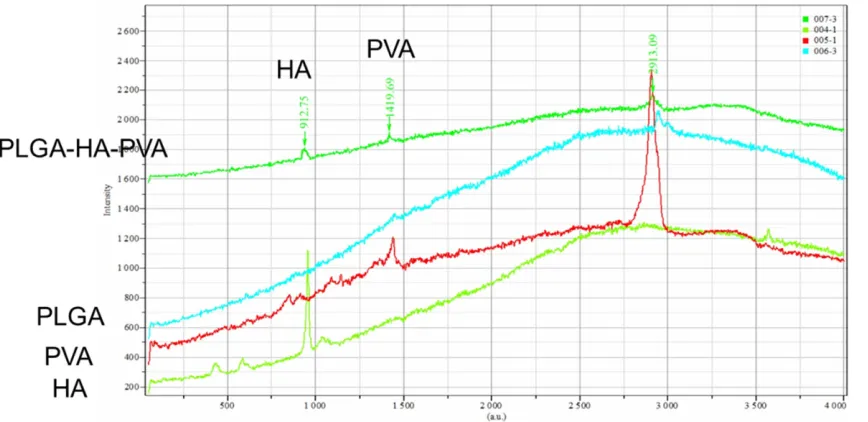
Raman spectra analysis
According to spectra analysis (Figure 1), the location of a peak corresponded to a compo-nent. The peaks of nano-HA, PVA and PLGA were located in 912.75 cm-1, 1419.69 cm-1 or
2910 cm-1, and 2950 cm-1 respectively. In
the analysis of PLGA/PVA/nano-HA composite scaffolds, the characteristic peaks of each sub-stance were appeared, and there was no other peak, and due to the overlapping of peaks, the peaks of PVA and PLGA were not fully embodied (Figure 1).
The results showed that the PLGA/PVA/nano-HA composites prepared by solvent extraction/ evaporation technique and freeze-thaw cycling methods have no visible evidence of impurity. During the entire making process of compos-ites, not any other chemicals are required beyond dichloromethane, a volatile liquid. By stirring constantly, dichloromethane was vola- tileout.
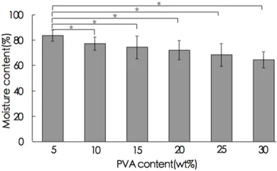
Swelling properties
As shown (Figure 2), the swelling properties could be compared among there groups. Data showed that the moisture content of each group was more than 60%. Through statistical
analysis, the moisture content of group A was greater than the other groups. In this study, the PVA hydrogel was physically cross-linked, in the form of entanglements or hydroxyls among molecular and secondary forces, such as hydro-gen bonding, Van der Waals interactions, by freeze-thaw method rather than chemical crosslink, then H2O and other small molecules were easily bonded with hydrophilic -OH group [31, 32]. Through repeating freeze-thaw cycle, the overall crystallinity was increased and the volume of amorphous region filling with water was decreased, then the microstructure of composites was changed into a fibrillar net-work. As the concentration of PVA increased, the volume of amorphous region decreased, which leaded to decreased swelling proper- ties.
ESEM detection
By analyzing the pictures (Figure 3), we could see obvious porous structures in the PLGA/ PVA/nano-HA scaffolds, which mainly consti-tuted by PVA. In addition, the PLGA particles attached to the walls of the pores, and nano-HA distributed evenly inside the scaffolds.
[image:5.612.92.287.73.194.2]In the process of freeze-thaw, molecular of PVA was redistributed and gathered to create the polymer-rich regions, which were separated by watery polymer-poor regions [30]. And the watery regions were the sites of pores. After repeating freeze-thaw cycle and addition of nano-HA, the pore structures were increasingly stable [30], which facilitated the adherence of cells. As bio-inert polymers, the hydrophilic PVA hydrogels went against cell adhesion and growth [33]. In contrast, the characteristic of hydrophobic PLGA particles facilitated the inte-gration with surrounding cartilage [18, 34]. In this study, the PLGA particles were prepared through solvent extraction/evaporation tech-nique. We evaluated the benefits of adding the micro particles to the hydrogels by the following cell experiments. There are reports that nano-HA has been used to improve the bioactivities and mechanical properties of the composites [20, 35]. Because of its high dispersibility and structure stability, nano-HA was distributed evenly inside the scaffolds. It is reported that the nano-HA/PVA composites has excellent bio-mechanics when the concentration of nano-HA is 6 wt% [20, 21]. The limitation of this paper was that the mechanical properties of the com-posites were not done yet.
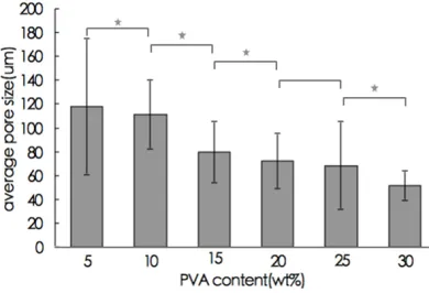
The results of pore size and PLGA particle diam-eter were showed in Figures 4 and 5.
With the increasing of PVA content, the average pore size of scaffolds was decreased, present-ing A>B>C>D and E>F (P<0.05), and there was no statistical difference (P = 0.15) between group D and group E. As the concentration of PVA increased, the proportion of polymer-rich regions increased while polymer-poor regions decreased [30, 32], which leaded to the forma-tion of smaller pores. In our research, the
aver-age pore size of group A was 117.74 μm, group B 113.30 μm, and group C 79.78 μm. It was known that pore size was a very important parameter of scaffolds, which should be large enough to benefit cell migration and permeabil-ity of nutrient while small enough to provide sufficient areas for cell attachment [36-38], meanwhile the optimal pore size of a scaffold was inconclusive, and in response to cell types [39]. Consequently, in follow-up experiments, we wanted to explore more detail about what size scaffolds were appropriate to the growth of cells.
With the increasing of PVA content, the PLGA particle diameter changed in the trend of small, but it was not statistical difference (P>0.05). In the future, loaded with cytokines, we assured that the PLGA particles might be a promising delivery system in situ.
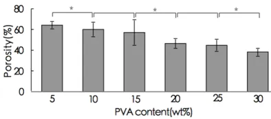
Determination of porosity
[image:6.612.90.524.71.342.2]The results of porosity were showed in Figure 6. With the increasing of PVA content, the porosity of scaffolds was decreased, presenting A>B, C>D and E>F (P<0.05), besides there was no
Figure 3. ESEM images of PLGA/PVA/nano-HA composite hydrogels. Scale bars: 200 μm. A: 5 wt% PVA+30 wt% PLGA+5 wt% nano-HA; B: 10 wt% PVA+30 wt% PLGA+5 wt% nano-HA; C: 15 wt% PVA+30 wt% PLGA+5 wt% nano-HA; D: 20 wt% PVA+30 wt% PLGA+5 wt% nano-HA; E: 25 wt% PVA+30 wt% PLGA+5 wt% nano-HA; F: 30 wt% PVA+30 wt% PLGA+5 wt% nano-HA.
[image:6.612.92.287.416.549.2]statistical difference between group B and group C (P = 1.64), and between group D and group E (P = 0.64). Results showed that when the concentration of PVA was less than 15 wt%, the porosity of composites was between 57% and 64%. As reported, a high porosity repre-sented large area/volume ratios, which facili-tated cell adhesion, while porosity and mechan-ical properties were often conflicting [39]. In our study, the relatively superior scaffolds, lying in the first three groups, were used for further cell experiment in vitro. Honestly, the porosity was inadequate comparing with other papers [40], the reason might be that the addition of PLGA particles occupied the position of pores. With the degradation of PLGA, the porosity would gradually increase.
Cellular adhesion ability and proliferation
The morphology and growth feature of human MSCs were as follow (Figure 7).
On the first day after resuscitation, the cultured cells grew against the wall of flask, with spin-dle-shaped morphology; and the number of cells increased gradually; on the eighth day, cells were spindle shaped and whorled or paral-lel along longitudinal axis.
The results of cellular adhesion ability were showed in Figure 8.
The adhesion ability in the experimental groups was significantly stronger than that in the con-trol one (P<0.05), while there was no statistical difference between the three experimental groups (P>0.05). This study found that the addi-tion of PLGA dramatically favored the adhesion of human MSCs. As mentioned above, the hydrophobic PLGA particles and nano-HA facili-tated the integration with surrounding cartilage and improved the porosity of scaffolds [18, 34, 41]. Through the combined effort of PLGA and nano-HA, the cellular attachment ability signi- ficantly increased. Since the mount of PLGA and nano-HA were identical, there was almost no difference among the three experimental groups.
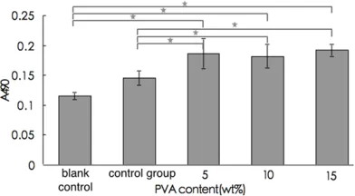
The results of cellular proliferation were showed in Figure 9.
The proliferation in the experimental groups was stronger than that in the control one (P<0.05), further, the proliferation of the latter was stronger than that of the blank control one (P<0.05), in addition, there were no statistical difference between the three experimental groups (P>0.05). Some researchers have sh- own that the morphology and mechanical pro- perty of scaffolds, such as elastic modulus, affected how the seeded cells behave [42-44]. The results showed that the existence of these scaffolds benefited the proliferation of cells; furthermore the PLGA/PVA/nano-HA hydrogels were better than the PVA/nano-HA hydrogels. The reason for this phenomenon might be that the elastic modulus in the PLGA/PVA/nano-HA hydrogels was more suitable for proliferation than that in the PVA/nano-HA hydrogels. Accordingly, we might think that the increase of pore size and porosity helped to improve cellu-lar proliferation.
The results of these tests led us to the conclu-sion that the addition of PLGA micro particles favored the adhesion and proliferation of seed-ed human MSCs.
Chondrogenesis of MSCs in hydrogels
HE staining results showed that plenty cells were observed in the pores of scaffolds (Figure 10).
[image:7.612.90.290.72.156.2]The results of HE staining demonstrated that the cells proliferated well and secreted extra-cellular matrix, mimicking chondroid tissue. In
Figure 5. PLGA particle determination, determined using ESEM and Image J software.
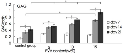
[image:7.612.90.287.210.297.2]addition, the amount of matrix in experimental group was more than that in control group. Results of Western blotting showed that the expression of COL2 and GAG could be detected in experimental and control group (Figures 11 and 12).
Main results were as follow: (1) The relative expression of COL2 and GAG increased over
time (P<0.05); (2) When co-cultured for 21 days, the expression of COL2 and GAG in exper-imental group was higher than control group (P<0.05), meanwhile the expression in group C was higher than the other two groups (P<0.05), and the group B higher than group A (P<0.05). This finding assured that the PLGA/PVA/nano-HA hydrogels, compared with PVA/nano-PLGA/PVA/nano-HA hydrogels, did better in improving the chondro-genic differentiation of human MSCs. The suc-cess of this novel composite was related to the addition of PLGA micro particles, which improved the biocompatibility of scaffolds. Meanwhile the amount of COL2 and GAG was higher in group C than that in the other two experimental groups. According to the above, the pore size and porosity in-group C was the least among the experimental groups. So we concluded that the chondrogenic differentia-tion of human MSCs could not be improved just by increasing the pore size and porosity. It is reported that the elastic modulus of scaffolds affects the differentiation of stem cells [45-47]. So we assured that the elastic modulus of the relatively small pore size and porosity might be more appropriate for differentiation. But more research is needed to explore the most suit-able elastic modulus of this composite. Conclusions
The morphological characterizations of this novel PLAG/PVA/nano-HA composite scaffolds, fabricating by solvent extraction/evaporation technique and freeze-thaw cycling method were changed in relation to PVA content. When PVA content was among 5-15 wt%, the pore size and porosity of the composites were
[image:8.612.90.526.72.219.2]appropri-Figure 7. Morphological and growth feature of human MSCs by inverted microscope, magnification ×400. A: 2 hours after resuscitation B: 8 days of culture.
Figure 8. Cell adhesion rate of PLGA/PVA/nano-HA composite hydrogels, the scaffolds in the control group were only composed of nano-HA 5 wt% and PVA 20 wt% (★P<0.05).
[image:8.612.90.287.272.366.2] [image:8.612.92.288.442.550.2]ate. The results of cell culture in vitro showed that the composites not only favored the adhe-sion and proliferation of seeded human MSCs, but also promote the differentiation toward chondrocytes. In addition, when the mass frac-tion of PLGA was 30 wt%, and nano-HA 5 wt%, the relatively suitable proportion of PVA was 15 wt% considering morphological properties and short-term practicability in vitro. Overall, all these results suggested that the feasibility of these novel composites as promising scaffolds in cartilage tissue engineering.
Acknowledgements
Research is supported by the national natural science foundation of China (NO. 81371934, NO. 81501860).
Disclosure of conflict of interest
None.
[image:9.612.90.523.71.398.2]Address correspondence to: Dr. Yihe Hu, Depart- ment of Orthopedics, Xiangya Hospital of Central
[image:9.612.92.286.443.543.2]Figure 10. HE staining of the composites seeding human MSCs for 21 days, magnification ×100.
Figure 11. Relative expression of COL2 in scaffolds (★P<0.05).
[image:9.612.90.288.597.683.2]South University, 87# Xiangya Road, Changsha,
Hunan, China. E-mail: [email protected]
References
[1] Castro NJ, Hacking SA, Zhang LG. Recent prog-ress in interfacial tissue engineering approach-es for osteochondral defects. Ann Biomed Eng 2012; 40: 1628-1640.
[2] Mauck RL, Wang CC, Oswald ES, Ateshian GA, Hung CT. The role of cell seeding density and nutrient supply for articular cartilage tissue en-gineering with deformational loading. Osteoar- thritis Cartilage 2003; 11: 879-890.
[3] Rajpurohit R, Koch CJ, Tao Z, Teixeira CM, Shapiro IM. Adaptation of chondrocytes to low oxygen tension: relationship between hypoxia and cellular metabolism. J Cell Physiol 1996; 168: 424-432.
[4] Meinhart J, Fussenegger M, Hobling W. Sta- bilization of fibrin-chondrocyte constructs for cartilage reconstruction. Ann Plast Surg 1999; 42: 673-678.
[5] Makris EA, Gomoll AH, Malizos KN, Hu JC, Athanasiou KA. Repair and tissue engineering techniques for articular cartilage. Nat Rev Rheumatol 2015; 11: 21-34.
[6] Holmes B, Zhu W, Li J, Lee JD, Zhang LG. Development of novel three-dimensional print-ed scaffolds for osteochondral regeneration. Tissue Eng Part A 2015; 21: 403-415.
[7] Seo SJ, Mahapatra C, Singh RK, Knowles JC, Kim HW. Strategies for osteochondral repair: Focus on scaffolds. J Tissue Eng 2014; 5: 2041731414541850.
[8] Zhang L, Hu J, Athanasiou KA. The role of tis-sue engineering in articular cartilage repair and regeneration. Crit Rev Biomed Eng 2009; 37: 1-57.
[9] Magalhaes J, Sousa RA, Mano JF, Reis RL, Blanco FJ, San Roman J. Synthesis and cha- racterization of sensitive hydrogels based on semi-interpenetrated networks of poly [2-eth-yl-(2-pyrrolidone) methacrylate] and hyaluronic acid. J Biomed Mater Res A 2013; 101: 157-166.
[10] Iwasa J, Engebretsen L, Shima Y, Ochi M. Clinical application of scaffolds for cartilage tissue engineering. Knee Surg Sports Trau- matol Arthrosc 2009; 17: 561-577.
[11] Noguchi T, Yamamuro T, Oka M, Kumar P, Kotoura Y, Hyon S, Ikada Y. Poly (vinyl alcohol) hydrogel as an artificial articular cartilage: evaluation of biocompatibility. J Appl Biomater 1991; 2: 101-107.
[12] Kobayashi M, Chang YS, Oka M. A two year in vivo study of polyvinyl alcohol-hydrogel (PVA-H) artificial meniscus. Biomaterials 2005; 26: 3243-3248.
[13] Gu ZQ, Xiao JM, Zhang XH. The development of artificial articular cartilage--PVA-hydrogel. Biomed Mater Eng 1998; 8: 75-81.
[14] Lee SY, Pereira BP, Yusof N, Selvaratnam L, Yu Z, Abbas AA, Kamarul T. Unconfined compres-sion properties of a porous poly (vinyl alcohol)-chitosan-based hydrogel after hydration. Acta Biomater 2009; 5: 1919-1925.
[15] Maher SA, Doty SB, Torzilli PA, Thornton S, Lowman AM, Thomas JD, Warren R, Wright TM, Myers E. Nondegradable hydrogels for the treatment of focal cartilage defects. J Biomed Mater Res A 2007; 83: 145-155.
[16] Spiteri CG, Pilliar RM, Kandel RA. Substrate porosity enhances chondrocyte attachment, spreading, and cartilage tissue formation in vitro. J Biomed Mater Res A 2006; 78: 676-683.
[17] Spiller KL, Laurencin SJ, Charlton D, Maher SA, Lowman AM. Superporous hydrogels for carti-lage repair: Evaluation of the morphological and mechanical properties. Acta Biomater 2008; 4: 17-25.
[18] Spiller KL, Holloway JL, Gribb ME, Lowman AM. Design of semi-degradable hydrogels based on poly(vinyl alcohol) and poly (lactic-co-glycolic acid) for cartilage tissue engineering. J Tissue Eng Regen Med 2011; 5: 636-647.
[19] Xu F, Li Y, Deng Y, Xiong J. Porous nano-hydroxy-apatite/poly (vinyl alcohol) composite hydrogel as artificial cornea fringe: characterization and evaluation in vitro. J Biomater Sci Polym Ed 2008; 19: 431-439.
[20] Pan Y, Xiong D. Study on compressive mech- anical properties of nanohydroxyapatite rein-forced poly(vinyl alcohol) gel composites as biomaterial. J Mater Sci Mater Med 2009; 20: 1291-1297.
[21] Pan Y, Xiong D, Gao F. Viscoelastic behavior of nano-hydroxyapatite reinforced poly(vinyl alco-hol) gel biocomposites as an articular carti-lage. J Mater Sci Mater Med 2008; 19: 1963-1969.
[22] Dziedzic K, Zalewski M, Gadek A, Drukala J. [Chondrocytes application in regenerative me- dicine]. Przegl Lek 2014; 71: 334-339. [23] Cheng T, Maddox NC, Wong AW, Rahnama R,
Kuo AC. Comparison of gene expression pat-terns in articular cartilage and dedifferentiat-ed articular chondrocytes. J Orthop Res 2012; 30: 234-245.
[24] Ahmed TA, Hincke MT. Mesenchymal stem cell-based tissue engineering strategies for repair of articular cartilage. Histol Histopathol 2014; 29: 669-689.
[26] Gupta PK, Das AK, Chullikana A, Majumdar AS. Mesenchymal stem cells for cartilage repair in osteoarthritis. Stem Cell Res Ther 2012; 3: 25. [27] Griffon DJ, Abulencia JP, Ragetly GR, Fredericks
LP, Chaieb S. A comparative study of seeding techniques and three-dimensional matrices for mesenchymal cell attachment. J Tissue Eng Regen Med 2011; 5: 169-179.
[28] Fisher OZ, Khademhosseini A, Langer R, Peppas NA. Bioinspired materials for control-ling stem cell fate. Acc Chem Res 2010; 43: 419-428.
[29] Klose D, Siepmann F, Elkharraz K, Krenzlin S, Siepmann J. How porosity and size affect the drug release mechanisms from PLGA-based microparticles. Int J Pharm 2006; 314: 198-206.
[30] Gupta S, Goswami S, Sinha A. A combined ef-fect of freeze--thaw cycles and polymer con-centration on the structure and mechanical properties of transparent PVA gels. Biomed Mater 2012; 7: 015006.
[31] Wan WK, Campbell G, Zhang ZF, Hui AJ, Boughner DR. Optimizing the tensile proper-ties of polyvinyl alcohol hydrogel for the con-struction of a bioprosthetic heart valve stent. J Biomed Mater Res 2002; 63: 854-861. [32] Millon LE, Padavan DT, Hamilton AM, Boughner
DR, Wan W. Exploring cell compatibility of a fi-bronectin-functionalized physically crosslinked poly (vinyl alcohol) hydrogel. J Biomed Mater Res B Appl Biomater 2012; 100: 1-10. [33] Nuttelman CR, Mortisen DJ, Henry SM, Anseth
KS. Attachment of fibronectin to poly (vinyl al-cohol) hydrogels promotes NIH3T3 cell adhe-sion, proliferation, and migration. J Biomed Mater Res 2001; 57: 217-223.
[34] Zhou G, Liu W, Cui L, Wang X, Liu T, Cao Y. Repair of porcine articular osteochondral de-fects in non-weightbearing areas with autolo-gous bone marrow stromal cells. Tissue Eng 2006; 12: 3209-3221.
[35] Na K, Kim S, Sun BK, Woo DG, Chung HM, Park KH. Blended construct consisting of thermo-reversible hydrogels and heparinized nanopar-ticles for increasing the proliferation activity of the rabbit chondrocyte in vivo test. Biotechnol Lett 2007; 29: 1447-1452.
[36] O’Brien FJ, Harley BA, Waller MA, Yannas IV, Gibson LJ, Prendergast PJ. The effect of pore size on permeability and cell attachment in collagen scaffolds for tissue engineering. Technol Health Care 2007; 15: 3-17.
[37] O’Brien FJ, Harley BA, Yannas IV, Gibson LJ. The effect of pore size on cell adhesion in col-lagen-GAG scaffolds. Biomaterials 2005; 26: 433-441.
[38] Matsiko A, Gleeson JP, O’Brien FJ. Scaffold mean pore size influences mesenchymal stem cell chondrogenic differentiation and matrix deposition. Tissue Eng Part A 2015; 21: 486-497.
[39] Zeltinger J, Sherwood JK, Graham DA, Mueller R, Griffith LG. Effect of pore size and void frac-tion on cellular adhesion, proliferafrac-tion, and matrix deposition. Tissue Eng 2001; 7: 557-572.
[40] Freed LE, Vunjak-Novakovic G, Biron RJ, Eagles DB, Lesnoy DC, Barlow SK, Langer R. Bio- degradable polymer scaffolds for tissue engi-neering. Biotechnology (N Y) 1994; 12: 689-693.
[41] Zheng L, Jiang X, Chen X, Fan H, Zhang X. Evaluation of novel in situ synthesized nano-hydroxyapatite/collagen/alginate hydrogels for osteochondral tissue engineering. Biomed Mater 2014; 9: 065004.
[42] Buxton PG, Bitar M, Gellynck K, Parkar M, Brown RA, Young AM, Knowles JC, Nazhat SN. Dense collagen matrix accelerates osteogenic differentiation and rescues the apoptotic re-sponse to MMP inhibition. Bone 2008; 43: 377-385.
[43] Bryant SJ, Anseth KS. Hydrogel properties in-fluence ECM production by chondrocytes pho-toencapsulated in poly(ethylene glycol) hydro-gels. J Biomed Mater Res 2002; 59: 63-72. [44] Boontheekul T, Hill EE, Kong HJ, Mooney DJ.
Regulating myoblast phenotype through con-trolled gel stiffness and degradation. Tissue Eng 2007; 13: 1431-1442.
[45] Engler AJ, Sen S, Sweeney HL, Discher DE. Matrix elasticity directs stem cell lineage spec-ification. Cell 2006; 126: 677-689.
[46] Jung H, Park JS, Yeom J, Selvapalam N, Park KM, Oh K, Yang JA, Park KH, Hahn SK, Kim K. 3D tissue engineered supramolecular hydro-gels for controlled chondrogenesis of human mesenchymal stem cells. Biomacromolecules 2014; 15: 707-714.