Original Article
Diagnostic significance of the BRAF V600E mutation in
conventional papillary thyroid carcinomas
Bo Zhang1, Chun-Wei Xu1, Yong-Fang Wu1, Qiu-Hong Man1, Ye-Ying Song1, Jing-Jing Wang1, Huai-Tao Wang1,
Hai-Yan Wang1, Xiao-Bing Li1, Hao Zhang2, Ting Ye2, Zhe Zhang2, Cong-Li Cai2
1Department of Pathology, Affiliated Hospital of Academy of Military Medical Sciences, Beijing 100071, People’s
Republic of China; 2Wuhan YZY Medical Science & Technology Co., Ltd, Wuhan 430075, Hubei, People’s Republic
of China
Received October 20, 2015; Accepted March 29, 2016; Epub May 15, 2016; Published May 30, 2016
Abstract: Papillary thyroid carcinoma (PTC) is the most common histological thyroid cancer, accounting for approxi-mately 80% of thyroid cancers. Although PTC is highly curable, some patients encounter high rates of morbidity and mortality. BRAF V600E mutations are the most common genetic mutation in PTC and the relationship between
BRAF V600E mutations and PTC has recently been a focus of research. The aim of the present study was to deter-mine the prevalence of BRAF V600E mutation in PTC tissue, pericarcinous tissue, and benign tumor samples, and explore the correlation between BRAF V600E mutations and different clinicopathological features. In the present study, 120 conventional PTC samples, 30 pericarcinous tissue, and 25 benign nodule samples were collected, and the BRAF V600E mutation was tested using the BRAF Gene V600E Mutation Detection Kit. Statistical analyses were performed for the data on BRAF V600E mutation in three kinds of tissue and in different clinicopathological categories of PTC. Finally, results revealed that the prevalence of BRAF V600E mutation in exact cancerous tissue of 120 PTC samples was 88.3%, and BRAF V600E mutations were absent in the pericarcinous tissue and benign nodule samples. There were no significant differences in the prevalence of BRAF V600E mutation depending on the age, gender, tumor size, lymph node metastasis, and lesion location (P > 0.05). Consequently, the BRAF V600E mu-tation was found to be highly prevalent in conventional PTC samples due to more accurate sample processing and the use of different detection methods, and absolutely negative in pericarcinous tissue and benign nodule samples. Therefore, BRAF V600E mutation could suggest a useful predictor of tumorigenesis in PTC, and such mutation may also affect local carcinoma development. There is no evidence that the BRAF V600E mutation significantly reflects aggressive characteristics and poor prognosis of patients with PTC.
Keywords: Papillary thyroid carcinoma, BRAF mutation, diagnostic significance, ARMS-qPCR
Introduction
Thyroid cancer is one of the most common endocrine malignancies with complicated pro-cesses, and 5-10% of thyroid nodules are thy-roid carcinomas [1, 2]. Recently, the incidence of thyroid cancer as well as the tumor-targeted therapy has increased rapidly; in China, the incidence of thyroid cancer increased annually by 14.51%, whereas the mortality increased by 1.42% [3]. Thyroid cancer can be classified as papillary thyroid cancer (PTC), follicular thyroid cancer (FTC), anaplastic thyroid cancer (ATC), and medullary carcinoma based on the histo-logical characteristics [1, 4]. Among the four histological types, PTC is the most common
thy-roid cancer, accounting for more than 80% of all thyroid malignancies [1, 5]. PTC is highly cur-able through surgery and roentgenotherapy and has a good prognosis compared to the worst prognosis of ATC [6]. Some patients face the risk of death from the occurrence of PTC when it becomes surgically inoperable and radioiodine avidity is not available [7].
funda-mental role in cell proliferation, differentiation, apoptosis, and survival [8-10]. Recurrence of PTC involves a series of genetic events such as RAS/RAF gene mutation, RET/PTC rearrange-ment, and p53 inactivation, and these muta-tions are found in most PTC cases and rarely overlap in the same tumor [11-13].
BRAF kinase, encoded by the BRAF gene, also referred to as proto-oncogene B-Raf and v-Raf murine sarcoma viral oncogene homolog B, is a member of the RAF family of serine-threonine kinases. The BRAF gene is the strongest activa-tor downstream of the MAPK pathway and
Materials and methods
Preparation of cancerous, pericarcinous, and benign tissue samples
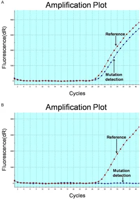
All samples used for the present study were obtained from the Pathology Department at the Affiliated Hospital of the Academy of Military Medical Sciences. We selected 120 patients who were diagnosed with conventional papil-lary thyroid cancer from 2011 to 2014. All the tumors showed typical papillary carcinoma his-tology. Then, we analyzed the histology of the cancerous tissue microscopically using hema-Figure 1. Amplification plot of BRAF V600E-positive (A) and -negative (B)
results in Mx3000P. Results were analyzed using fluorescence quantitative PCR, The reference curve reflects the DNA quality in samples; the mutation-detection curve performs an obvious amplification curve and with the delta Ct (Ctmutation-Ctreference) < 9 in positive results, the mutation-detection curve showed a straight line when no mutation signal was collected.
tumorigenesis occurs when
BRAF is aberrantly activated [14-16]. BRAF mutations exist in 66% of malignant melano-mas and 15% of colorectal cancer [17]; however, the BRAF protein may be indepen-dent of the RAS protein in can-cer occurrence. BRAF muta-tions showed a high preva-lence in PTC, among which the T1799A point BRAF muta-tion is the most common, ac- counting for more than 90% of all BRAF mutations [18-20]. Based on the important role of BRAF mutations in papillary carcinomas, much research has focused on BRAF gene mutations and clinical patho-logical parameters of papillary carcinomas.
In the present study, we ana-lyzed BRAF V600E mutations in 120 PTC tissues, 30 peri-carcinous tissues, and 25 benign tumor (adenoma and hyperplasia) samples. This article will focus on the preva-lence of BRAF mutations in 175 tissues and the discrep-ancy in the presence of BRAF
[image:2.612.92.371.74.470.2]toxylin and eosin (HE) staining, the exact can-cerous part was marked in the slice. Corresponding formalin-fixed paraffin blocks were prepared and sliced into appropriate sec-tions. Cancerous tissues were marked in paraf-fin sections and compared with the marked HE stained tissues. The cancerous tissue was scraped into 1.5 ml clean test tubes for DNA extraction. All cancerous samples used were large, excluding fine needle aspiration and nee-dle biopsy tissues. The PTC patients included 94 females and 26 males ranging from 17 to 70 years old, with PTC tumor with sizes ranging from 0.3 to 4.5 cm. Pericarcinous samples were randomly collected from 30 patients with PTC. Pericarcinous tissues were also verified microscopically. We also collected 25 benign tumor samples including hyperplastic nodules associated with Hashimoto’s thyroiditis.
Genomic DNA isolation
Genomic DNA was isolated using the QIAamp DNA FFPE Tissue Kit (Qiagen, Germany, Cat.56404). Samples embedded in paraffin were treated by dimethylbenzene dewaxing, and washed in ethanol. Dried samples were incubated in buffer ATL and proteinase K at 56°C overnight. DNA was purified and eluted, and the final concentration was measured using a Nanodrop 2000 spectrophotometer. DNA quality was evaluated by measuring the A260/A280 ratio.
Detection of the BRAF V600E mutation
All DNA samples were diluted to 10 ng/µl, and detection of the BRAF V600E mutation was performed using the Human BRAF Gene V600E Mutation Detection Kit provided by the Wuhan
YZY Medical Science & Technology Co., Ltd. The kit was developed using an Amplification Re- fractory Mutation System (ARMS)-qPCR detect-ing method. The reaction system was per-formed in two PCR tubes using 20 ng of DNA in a 25 µL reaction volume containing the refer-ence and V600E mutation detection reagent (containing PCR buffer, dNTPs, specific prim-ers, specific probes, and Taq polymerase). The amplification procedure was subjected to 40 cycles involving uracil-N-glycosylase treatment at 37°C for 10 min, initial denaturation at 95°C for 5 min, denaturation at 95°C for 15 s, and annealing and extension at 60°C for 1 min. The PCR was processed in the Agilent StrataGene Mx3000P QPCR System. The detection limit of this Kit was 1% BRAF V600E mutation in 20 ng genomic DNA; positive and negative results could be explicitly determined according to the user instructions.
Statistical analyses
BRAF V600E-positive and -negative papillary thyroid cancer numbers were compared for each category containing gender, age, tumor size, tumor location, and clinical stage. Com- parison between groups was performed using SPSS 13.0 statistical software and a χ2 test
was used for the comparisons. P < 0.05 was considered to be statistically different.
Results
Basic clinicopathological features of patients
[image:3.612.89.524.72.180.2]ried lymph node metastasis. Among 25 indi-viduals with benign nodules, 4 were male and 21 were female with a female: male ratio of 5:3. The range of age was 33~62.
Pathological characteristics of three kinds of tissues
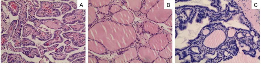
All samples diagnosed with PTC and benign nodules were traced using HE staining, and typical staining results of three tissue types are shown in Figure 2.
BRAF V600E mutations in three tissue types
Among all the procedures performed in Mx- 3000P, the positive and negative controls in
size (P = 0.821), lymph node metastasis (P = 0.352), and lesion location (P = 0.399) (Table 2).
Discussion
The BRAF V600E mutation is the most com-mon genetic mutation in PTC and generally occurs in approximately 29-83% of all cases [20]. In the present study, we analyzed the prevalence of the BRAF V600E mutation in dif-ferent tissues, and especially in PTC, we report-ed an excereport-edingly high prevalence of the BRAF
V600E mutation (88.3%). Moreover, no BRAF
mutation was detected in benign nodules and pericarcinous tissue, which raised the
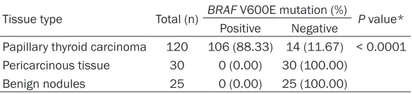
possibil-Table 1.BRAF V600E mutation in three different tissues
Tissue type Total (n) BRAF V600E mutation (%) P value* Positive Negative
Papillary thyroid carcinoma 120 106 (88.33) 14 (11.67) < 0.0001 Pericarcinous tissue 30 0 (0.00) 30 (100.00)
Benign nodules 25 0 (0.00) 25 (100.00)
*Papillary thyroid carcinoma Vesus Pericarcinous tissue P < 0.001; Papillary thyroid carcinoma Vesus Benign nodules P < 0.001; Pericarcinous tissue Vesus benign nodules P = 1.000.
Table 2. Correlation of BRAF V600E mutation with differ-ent clinicopathological characteristics
Clinical features Total (n) BRAF V600E mutation (%) valueP Positive Negative
Gender 1.000
Male 26 23 (88.46) 3 (11.54) Female 94 83 (88.30) 11 (11.70)
Age 0.755
≥ 50 31 27 (87.10) 4 (12.90) < 50 89 79 (88.76) 10 (11.24)
Tumor size, cm 0.821
≥ 1.0 55 49 (89.09) 6 (10.91) < 1.0 65 57 (87.69) 8 (12.31)
Lymph metastasis 0.352 Yes 36 30 (83.33) 6 (16.67)
No 84 76 (90.48) 8 (9.52)
Tumor stage 0.536
I and II 86 77 (89.53) 9 (10.47) III and IV 34 29 (85.29) 5 (14.71)
Tumor lesion 0.399
Left 59 54 (91.53) 5 (8.47) Right 45 39 (86.67) 6 (13.33) Both 13 10 (76.92) 3 (23.08) Isthmus 3 3 (100.00) 0 (0.00)
conventional PTC tumors, and 14 cases were BRAF V600E-negative. No
BRAF V600E mutations were detected in 30 cases of pericarcinous tissues and 25 cases of benign tumor tissues. Amplification plot of one BRAF V600E positive and negative result is shown in
Figure 1.
The prevalence of BRAF V600E muta-tions in PTC is 88.3%, and 0% in peri-carcinous tissue and benign nodules; therefore, the difference between PTC and other two groups is statistically sig-nificant (P < 0.05).
Analysis of BRAF V600E prevalence in different categories in PTC
As described above, different clinico-pathological features involving age, tumor size, lymph node metastases, gender, tumor stage were classified and analyzed for the correlation with the status of the BRAF V600E muta-tion. No statistical significance were shown in the prevalence of BRAF
[image:4.612.91.383.85.152.2]ity that BRAF V600E acts as a determinant fac-tor in PTC tumorigenesis. In fact, there are some differences in the prevalence of BRAF
V600E mutations in different subtypes of PTC. Lee et al. [21] exhibited different distributions of BRAF V600E mutations in different histologi-cal subtypes. They found that BRAF V600E mutations are most frequent in tall cell variants of PTC (79.1%), and then in conventional PTC (59.1%). In the present study, we observed a higher prevalence of BRAF V600E mutations in PTC. Several reasons can be elucidated for this striking distribution: no fine-needle aspiration (FNA) biopsy tissue of papillary thyroid cancer was used, and inaccurate detection results could be obtained from minute tissue samples and operation instability, and in contrast, large mass tissues diagnosed pathologically were less susceptible; exact cancerous tissues were detected and scraped from the slice, con-tributing a significant role in reducing false-negative result from the pericarcinous portion. Interestingly, pericarcinous tissues in group B have been proven as negative.
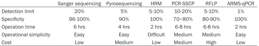
Detection of somatic mutations in tumor tis-sues including direct sequencing, pyrosequenc-ing, PCR-RFLP (polymerase chain reaction restriction fragment length polymorphism), SSCP (Single-Strand Conformation Polymorph- ism), was most used in current research [22-30]. Sensitivity, specificity, and other indexes of different detection methods were compared (Table 3), and direct sequencing was demon-strated to be a more reliable method with a sensitivity of 83% and specificity of 96% than PCR-RFLP with a sensitivity of 78.6% and spec-ificity of 80% [30, 31]. The mutation detecting methods used in the present study, ARMS-qPCR is a conventional technology for measur-ing somatic mutation in tumors [32]. Kits can reach a detection limit at 0.5-1% mutation in 10 ng genomic DNA, which is exceedingly more sensitive than the methods mentioned above. According to the manufacturer’s instructions,
several samples harbored a BRAF V600E muta-tion less than 5% (6 < Ctmutation-Ctreference < 9), which could be assumed as negative by other detection methods. Consequently, we identi-fied a higher prevalence of BRAF V600E muta-tions in the present study.
The prognostic value of BRAF V600E mutations in PTC has been controversial in different stud-ies recently, Ito et al. [33] investigated BRAF
V600E mutations in 631 patients with PTC hav-ing median follow-up periods of 83 months, and they finally indicated that BRAF V600E mutations did not significantly reflect the aggressive characteristics and poor prognosis of patients with PTC in Japan. Also, Kim et al.
[34] studied 60 patients with conventional micro-PTC, and BRAF V600E mutations were detected in 31 of 60 PTC patients (52%), and the age distribution, tumor size, extrathyroid extension, and staging did not differ significant-ly between patients with and without the BRAF
V600E mutation. On the contrary, many reports concluded that BRAF V600E mutations show poor prognostic relationships in positive PTC patients. In the present study, we discussed the mutation status in different groups classi-fied by clinical features of PTC. It was observed that BRAF V600E mutations did not significant-ly correlate with age, gender, tumor size, significant-lymph node metastasis, and different lesion locations (P > 0.05). Among 14 BRAF V600E-negative patients, we studied the correlation with clini-cal features, and determined that BRAF V600E-negative individuals did not show significant differences with age, gender, tumor size, lymph node metastasis, and lesion locations (P > 0.05).
In the present study, BRAF V600E mutation prevalence in PTC and benign tumors was sig-nificantly different. Samples harboring V600E mutations were all cancerous tissue and no
BRAF mutations were found in benign tissue.
[image:5.612.90.525.86.164.2]BRAF V600E mutations showed extremely high
Table 3. Comparison of different detection methods used in somatic mutation
specificity in non-cancerous samples including pericarcinous tissues and benign tumor tis-sues. The detection of other genetic alterations in the MAPK pathway including BRAF V600K,
BRAF V600D, Kras, and Nras mutation was also performed in 14 V600E-negative samples. Similarly to V600E mutations with a 1% detec-tion limit, the mutadetec-tions mendetec-tioned above were negative in the 14 samples (data not shown), which raised compelling evidence that the con-stitutive activation of the RAS/RAF gene in the MAPK pathway plays a predominant role in the pathogenesis of PTC. Therefore, individuals with conventional PTC tumorigenesis could har-bor other mutations such as the RET/PTC rear-rangement in the MAPK pathway or driver gene alterations in other signal pathways. These sug-gestions may assist pathologist to determine whether cancer occurred due to aberrantly activated BRAF proteins. Testing for the BRAF
mutation may be a useful predictor of tumori-genesis in PTC.
Acknowledgements
This work is supported by the pathology depart-ment of Affiliated Hospital of Academy of Military Medical Sciences.
Disclosure of conflict of interest
None.
Authors’ contribution
HZ performed the molecular genetic studies, nucleic acid extraction and detection, and drafted the manuscript. CWX, YFW, QHM, YYS, JJW, HTW, HYW, and XBL selected and pre-pared the samples used in this study, and ana-lyzed their clinical features. HZ, TY, and ZZ designed the study and performed the statisti-cal analyses. BZ and CLC conceived the study, and contributed to the manuscript design, coor-dination, and drafting. All authors read and approved the final manuscript.
Address correspondence to: Dr. Cong-Li Cai, Wuhan YZY Medical Science & Technology Co., Ltd, Wuhan 430075, Hubei, People’s Republic of China. E-mail: [email protected]
References
[1] Xing M. BRAF mutation in thyroid cancer. Endocr Relat Cancer 2005; 12: 245-262.
[2] Hegedus L. Clinical practice. The thyroid nod-ule. N Engl J Med 2004; 351: 1764-1771. [3] Wang Y and Wang W. Increasing Incidence of
Thyroid Cancer in Shanghai, China, 1983-2007. Asia Pac J Public Health 2015; 27: NP223-9.
[4] Chan JK. Papillary carcinoma of thyroid: classi-cal and variants. Histol Histopathol 1990; 5: 241-257.
[5] Ain KB. Papillary thyroid carcinoma. Etiology, assessment, and therapy. Endocrinol Metab Clin North Am 1995; 24: 711-760.
[6] Fernandes JK, Day TA, Richardson MS and Sharma AK. Overview of the management of differentiated thyroid cancer. Curr Treat Options Oncol 2005; 6: 47-57.
[7] Xing M, Westra WH, Tufano RP, Cohen Y, Rosenbaum E, Rhoden KJ, Carson KA, Vasko V, Larin A, Tallini G, Tolaney S, Holt EH, Hui P, Umbricht CB, Basaria S, Ewertz M, Tufaro AP, Califano JA, Ringel MD, Zeiger MA, Sidransky D and Ladenson PW. BRAF mutation predicts a poorer clinical prognosis for papillary thyroid cancer. J Clin Endocrinol Metab 2005; 90: 6373-6379.
[8] Lam KY, Lo CY and Leung PS. High prevalence of RET proto-oncogene activation (RET/PTC) in papillary thyroid carcinomas. Eur J Endocrinol 2002; 147: 741-745.
[9] Vecchio G and Santoro M. Oncogenes and thy-roid cancer. Clin Chem Lab Med 2000; 38: 113-116.
[10] Xu X, Quiros RM, Gattuso P, Ain KB and Prinz RA. High prevalence of BRAF gene mutation in papillary thyroid carcinomas and thyroid tumor cell lines. Cancer Res 2003; 63: 4561-4567. [11] Kimura ET, Nikiforova MN, Zhu Z, Knauf JA,
Nikiforov YE and Fagin JA. High prevalence of BRAF mutations in thyroid cancer: genetic evi-dence for constitutive activation of the RET/ PTC-RAS-BRAF signaling pathway in papillary thyroid carcinoma. Cancer Res 2003; 63: 1454-1457.
[12] Soares P, Trovisco V, Rocha AS, Lima J, Castro P, Preto A, Maximo V, Botelho T, Seruca R and Sobrinho-Simoes M. BRAF mutations and RET/ PTC rearrangements are alternative events in the etiopathogenesis of PTC. Oncogene 2003; 22: 4578-4580.
[13] Frattini M, Ferrario C, Bressan P, Balestra D, De Cecco L, Mondellini P, Bongarzone I, Collini P, Gariboldi M, Pilotti S, Pierotti MA and Greco A. Alternative mutations of BRAF, RET and NTRK1 are associated with similar but distinct gene expression patterns in papillary thyroid cancer. Oncogene 2004; 23: 7436-7440. [14] Sebolt-Leopold JS and Herrera R. Targeting the
[15] Kohno M and Pouyssegur J. Targeting the ERK signaling pathway in cancer therapy. Ann Med 2006; 38: 200-211.
[16] Mercer KE and Pritchard CA. Raf proteins and cancer: B-Raf is identified as a mutational tar-get. Biochim Biophys Acta 2003; 1653: 25-40. [17] Davies H, Bignell GR, Cox C, Stephens P, Edkins
S, Clegg S, Teague J, Woffendin H, Garnett MJ, Bottomley W, Davis N, Dicks E, Ewing R, Floyd Y, Gray K, Hall S, Hawes R, Hughes J, Kosmidou V, Menzies A, Mould C, Parker A, Stevens C, Watt S, Hooper S, Wilson R, Jayatilake H, Gusterson BA, Cooper C, Shipley J, Hargrave D, Pritchard-Jones K, Maitland N, Chenevix-Trench G, Riggins GJ, Bigner DD, Palmieri G, Cossu A, Flanagan A, Nicholson A, Ho JW, Leung SY, Yuen ST, Weber BL, Seigler HF, Darrow TL, Paterson H, Marais R, Marshall CJ, Wooster R, Stratton MR and Futreal PA. Mutations of the BRAF gene in human cancer. Nature 2002; 417: 949-954.
[18] Riesco-Eizaguirre G and Santisteban P. New insights in thyroid follicular cell biology and its impact in thyroid cancer therapy. Endocr Relat Cancer 2007; 14: 957-977.
[19] Oler G, Ebina KN, Michaluart P Jr, Kimura ET and Cerutti J. Investigation of BRAF mutation in a series of papillary thyroid carcinoma and matched-lymph node metastasis reveals a new mutation in metastasis. Clin Endocrinol (Oxf) 2005; 62: 509-511.
[20] Sapio MR, Posca D, Troncone G, Pettinato G, Palombini L, Rossi G, Fenzi G and Vitale M. Detection of BRAF mutation in thyroid papillary carcinomas by mutant allele-specific PCR am-plification (MASA). Eur J Endocrinol 2006; 154: 341-348.
[21] Lee JH, Lee ES and Kim YS. Clinicopathologic significance of BRAF V600E mutation in papil-lary carcinomas of the thyroid: a meta-analy-sis. Cancer 2007; 110: 38-46.
[22] Jancik S, Drabek J, Berkovcova J, Xu YZ, Stankova M, Klein J, Kolek V, Skarda J, Tichy T, Grygarkova I, Radzioch D and Hajduch M. A comparison of Direct sequencing, Pyrose- quencing, High resolution melting analysis, TheraScreen DxS, and the K-ras StripAssay for detecting KRAS mutations in non small cell lung carcinomas. J Exp Clin Cancer Res 2012; 31: 79.
[23] Young EC, Owens MM, Adebiyi I, Bedenham T, Butler R, Callaway J, Cranston T, Crosby C, Cree IA, Dutton L, Faulkes C, Faulkner C, Howard E, Knight J, Huang Y, Lavender L, Lazarou LP, Liu H, Mair D, Milano A, Sandell S, Skinner A, Wallace A, Williams M, Spivey V, Goodall J, Frampton J, Ellard S; Clinical Molecular Genetics Society (CMGS) Scientific Subcom-
mittee. A comparison of methods for EGFR mu-tation testing in non-small cell lung cancer. Diagn Mol Pathol 2013; 22: 190-195.
[24] Tsiatis AC, Norris-Kirby A, Rich RG, Hafez MJ, Gocke CD, Eshleman JR and Murphy KM. Comparison of Sanger sequencing, pyrose-quencing, and melting curve analysis for the detection of KRAS mutations: diagnostic and clinical implications. J Mol Diagn 2010; 12: 425-432.
[25] Pinto P, Rocha P, Veiga I, Guedes J, Pinheiro M, Peixoto A, Pinto C, Fragoso M, Sanches E, Araujo A, Alves F, Coutinho C, Lopes P, Henrique R and Teixeira MR. Comparison of methodolo-gies for KRAS mutation detection in metastatic colorectal cancer. Cancer Genet 2011; 204: 439-446.
[26] Ihle MA, Fassunke J, Konig K, Grunewald I, Schlaak M, Kreuzberg N, Tietze L, Schildhaus HU, Buttner R and Merkelbach-Bruse S. Comparison of high resolution melting analy-sis, pyrosequencing, next generation sequenc-ing and immunohistochemistry to convention-al Sanger sequencing for the detection of p. V600E and non-p.V600E BRAF mutations. BMC Cancer 2014; 14: 13.
[27] Georgieva M, Krasteva M, Angelova E, Ralchev K, Dimitrov V, Bozhimirov S, Georgieva E and Berger MR. Analysis of the K-ras/B-raf/Erk sig-nal cascade, p53 and CMAP as markers for tumor progression in colorectal cancer pa-tients. Oncol Rep 2008; 20: 3-11.
[28] Cho NY, Choi M, Kim BH, Cho YM, Moon KC and Kang GH. BRAF and KRAS mutations in prostatic adenocarcinoma. Int J Cancer 2006; 119: 1858-1862.
[29] Jin YM, Li BJ, Qu B and Du YJ. BRAF, K-ras and BAT26 mutations in colorectal polyps and stool. World J Gastroenterol 2006; 12: 5148-5152.
[30] Gulija TK, Ivancic-Jelecki J, Santak M and Forcic D. Comparative analysis of CE-SSCP to standard RFLP-CE-FLA method in quantifica-tion of known viral variants within an RNA virus quasispecies. Electrophoresis 2011; 32: 1852-1859.
[31] Dote H, Tsukuda K, Toyooka S, Yano M, Pass HI and Shimizu N. Mutation analysis of the BRAF codon 599 in malignant pleural mesothelioma by enriched PCR-RFLP. Oncol Rep 2004; 11: 361-363.
[32] Huang T, Zhuge J and Zhang WW. Sensitive detection of BRAF V600E mutation by Ampli- fication Refractory Mutation System (ARMS)-PCR. Biomark Res 2013; 1: 3.
Takamura Y, Miya A, Kobayashi K, Matsuzuka F and Miyauchi A. BRAF mutation in papillary thyroid carcinoma in a Japanese population: its lack of correlation with high-risk clinicopath-ological features and disease-free survival of patients. Endocr J 2009; 56: 89-97.