Original Article
Prognostic significance of cancer stem cell marker
CD133 expression in breast cancer
Linjun Han1, Xianshu Gao2, Xiaobin Gu2, Wei Guo1, Mingwei Ma2, Xin Qi2, Ming Cui2, Mu Xie2, Yun Bai2, Chuan Peng2, Xiaoying Li2
1Hebei North University, Zhangjiakou, Hebei, China; 2Department of Radiation Oncology, Peking University First Hospital, Peking University, Beijing, China
Received December 13, 2016; Accepted January 19, 2017; Epub March 15, 2017; Published March 30, 2017
Abstract: CD133 has been commonly used as a cancer stem cell (CSC) marker in breast cancer. However, the correlation between CD133 expression, and clinicopathological characteristics and prognosis in breast cancer, remains inconsistent. This study was designed to explore the relationship between CD133 and clinicopathological characteristics, as well as overall survival (OS), through meta-analysis. An electronic search was conducted utilizing the databases of PubMed, Embase, and the Web of Science, up to October 21, 2016. Pooled odds ratios (ORs), and hazard ratios (HRs) with 95% confidence intervals (CIs), were calculated. Publication bias was estimated using Begg’s test and Egger’s test. A total of 11 studies involving 1447 patients were included in this meta-analysis. The data showed that CD133 expression was correlated with a G3 tumor grade (OR=1.82, 95% CI=1.4-2.36, P<0.001), the presence of lymph node metastasis (OR=2.21, 95% CI=1.75-2.79, P<0.001), negative PR status (OR=0.62, 95% CI=0.47-0.81, P=0.001), negative ER status (OR=0.4, 95% CI=0.19-0.86, P=0.018), advanced TNM stage (OR=2.74, 95% CI=2.05-3.66, P<0.001) and positive HER2 status (OR=2.00, 95% CI=1.04-3.85, P=0.039). Fur-thermore, CD133 expression was correlated with poor OS (HR=2.04, 95% CI=1.32-3.14, P<0.001). There was no significant publication bias in this meta-analysis. The present meta-analysis demonstrated that CD133 expression was correlated with several clinicopathological characteristics and a poor prognosis. CD133 can be considered as an effective tool for pathological diagnosis and prognostic prediction in breast cancer.
Keywords: Meta-analysis, CD133, breast cancer, risk factors
Introduction
Breast cancer is the most commonly diagnos- ed cancer and the leading cause of cancer related death in women worldwide [1]. Breast cancer poses a severe threat to women’s health, both in developed countries and in developing countries [1]. Over the past several decades, advances in surgical techniques and targeted therapy for this disease have occurr- ed; however, the prognosis of breast cancer remains unsatisfactory [2]. A variety of prog-nostic factors, including TNM stage, estrogen receptor (ER) status, and histological grade, are proposed and implemented in clinical practice [3]. Unfortunately, these biomarkers provide limited prognostic value, and lack accuracy. Therefore, more reliable and efficient prognos-tic factors for breast cancer are required, in order to stratify high-risk populations.
characteristics, and the prognosis for breast cancer.
Materials and methods
Literature search
This study was designed and carried out in accordance with the Preferred Reporting Items for Systematic Reviews and Meta-Analyses (PRISMA) statement [21]. The databases of PubMed, Embase, and the Web of Science were thoroughly searched up to October 21, 2016. The following search terms were used: “CD133”, “prominin-1”, “AC133”, “breast can-cer”, “breast carcinoma”, and “breast neo-plasms” [MeSH Terms]. Moreover, reference lists of relevant original articles were manually searched for additional studies.
Selection criteria
The inclusion criteria were: (1) the diagnosis of breast cancer was based on pathological exam-ination; (2) CD133 expression was detected using immunohistochemical staining (IHC); (3) studies reported the association between CD133 and overall survival (OS) and/or clinico-pathological features, or the data of OS could
ered high quality studies. Two researchers (LJH and XBG) independently extracted data from the eligible studies. The extracted data comprised first author, publication year, coun-try, age of patients, sample size, detection method, TNM stage, research period, and per-cent of samples positive for CD133. Any dis-crepancies between the two investigators were resolved by discussion with a third investigator (XSG).
Statistical analysis
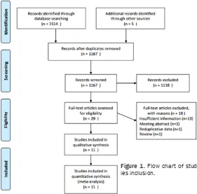
This meta-analysis was conducted with STATA version 12.0 (StataCorp LP, Texas, USA). P< 0.05 was considered as statistically significant. Odds ratios (ORs) with 95% confidence inter-vals (CIs) were utilized to evaluate the associa-tion between CD133 expression and clinico-pathological characteristics. The relevant clin- icopathological features included tumor grade, lymph node metastasis, tumor size, TNM stage, age, progesterone receptor (PR) status, ER sta-tus, and human epidermal growth factor recep-tor 2 (HER2) status. Hazard ratios (HRs) and 95% CIs of OS were used to evaluate pooled HR. If HR and 95% CI were not reported in the text, then they were calculated from Kaplan-Meier curves, according to the method intro-Figure 1. Flow chart of
stud-ies inclusion.
be calculated using Parm- ar’s method [22]; (4) studies were published in English or Chinese; and (5) if multiple studies were conducted on the same patient population, the most comprehensive stu- dy was selected. The exclu-sion criteria were: (1) studies with insufficient data; (2) re- views, meeting abstracts, case reports, and letters; and (3) duplicated studies. Quality assessment and data extraction
[image:2.612.93.374.76.353.2]consid-duced by Parmar [22]. Heterogeneity between studies was assessed using the I2 test and
Cochran’s Q test. If I2 was >50% or the result
of the Q test gave a P-value of <0.1, indicating significant heterogeneity, the random-effect model was conducted; otherwise, the fixed-effect model was adopted. Publication bias was assessed using Begg’s test and Egger’s test.
Results
Search results
The process of study selection is detailed in
Figure 1. A total of 1514 records from data-base searches and 5 records from other sourc-es were identified. After duplicate records were removed, 1167 records were screened by inspection of title and/or abstract. On this basis, 1138 records were excluded, and 29 full-text studies were evaluated for eligibility. Eighteen studies were further excluded for the following reasons: 13 studies provided insuffi-cient data, 3 studies were meeting abstracts, 1 study presented duplicate data, and 1 study was a review. Finally, 11 studies [10-20] were included in the meta-analysis.
Characteristics of included studies
The characteristics of the included studies are shown in Table 1. The included studies were published from 2009 to 2015. The total num-ber of samples was 1447, ranging from 49 to 325 per study. Ten studies [10, 11, 13-20] were in English and one [12] was in Chinese. Seven
studies [10, 12-14, 17-19] were performed in Asia, two [11, 16] were conducted in Europe, one [15] was carried out in Oceania and one [20] was conducted in Africa. All studies used IHC to detect CD133 expression, and the per-centage of positive CD133 expression varied from 18.8% to 74.4%. All 11 studies [10-20] reported an association between CD133 and clinicopathological features, and 4 studies [13, 14, 17, 18] reported a correlation between CD133 and OS. The NOS scores of the included studies ranged from 6 to 8, indicating that all eligible studies were high quality studies. Correlation of CD133 expression with clinico-pathological factors
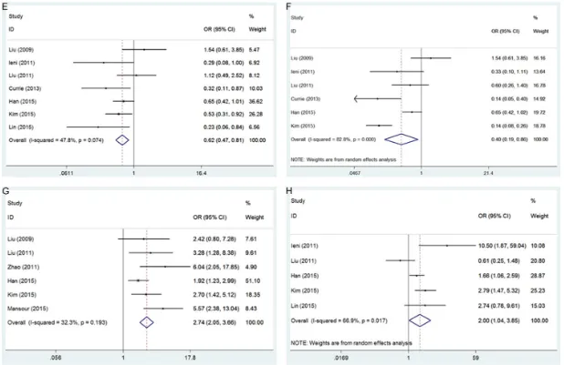
The relationship between CD133 expression and 8 clinicopathological factors was investi-gated. These 8 factors were; tumor grade (G3 vs. G1+G2), lymph node metastasis (positive vs. negative), tumor size (≥2 cm vs. <2 cm), age (≥50 years vs. <50 years), PR status (positive vs. negative), ER status (positive vs. negative), TNM stage (III+IV vs. I+II), and HER2 status (positive vs. negative). As shown in Figure 2
[image:3.612.91.527.85.253.2]and Table 2, CD133 overexpression was asso-ciated with G3 tumor grade (OR=1.82, 95% CI=1.4-2.36, P<0.001), presence of lymph node metastasis (OR=2.21, 95% CI=1.75-2.79, P<0.001), negative PR status (OR=0.62, 95% CI=0.47-0.81, P=0.001), negative ER status (OR=0.4, 95% CI=0.19-0.86, P=0.018), advan- ced TNM stage (OR=2.74, 95% CI=2.05-3.66, P<0.001), and positive HER2 status (OR=2.00, 95% CI=1.04-3.85, P=0.039). However, there was no significant correlation between CD133
Table 1. Characteristics of 11 included studies
Study Year Region Age mean (range) patientsNo. of Detectionmethod stageTNM CD133+(%) Research period NOS score
Liu 2009 China 49 (28-71) 74 IHC I-III 52.7 2004 7
Ieni 2011 Italy 61.8 (41-85) 49 IHC I-II 34.7 1998-2007 8
Liu 2011 China NR 121 IHC I-IV 74.4 2006-2008 7
Zhao 2011 China 47 (25-92) 67 IHC I-III 43.4 2003-2008 7
Aomatsu 2012 Japan 55 (26-78) 102 IHC II-III 46.1 2004-2009 8
Currie 2013 New Zealand NR 89 IHC NR 25 2003-2005 6
Collina 2015 Italy 57 (24-93) 160 IHC I-IV 18.8 2003-2009 8
Han 2015 China 45.6 (21-74) 325 IHC I-IV 48.6 2004-2008 8
Kim 2015 Korea 49 (25-85) 291 IHC I-III 24.7 2005-2010 7
Lin 2015 Taiwan 52.4 49 IHC NR 30.6 2001-2013 6
Mansour 2015 Egypt 49.1 (28-70) 120 IHC I-III 53.3 2006-2013 7
expression and tumor size (P=0.083) or age (P=0.41).
Relationship between CD133 expression and OS
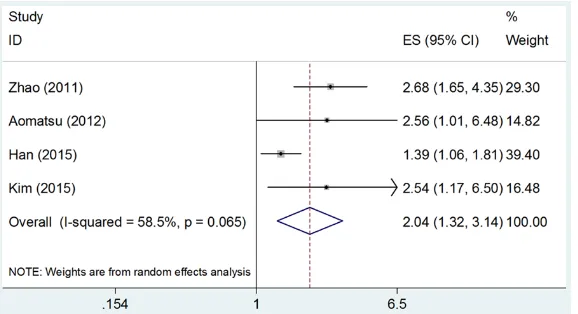
Four studies [13, 14, 17, 18] investigated the impact of CD133 expression on OS. Since sig-nificant heterogeneity was found (I2=58.5%,
P=0.065), a random-effect model was used. The meta-analysis results demonstrated that CD133 overexpression was significantly associ-ated with poor OS (HR=2.04, 95% CI=1.32-3.14, P<0.001) (Figure 3).
Publication bias
Begg’s rank correlation test and Egger’s regres-sion test were used to examine potential publi-cation bias in our meta-analysis. The results were P=1 for Begg’s test, and P=0.186 for Egger’s test, for OS analysis (Figure 4). These results showed that there was no evidence of
a meta-analysis. The results showed that CD133 expression was associated with a high-er tumor grade, occurrence of lymph node metastasis, negative PR status, negative ER status, advanced TNM stage, and positive HER2 status. With respect to the association between CD133 expression and OS, the data showed that CD133 overexpression was an indicator of a poorer OS (HR=2.04, 95% CI= 1.32-3.14, P<0.001). Taken together, this me- ta-analysis demonstrated that CD133 expres-sion is a potential marker for a panel of clinico-pathological factors and linked to a poor prog-nosis for breast cancer.
[image:6.612.87.524.85.220.2]CD133 is a commonly used cell surface marker of CSCs in a wide spectrum of cancers [4]. CD133 is expressed in normal tissues, includ-ing hematopoietic stem and progenitor cells [24], fetal neural stem cells [25], renal stem cells [26], and prostate stem cells [27]. Furthermore, CD133 is also used as a marker
Table 2. Association between CD133 expression and clinicopathological features in breast cancer
Factors studiesNo. of Effectsmodel OR (95% CI) p I2Heterogeneity (%) P
h
Tumor grade (G3 vs. G1+G2) 10 Fixed 1.82 (1.4-2.36) <0.001 18.1 0.276
Lymph node metastasis (positive vs. negative) 10 Fixed 2.21 (1.75-2.79) <0.001 49.5 0.037
Tumor size (≥2 cm vs. <2 cm) 9 Random 1.59 (0.94-2.67) 0.083 67.3 0.002
Age (≥50 years vs. <50 years) 8 Random 0.84 (0.55-1.27) 0.41 52.8 0.038
PR status (positive vs. negative) 7 Fixed 0.62 (0.47-0.81) 0.001 47.8 0.074
ER status (positive vs. negative) 6 Random 0.4 (0.19-0.86) 0.018 82.8 <0.001
TNM stage (III+IV vs. I+II) 6 Fixed 2.74 (2.05-3.66) <0.001 32.3 0.193
HER2 status (positive vs. negative) 5 Random 2.00 (1.04-3.85) 0.039 66.9 0.017
PR: progesterone receptor; ER: estrogen receptor; HER2: human epidermal growth factor receptor 2.
Figure 3. Forrest plot of HR for the relation of CD133 expression with OS in breast cancer.
significant publication bias existing in this meta-analysis.
Discussion
[image:6.612.91.380.250.407.2]for CSC isolation in a variety of malignant tumors [28-30]. CD133 is a cell surface glyco-protein whose expression decreases with cel-lular differentiation. A number of studies have demonstrated that CD133+ cells possess CSC properties, including greater colony-forming efficiency, self-renewal capacity, and higher tumorigenicity in xenografts [31, 32]. Although the normal function of CD133 remains unclear, CD133 has been investigated for use as a prognostic biomarker in a range of different cancers. We also noted that several meta-anal-ysis studies have investigated the prognostic role of CD133 in different cancers, including ovarian cancer [33], glioma [34], gastric cancer [35], esophageal carcinoma [36], colorectal cancer [37], and renal cell carcinoma [38]. The results from other cancer types showed that
rence of lymph node metastasis, negative PR status, negative ER status, advanced TNM stage, positive HER2 status, and poor OS, in breast cancer. CD133 may be considered a useful tool for pathological diagnosis and prog-nostic prediction in breast cancer. Owing to the aforementioned limitations, further large-scale studies, recruiting populations of various eth-nicities, are needed to confirm our results.
Disclosure of conflict of interest
None.
[image:7.612.92.375.72.452.2]Address correspondence to: Xianshu Gao, Depart- ment of Radiation Oncology, Peking University First Hospital, Peking University, Beijing, China. Tel: +86-10-83575239; Fax: +86-10-66551788; E-mail: doc-torgaoxs@126.com
Figure 4. (A) Begg’s funnel plot and (B) Egger’s regression asymmetry plot for CD133 overexpression and OS.
CD133 overexpression was correlated with poor survival outcomes, which was consis-tent with our results. Fur- thermore, there has to date been no meta-analysis inves-tigating the prognostic value of CD133 in breast cancer. To the best of our knowledge, this meta-analysis is the first study to comprehensively and systematically evaluate the association between CD133 expression and prognosis in breast cancer.
occur-References
[1] Torre LA, Bray F, Siegel RL, Ferlay J, Lortet-Tieu-lent J and Jemal A. Global cancer statistics, 2012. CA Cancer J Clin 2015; 65: 87-108. [2] Lin SX, Chen J, Mazumdar M, Poirier D, Wang
C, Azzi A and Zhou M. Molecular therapy of breast cancer: progress and future directions. Nat Rev Endocrinol 2010; 6: 485-493. [3] Rakha EA, Reis-Filho JS and Ellis IO.
Combina-torial biomarker expression in breast cancer. Breast Cancer Res Treat 2010; 120: 293-308. [4] Pattabiraman DR and Weinberg RA. Tackling
the cancer stem cells-what challenges do they pose? Nat Rev Drug Discov 2014; 13: 497-512.
[5] Singh SK, Hawkins C, Clarke ID, Squire JA, Bay-ani J, Hide T, Henkelman RM, Cusimano MD and Dirks PB. Identification of human brain tu-mour initiating cells. Nature 2004; 432: 396-401.
[6] Chen K, Li Z, Jiang P, Zhang X, Zhang Y, Jiang Y, He Y and Li X. Co-expression of CD133, CD44v6 and human tissue factor is associated with metastasis and poor prognosis in pancre-atic carcinoma. Oncol Rep 2014; 32: 755-763. [7] Herpel E, Jensen K, Muley T, Warth A, Schna-bel PA, Meister M, Herth FJ, Dienemann H, Thomas M and Gottschling S. The cancer stem cell antigens CD133, BCRP1/ABCG2 and CD117/c-KIT are not associated with progno-sis in resected early-stage non-small cell lung cancer. Anticancer Res 2011; 31: 4491-4500. [8] Yilmaz G, Akyol G, Cakir A and Ilhan M. Investi-gation of diagnostic utility and expression pro-files of stem cell markers (CD133 and CD90) in hepatocellular carcinoma, small cell dyspla-sia, and cirrhosis. Pathol Res Pract 2014; 210: 419-425.
[9] Zhang J, Guo X, Chang DY, Rosen DG, Mercado-Uribe I and Liu J. CD133 expression associated with poor prognosis in ovarian cancer. Mod Pathol 2012; 25: 456-464.
[10] Liu Q, Li JG, Zheng XY, Jin F and Dong HT. Ex-pression of CD133, PAX2, ESA, and GPR30 in invasive ductal breast carcinomas. Chin Med J (Engl) 2009; 122: 2763-2769.
[11] Ieni A, Giuffre G, Adamo V and Tuccari G. Prog-nostic impact of CD133 immunoexpression in node-negative invasive breast carcinomas. An-ticancer Res 2011; 31: 1315-1320.
[12] Liu T, Li X, Wang L, Chen Y, Pan X, Song S, Yang L, Cui J and Yang J. Distribution and clinico-pathologic significance of CD133+ cells in be-nign and malignant breast lesions. Chin J Clin Oncol 2011; 38: 1196-1200.
[13] Zhao P, Lu Y, Jiang X and Li X. Clinicopathologi-cal significance and prognostic value of CD133
expression in triple-negative breast carcinoma. Cancer Sci 2011; 102: 1107-1111.
[14] Aomatsu N, Yashiro M, Kashiwagi S, Takashi-ma T, Ishikawa T, Ohsawa M, Wakasa K and Hirakawa K. CD133 is a useful surrogate mark-er for predicting chemosensitivity to neoadju-vant chemotherapy in breast cancer. PLoS One 2012; 7: e45865.
[15] Currie MJ, Beardsley BE, Harris GC, Gunning-ham SP, Dachs GU, Dijkstra B, Morrin HR, Wells JE and Robinson BA. Immunohistochem-ical analysis of cancer stem cell markers in in-vasive breast carcinoma and associated duc-tal carcinoma in situ: relationships with markers of tumor hypoxia and microvasculari-ty. Hum Pathol 2013; 44: 402-411.
[16] Collina F, Di Bonito M, Li Bergolis V, De Lauren-tiis M, Vitagliano C, Cerrone M, Nuzzo F, Cantile M and Botti G. Prognostic value of cancer stem cells markers in triple-negative breast cancer. Biomed Res Int 2015; 2015: 158682.
[17] Han Z, Chen Z, Zheng R, Cheng Z, Gong X and Wang D. Clinicopathological significance of CD133 and CD44 expression in infiltrating ductal carcinoma and their relationship to an-giogenesis. World J Surg Oncol 2015; 13: 56. [18] Kim SJ, Kim YS, Jang ED, Seo KJ and Kim JS.
Prognostic impact and clinicopathological cor-relation of CD133 and ALDH1 expression in invasive breast cancer. J Breast Cancer 2015; 18: 347-355.
[19] Lin CH, Liu CH, Wen CH, Ko PL and Chai CY. Differential CD133 expression distinguishes malignant from benign papillary lesions of the breast. Virchows Arch 2015; 466: 177-184. [20] Mansour SF and Atwa MM. Clinicopathological
significance of CD133 and ALDH1 cancer stem cell marker expression in invasive ductal breast carcinoma. Asian Pac J Cancer Prev 2015; 16: 7491-7496.
[21] Moher D, Liberati A, Tetzlaff J and Altman DG. Preferred reporting items for systematic re-views and meta-analyses: the PRISMA state-ment. Ann Intern Med 2009; 151: 264-269, w264.
[22] Parmar MK, Torri V and Stewart L. Extracting summary statistics to perform meta-analyses of the published literature for survival end-points. Stat Med 1998; 17: 2815-2834. [23] Stang A. Critical evaluation of the
Newcastle-Ottawa scale for the assessment of the quality of nonrandomized studies in meta-analyses. Eur J Epidemiol 2010; 25: 603-605.
[25] Uchida N, Buck DW, He D, Reitsma MJ, Masek M, Phan TV, Tsukamoto AS, Gage FH and Weissman IL. Direct isolation of human central nervous system stem cells. Proc Natl Acad Sci U S A 2000; 97: 14720-14725.
[26] Bussolati B, Bruno S, Grange C, Buttiglieri S, Deregibus MC, Cantino D and Camussi G. Iso-lation of renal progenitor cells from adult hu-man kidney. Am J Pathol 2005; 166: 545-555. [27] Richardson GD, Robson CN, Lang SH, Neal DE,
Maitland NJ and Collins AT. CD133, a novel marker for human prostatic epithelial stem cells. J Cell Sci 2004; 117: 3539-3545. [28] Singh SK, Clarke ID, Terasaki M, Bonn VE,
Hawkins C, Squire J and Dirks PB. Identifica-tion of a cancer stem cell in human brain tu-mors. Cancer Res 2003; 63: 5821-5828. [29] Hermann PC, Huber SL, Herrler T, Aicher A,
Ell-wart JW, Guba M, Bruns CJ and Heeschen C. Distinct populations of cancer stem cells de-termine tumor growth and metastatic activity in human pancreatic cancer. Cell Stem Cell 2007; 1: 313-323.
[30] Collins AT, Berry PA, Hyde C, Stower MJ and Maitland NJ. Prospective identification of tu-morigenic prostate cancer stem cells. Cancer Res 2005; 65: 10946-10951.
[31] Wright MH, Calcagno AM, Salcido CD, Carlson MD, Ambudkar SV and Varticovski L. Brca1 breast tumors contain distinct CD44+/CD24- and CD133+ cells with cancer stem cell char-acteristics. Breast Cancer Res 2008; 10: R10. [32] Schmitt F, Ricardo S, Vieira AF, Dionisio MR
and Paredes J. Cancer stem cell markers in breast neoplasias: their relevance and distri-bution in distinct molecular subtypes. Virchows Arch 2012; 460: 545-553.
[33] Zhou Q, Chen A, Song H, Tao J, Yang H and Zuo M. Prognostic value of cancer stem cell marker CD133 in ovarian cancer: a meta-analysis. Int J Clin Exp Med 2015; 8: 3080-3088.
[34] Han M, Guo L, Zhang Y, Huang B, Chen A, Chen W, Liu X, Sun S, Wang K, Liu A and Li X. Clinicopathological and prognostic significan- ce of CD133 in glioma patients: a meta-analy-sis. Mol Neurobiol 2016; 53: 720-727. [35] Wen L, Chen XZ, Yang K, Chen ZX, Zhang B,
Chen JP, Zhou ZG, Mo XM and Hu JK. Prognos-tic value of cancer stem cell marker CD133 expression in gastric cancer: a systematic re-view. PLoS One 2013; 8: e59154.
[36] Sui YP, Jian XP, Ma LI, Xu GZ, Liao HW, Liu YP and Wen HC. Prognostic value of cancer stem cell marker CD133 expression in esophageal carcinoma: a meta-analysis. Mol Clin Oncol 2016; 4: 77-82.
[37] Zhao Y, Peng J, Zhang E, Jiang N, Li J, Zhang Q, Zhang X and Niu Y. CD133 expression may be useful as a prognostic indicator in colorectal cancer, a tool for optimizing therapy and sup-portive evidence for the cancer stem cell hy-pothesis: a meta-analysis. Oncotarget 2016; 7: 10023-10036.