Department of Medicine
Clinical Epidemiology Unit (KEP)
Karolinska Institutet, Stockholm, Sweden
REPRODUCTIVE FACTORS WITH
RESPECT TO BREAST CANCER RISK
AND BREAST CANCER SURVIVAL
Mohammadhossein Hajiebrahimi
All previously published papers were reproduced with permission from the publisher. Published by Karolinska Institutet.
Printed by Åtta.45 Tryckeri AB
© Mohammadhossein Hajiebrahimi, 2014 ISBN 978-91-7549-666-5
Reproductive factors with respect to breast cancer risk
and breast cancer survival
THESIS FOR DOCTORAL DEGREE (Ph.D.)
From karolinska Institutet to be publicly defended in Medicine Hall, Plan 4, A6, Karolinska Hospital, Solna, Friday Oct. 24th 2014. At 9:00
By
Mohammadhossein Hajiebrahimi
Principal Supervisor: Shahram Bahmanyar Karolinska Institutet Department of MedicineDivision of clinical Epidemiology Unit (KEP)
Co-supervisor(s):
Sven Cnattingius Karolinska Institutet Department of Medicine
Division of clinical Epidemiology Unit (KEP) Mats Lambe
Karolinska Institutet
Department of Medical Epidemiology and statistics (MEB)
Anastasia Nyman Iliadou Karolinska Institutet
Department of Medical Epidemiology and statistics (MEB)
Opponent:
Prof. Jonas Manjer University of Lund Department of surgery
Examination Board:
Ass. Prof. Lisa Ryden University of Lund Department of surgery
Prof. Kamila Czene Karolinska Institutet
Department of Medical Epidemiology and statistics (MEB)
Ass. Prof. Jana de Boniface Karolinska Institutet
Dedication
ABSTRACT
Aims:
The primary aim of this thesis was to examine the potential relationship between indirect markers of exposure to hormones during pregnancy and the risk of and survival from breast cancer, with special emphasis on young patients. Our specific objectives were as follows: to determine whether the association between placental weight and offspring size, on the one hand, and maternal mortality from breast cancer, on the other, are influenced by tumor characteristics; to examine the association between birth weight and risk of breast cancer in the female member of opposite-sexed twins; and to investigate whether familial factors influence previously reported association between reproductive factors and risk of breast cancer.Methods:
Based on the Swedish Quality Register of Breast Cancer, two different cohort studies were designed in the Stockholm-Gotland and Uppsala-Örebro regions, where records on characteristics of breast cancer have been collected since 1992. The first cohort was restricted to women who had a pregnancy between 1982 and 1989, and subsequently developed breast cancer. The cohort included 1,067 subjects and 180 deaths, and was conducted to investigate if placental weight is associated with maternal risk of dying from breast cancer, taking tumor characteristics into account. In the second study, we studied the possible association between birth weight and maternal risk of death from breast cancer, also taking tumor characteristics into account. We included 6,019 women who had a pregnancy between 1973 and 2008 and subsequently developed breast cancer, of whom 1017 died from the disease.Two case-control studies were also performed. In a nested case-control study, involving the female members of opposite sexed twin pairs, 543 cases and 2715 controls were included to investigate the potential association between offspring birth weight and risk of breast cancer, as well as a possible modifying effect of birth weight of the male twin sibling. Information on the twins (including birth weight, birth height, head circumference and gestational age of the females, and birth weight of the male co-twin) was extracted from the Swedish Twins Register and data on women diagnosed with breast cancer from the Swedish Cancer Register.
A second case-control study examined the potential modifying effect of familial factors on the association between reproductive factors and the risk of breast cancer. All women who delivered between 1973 and 2010 and had a full sister were selected as the study population, using the Swedish Medical Birth Register. Information on breast cancer was obtained from
Register. The cases examined included all parous women diagnosed with breast cancer between 1973 and 2010 who were 50 years old or younger and had at least one sister who also gave birth during this same period. The two control groups were sister controls (including the sister without breast cancer and closest in age to the case) and population controls (all parous women without breast cancer with at least a full sister except those in the sisters control group). In total, 8,349 cases, 8,349 sister controls, and 1,053,688 population controls were used.
Results:
Our findings indicate that the association between higher placental weight in connection with the most recent pregnancy and maternal risk of mortality from premenopausal breast cancer is dependent on the receptor status of the tumor. A positive association was more pronounced in the case of ER-/PR- tumors, but we did not find a dose– response association. Birth weight demonstrated no association with maternal mortality from premenopausal breast cancer, even in analyses stratified by the time that elapsed between pregnancy and cancer diagnosis, tumor stage, and receptor status. There was an inverse association between birth-weight-for-gestational age and mortality from premenopausal breast cancer among uniparous women. The nested case-control study of opposite-sexed twins did not reveal any statistically significant association between birth weight and risk of breast cancer. Furthermore, we observed no associations between other birth characteristics, including co-twin birth weight, and the risk of developing pre- or postmenopausal breast cancer.Our last study provided some evidence that the association between reproductive factors and maternal risk of breast cancer or between maternal factors and maternal risk of breast cancer may differ when using population or sister controls. We found that parity exhibited an inverse association to premenopausal breast cancer using population controls and was a risk factor using sister controls, suggesting a gene-environment interaction. Very preterm delivery (<31 weeks) was associated with a higher breast cancer risk using sister controls than when population controls were used, also suggesting a gene-environment interaction.
Conclusions:
We found some, but no strong evidence in support of the hypothesis that higher hormone levels during pregnancy are associated with mortality from premenopausal breast cancer. The hypothesis was supported when placental weight was employed as indirect indicator of estrogen levels during pregnancy, although birth weight showed no such association. The more pronounced effect of placental weight among ER-/PR- tumors suggests that premenopausal hormonal exposure might exert a greater impact on such tumors. The association between parity and risk of premenopausal breast cancer was modified by agene-environment interaction, as was the association between gestational age and the risk of breast cancer.
Keywords:
Breast cancer, Premenopausal, Postmenopausal, Placental weight, Birth size, Tumor characteristics, Estrogen Receptor, Progesterone Receptor, Twin, Opposite-sex, Sister control, Population controlLIST OF SCIENTIFIC PAPERS
This thesis was based on the following articles:
I. Hajiebrahimi MH, Bahmanyar S, Lambe M, Adolfsson J, Fornander T,
Wärnberg F, Cnattingius S. Placental weight and mortality in premenopausal breast cancer by tumor characteristics.Breast Cancer Res Treat 2013 Jan; 137(1):297-305.
II. Hajiebrahimi MH, Cnattingius S, Lambe M, Hsieh C-C, Ahlgren J,
Adolfsson J, Bahmanyar S. Birth size in the most recent pregnancy and maternal mortality in premenopausal breast cancer by tumor characteristics.Breast Cancer Res Treat 2014 Jun; 145(2):471-80.
III. Hajiebrahimi MH, Bahmanyar S, Öberg S, Nyman Iliadou A,
Cnattingius S. Breast cancer risk in opposite-sexed twins: influence of birth weight and co-twins birth weight.J Natl Cancer Inst. 2013 Dec 4; 105(23):1833-6.
IV. Hajiebrahimi MH, Cnattingius S, Lambe M, Bahmanyar S. Pregnancy
history and risk of breast cancer - a nested case control study on sisters discordant for breast cancer. In manuscript.
CONTENTS
Dedication ... . List of Abbreviations ... . 1. Introduction ... 1 2. Background ... 2 2.1 Epidemiology ... 22.2 Risk factors for breast cancer ... 5
2.3 Prognosis for women with breast cancer ... 16
2.4 Mechanism(s) underlying the development of breast cancer ... 27
3. Aim of the present study ... 29
4. Methodological considerations ... 30
4.1 Registries ... 30
4.1.1 The Medical Birth Register (MFR) (Studies І, ІІ, ІV)... 30
4.1.2 The Swedish Quality Registry of Breast Cancer (SQRBC) (Studies І, ІІ) ... 30
4.1.3 The Swedish Twin Registry (STR) (Study ІІІ) ... 30
4.1.4 The Swedish Cancer Register (SCR) (studies ІІІ, ІV) ... 31
4.1.5 The Swedish Cause of Death Register (SCDR) (studies І, ІІ, ІV) ... 31
4.1.6 The Multi-Generation Register (MGR) (Study ІV) ... 31
4.2 The individual Studies ... 32
4.2.1 Study І ... 32 4.2.2 Study ІІ ... 33 4.2.3 Study ІІІ ... 34 4.2.4 Study ІV ... 36 5. Results ... 37 5.1 Study І ... 37 5.2 Study ІІ ... 38 5.3 Study ІІІ ... 39 5.4 Study ІV ... 41 6. Discussion ... 43 6.1 Methodological considerations ... 43 6.1.1 Study designs ... 43
6.1.2 The validity of study ... 45
6.1.3 General discussion ... 50
7. Conclusion ... 54
8. Final remarks and future perspectives ... 55
9. Acknowledgments: ... 57
List of Abbreviations
AGA
Appropriate for Gestational Age
BMI
Body-Mass Index
BW/GA
Birth Weight for Gestational Age
CI
Confidence Interval
DZ
Dizygotic
ER
Estrogen Receptor
GA
Gestational Age
HR
Hazard Ratio
HER2
Human Epidermal Growth Factor Receptor-2
ICD
International Classification of Disease
IGF1
Insulin-like Growth Factor-1
LGA
Large for Gestational Age
LMP
Last Menstrual Period
MFR
Medical Birth Register
MGR
Multi-Generation Register
MZ
Monozygotic
OR
Odds Ratio
PI
Ponderal Index
PR
Progesterone Receptor
RCT
Randomized Clinical Trial
SCR
Swedish Cancer Register
SCDR
Swedish Cause of Death Register
SGA
Small for Gestational Age
SQRBC
Swedish Quality Register of Breast Cancer
STR
Swedish Twins Register
SHBG
Sex-Hormone Binding Globulin
TDLU
Terminal Duct Lobular Units
TNBC
Triple-Negative Breast Cancer
VLGA
Very Large for Gestational Age
VSGA
Very Small for Gestational Age
1. Introduction
Breast cancer is the most common form of cancer among women worldwide and its incidence is increasing in all countries, being highest in high income countries (1). In Sweden, for instance, one in every eight women is diagnosed with breast cancer during her lifetime (2). Despite the declining mortality from breast cancer in high-income countries in recent decades, this form of cancer still kills women more than any other form in all nations (3).
Estrogens and reproductive factors associated with exposure to estrogen e.g., low parity, early age at menarche, late age at menopause, and late age at the time of first pregnancy, are well-known risk factors for breast cancer. During the course of life, women are exposed to different levels of estrogen, especially high levels during their fetal life and when they become pregnant. Thus, exposure during these latter periods may be particularly important for the development of breast cancer. The impact of reproductive factors on the risk of breast cancer has been examined in many studies and some researchers propose that these factors may also play a role in breast cancer mortality.
Tumor characteristics, including stage, expression of hormone receptors and histopathology, are prognostic factors for the outcome of breast cancer, including mortality. Moreover, the expression of a group of hormone receptors may provide a better indicator than considering these receptors individually. Thus, at the twelfth St Gallen International Breast Cancer Conference an expert panel, introduced a new method for classification of breast cancer for therapeutic purposes based on a combination of the estrogen receptor (ER), Progesterone receptor (PR), and human epidermal growth factor-2 (HER2) (4). It seems likely that reproductive factors exert different impacts on the prognosis for breast tumors with different characteristics.
The primary aim of the present thesis was to examine associations between indirect markers of antenatal exposure to hormones and the risk of and survival from breast cancer. Taking tumor characteristics into account, associations between pregnancy and offspring characteristics and maternal mortality from breast cancer were focused on in two investigations. In another study, the possible relationship of birth characteristics and breast cancer risk in the female member of opposite-sexed twin pairs was explored. Finally, the possible influence of familial factors on the relationship between reproductive factors and the risk of breast cancer was examined.
2. Background
2.1 Epidemiology
2.1.1 Incidence
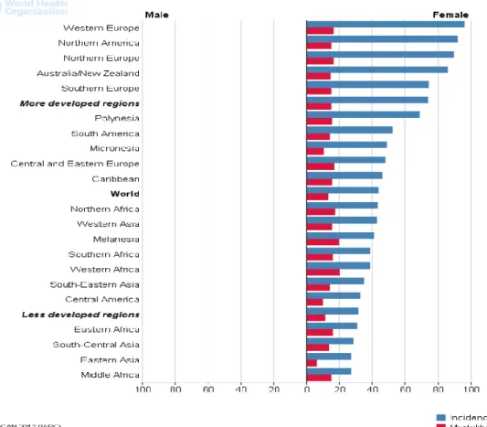
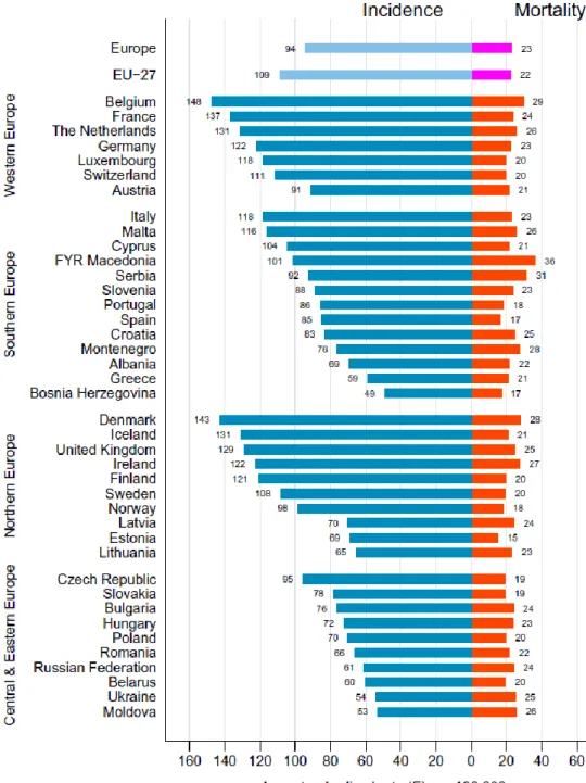
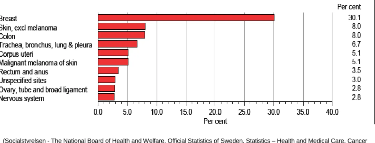
In 2012, breast cancer accounted for 25% of newly diagnosed cancers among women around the world (1), with the highest incidences in high-income countries (Figure 1) (5-7). In this same year, the age-standardized incidence of breast cancer in Sweden was approximately 108 per 100,000 population (Figure 2) (8), comprising 30% of all female cancers (Figure 3)(2) and afflicting approximately 1 of every 8 Swedish women during their lifetime.
Figure 1: Estimated age-standardized rates of the incidence and mortality from breast cancer (per 100,000 populations) in different regions in 2012
(Taken from Ferlay J, Soerjomataram I, Ervik M, Dikshit R, Eser S, Mathers C, Rebelo M, Parkin DM, Forman D, Bray, F. GLOBOCAN 2012 v1.0, Cancer Incidence and Mortality Worldwide: IARC, CancerBase No. 11 [Internet]. Lyon, France: International Agency for Research on Cancer; 2013. Available from: http://globocan.iarc.fr, accessed on 04.04.2014, with permission from the publisher)
Figure 2: Age-standardized incidence of and mortality from breast cancer per 100,000 population in Europe in 2012
Figure 3: The relative frequencies of different form of cancer in Sweden in 2010
The annual increase in breast cancer incidence in Sweden has been approximately 1.3% during the past 20 years (Figure 4)(2) .
Figure 4: Overall cancer incidence in Sweden since 1960
(Socialstyrelsen - The National Board of Health and Welfare, Official Statistics of Sweden, Statistics – Health and Medical Care, Cancer Incidence in Sweden 2010)
(Socialstyrelsen - The National Board of Health and Welfare, Official Statistics of Sweden, Statistics – Health and Medical Care, Cancer Incidence in Sweden 2010)
2.1.2 Mortality
In 2012, more than 14% of all cancer deaths in low-income countries were due to breast cancer (1). In spite of the fact that the relative rate of mortality is lower in high-income countries, the higher incidence in these countries (90/100,000 women) than in low-income countries (30/100,000 women) means that the overall mortality from breast cancer is almost equal (15/100,000 women) (3). In Europe, the risk of breast cancer mortality varies two-fold between countries (8). For example, the age-standardized mortality from breast cancer are 29 and 28 per 100,000 population in Belgium and Denmark, respectively, but only 15 and 17 per 100,000 in Estonia and Spain. In Sweden, the corresponding rate is 20 per 100,000 (Figure 2)(8), and the annual reduction during the last four decades is around 1% (9).
2.2 Risk factors for breast cancer
The incidence of breast cancer increases with age. Although one possible definition of aging is “the accumulation of cell mutations and tissue damage that leads to disease”, aging does not itself induce all such changes (10), but is rather thought to reflect of the occurrence different life-events. Thus, the risk of breast cancer may be influenced by genetic changes, exposures during prenatal and early life, reproductive factors and late-life exposures such as those associated with hormone replacement therapy and menopause. For example, in an older woman a low-grade breast cancer with good-prognosis in terms of progression might be caused by a lack of DNA genomic material on the long arm of chromosome 16 (11). Thus, the impact of risk factors such as age at menarche, the time of first pregnancy and of first full-term pregnancy that changes with age (12, 13) may be due to long-full-term exposure to, e.g., estrogens, rather than to increasing age itself. Even though the impact of certain risk factors for breast cancer such as a high body-mass index (BMI) increases with age, this might reflect physiological changes rather than simply increasing age.
2.2.1 Familial factors
2.2.1.1 BRCA1 and BRCA2
Mutations in genes such as BRCA1 and BRCA2 appear to be responsible for less than 10% of breast cancers (14, 15). Mutations in BRCA1, a tumor suppressor gene identified in 1990 and located on chromosome arm 17q (16), are rare in the general population (Table 1), but carried by approximately 5-10% of women diagnosed with breast cancer (17). Such mutations are also linked to an enhanced risk of ovary cancer.
Women in high-risk families (i.e., those with multiple cases of breast cancer across several generations) and who also carry a mutation in BRCA1 have a 80-85% risk of developing breast cancer during their lifetime, while the risk of women with the mutation but without such a family history is 55-70% (14). In comparison to sporadic breast cancer, tumors associated with a mutation in the BRCA1 gene are more poorly differentiated, of higher stage and grade, do not express hormone receptors and human epidermal growth factor receptor-2 (HER2) (Triple Negative Breast Cancer (TNBC)) and exhibit invasive ductal histology (15, 18, 19). Most, but not all women with such a mutation are diagnosed with breast cancer at a younger age.
Mutations in BRCA2, located on chromosome arm 13q, are known to be responsible for breast cancer in men, but are also linked to breast but not ovary cancer in women. Such tumors in women are well differentiated and express the estrogen receptor (ER)(15, 19). Although the risk of breast cancer is substantially higher among women who carry mutations in BRCA1 or BRCA2, penetrance is not 100% i.e., not every carrier develops breast cancer (17).
Table 1: The frequency of BRCA mutations among different populations
(Taken from Breast disease, 2006. 23(1), James P. Evans, C´ecile Skrzynia, Lisa Susswein and Megan Harlanc, Genetics and the young woman with breast cancer, p. 17-29, Copyright (2005,2006), with permission from IOS Press)
2.2.1.2 Twins
Compared with singletons, twins are more commonly born preterm and with a low birth weight, due to the anatomy of the uterus and placental capacity (20). A discordance in the birth weights of twin siblings is common, especially at older gestational ages.
In addition, twin pregnancies are associated with higher and more varied levels of estrogens than singleton pregnancies (21-23). These levels are also higher in dizygotic (DZ) than in monozygotic (MZ) twin pregnancies (24). Since the placenta is the main source of hormones during pregnancy, the higher levels of estrogen in DZ twin pregnancies is probably due to the presence of two placentas. Since estrogen is a well-known risk factor for breast cancer, the higher levels of this hormone during pregnancy might elevate the risk of breast cancer (25).
The female member of twins of opposite-sexes may also exposed to androgens produced by their male co-twin (26). Glinianaia and colleagues (27) have shown that the birthweight of the female in twins of opposite sex is higher than when both twins are female. It is thought that androgens are responsible for the weight difference between male and female fetuses in connection with singelton pregnancies (26). Androgens compete with estrogens for binding to sex hormone binding globulin (SHBG) (26) and might thereby increase the level of bioavailable maternal estrogens, perhaps enhancing the risk of breast cancer.
2.2.1.3 Familial history
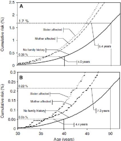
Having a first-degree relative (sister, mother or daughter) who develops breast cancer doubles a woman’s risk of breast cancer (28, 29), and this risk is higher if the relative is the sister than the mother (Figure 5) (29-31). More specifically, the meta-analysis by Pharoah and colleagues (29) revelad that the relative risk of developing breast cancer was 1.9 if any first-degree relative had breast cancer, 1.8 in the case of daughters, 2.0 for mother and 2.3 for sisters, and highest among women with both a sister and mother with breast cancer (28). The elevated risk associated with a family history of breast cancer declines with increasing age (31-35). Based on a large population-based set of Swedish data, Brandt and colleagues (31) concluded that women with a family history of breast cancer were diagnosed and died from this disease at a younger age than those without a family history (Figure 5).
2.2.2 Reproductive factors
Since measuring estrogen levels throughout a lifetime is difficult especially during the reproductive period, various indirect indicators have been developed, including age at menarche, age at first pregnancy, age at menopause, parity, gestational age and comorbidities related to pregnancy, such as pre-eclampsia, eclampsia, gestational hypertension and gestational diabetes. The relevance of such factors for the present work are discussed below.
(Taken from Brandt, A., et al., Age of onset in familial breast cancer as background data for medical surveillance. British Journal of Cancer, 2009. 102(1): 42-47, with permission from publisher)
Figure 5: Cumulative incidence of breast cancer and cumulative risk of death by breast cancer according to the type of family history. (A) Age at which women with a family history reach the cumulative risk of women lacking a family history at the age of 40 and 50 years for incidence. (B) Age at which women with a family history reach the cumulative risk of women lacking a family history at the age of 40 and 50 years for death from breast cancer.
2.2.2.1 Maternal characteristics
2.2.2.1.1 Age at first pregnancy
Later age at first pregnancy is a strong risk factor for breast cancer (12, 36, 37) and indeed, epidemiological studies have shown that each year of delay can increase the risk of breast cancer about 5% among premenopausal and around 3% among postmenopausal women (12). Being exposued to estrogen at a younger age could stimulate a higher proliferation of mammary cells which protect breast cells from being cancerous in future.
2.2.2.1.2 Age at last pregnancy
It has been reported that every 5-years increase in age at last pregnancy might elevate the risk of breast cancer by 5-8% (38, 39). However, Nechuta (40) did not find any association between these parameters.
2.2.2.1.3 Parity
Although one full-term pregnancy can reduce the overall risk of breast cancer during a woman’s lifetime (41, 42), childbirth has a dual effect. There appears to be a short-term increase and a long-term decrease in risk after each pregnancy (43-45), effects that recur with each repeated pregnancy (45-47). With each full-term pregnancy, the risk of premenopausal breast cancer is reduced by 3%, while the reduction in postmenopausal breast cancer is 12% (12).
The effects of parity, age at first pregnancy, and age at last pregnancy on breast cancer risk may interact. For instance, women with higher parity often experience their first pregnancy at a younger age or the long-term protective effect of pregnancy in women who have two children is more dominant among those who were pregnant at a younger age (47). Therefore, when investigating the influence of parity on risk of breast cancer, it is important to control for the age at the time of pregnancy, the period that elapses between pregnancy and the diagnosis of breast cancer, and the intervals between pregnancies.
2.2.2.1.4 Maternal weight, height and body-mass index
Maternal body characteristics, including weight and height, have been associated with breast cancer risk in different studies. Some researchers tend combine these variables, e.g., in the Body-Mass Index (BMI, calculated as weight (in kilogram) divided by height (in meter2), and
categorized as under weight (<18.5), normal weight (18.5 to <25), overweight (25 to <30) and obese (≥30). The association between BMI and breast cancer risk varies with other factors, such as age. For example several investigations have shown that obesity protects against breast cancer among premenopausal women (36, 48, 49), but enhances the risk among postmenopausal women (48-51). A meta-analysis of 13 studies revealed that for each unit increase in BMI breast cancer risk was reduced 2% in premenopausal and elevated 2% in postmenopausal women(52).
It has been proposed that premenopausal women with a high BMI have fewer ovary cycles which reduces their estrogen levels and in turn lowers their breast cancer risk (48, 53). In contrast, and despite cessation of ovary function in postmenopausal women, a higher BMI involves production of more estrogen by body fat tissue, which increases the risk. Indeed, postmenopausal women with a higher BMI exhibit higher levels of estrone, estradiol and free estradiol (50), as well as a lower level of SHBG, which enhance the bioavailablility of estrogen (48).
2.2.2.2 Characteristics of pregnancy
Pregnancy exerts a dual impact on breast cancer risk (43, 47), which increases during the first five years after pregnancy and declines thereafter (45, 47). Both the mother’s age at the time of delivery and parity can influence this association. Albrektsen and colleagues (47) found that the short-term risk of uniparous women who deliver their first child before 25 years is the same as for nulliparous women, but those who give birth after 30 years exhibit on elevated risk of at least 15 years after delivery. Mothers who have two children and their second delivery happens at an age younger than 25 demonstrate a transient risk increase for breast cancer shortly after delivery and this adverse effect is more prolonged among older mother (>30 years of age). Apparently, age at third delivery is less important.
2.2.2.2.1 Gestational age of the child at birth
Estimation the precise gestational age (GA) i.e., the duration of pregnancy (55) from fertilization to delivery is virtually impossible. There are three indirect measures of the GA: the time that elapses between the first day of the last menstrual period (LMP) and delivery; ultrasound; and neonatal estimation (56). GA of <37, 37-42 and >42 completed weeks are commonly categorized as pre-term, term and post-term, respectively. Pre-term GA is subcategorized as <28 (extremely pre-term), 28-31 (very pre-term) and 32-36 weeks (moderate pre-term) (57).
Since estrogen levels increase as pregnancy progresses, the GA provides an indirect indication of this level. For example, Mucci and co-workers (58) reported that estrogens level are higher during the 27th than the 16th week of pregnancy and, moreover, that this increased level is associated with a higher later risk of breast cancer in the female offspring. Other investigators have found elevated breast cancer risk among offspring delivered before 32 (59, 60) or after 40 weeks of pregnancy (36). Russo and colleagues (61) proposed that since mammary cells proliferate during the first and second trimesters of pregnancy and differentiate during the third, shortening pregnancy holds mammary cells in proliferation phase, which makes them more prone to malignancy.
2.2.2.2.2 Placental weight
The placenta, a fetomaternal multifunctional organ of pregnancy, has an average length of 22 cm and thickness of 2–2.5 cm, being thickest in the middle. Its mean weight at a gestational age 40 weeks is 678 (±134) or 690 (±135) grams for a female or male fetus, respectively (62). The main functions performed by the placenta are nutrition, transporting oxygen and nutrients to the fetus, excretion, immunity and regulation of the endocrine status, becoming the primary steroid-producing organ during pregnancy (63).
During pregnancy, the placenta produces four dominant hormones, i.e., human chorionic gonadotropin (hCG), human placental lactogen (hPL), estrogen and progesterone. Previous work has revealed that placental weight can serve as an indirect indicator of estrogen levels during pregnancy (25, 63-67) and that breast cancer risk increases with placental weight (36). Thus, women whose placenta weighed more than 700 grams had a 38% higher risk of developing breast cancer than those whose placental weight was less than 500 grams (36).
2.2.2.3 Characteristics of the offspring
2.2.2.3.1 Birth weight, height and head circumference
It has been proposed that breast cancer may actually originate in utero (68). The level of estrogen, a well-known risk factor for breast cancer (see above), in the mother’s blood is almost 200 times higher during than before pregnancy and it has been suggested that this exposure may be associated with the risk of breast cancer. Given the difficulty of directly and continuously measuring estrogen levels during pregnancy, some proxy measures have been considered. For instance epidemiological studies have reported that birth size (including weight, height and head circumference) are correlated with level of hormones, including estrogens during pregnancy (63, 69, 70).
Most investigations have found a positive association between birth weight and breast cancer risk in the female offspring (67, 71-79), particularly in premenopausal cancer (77, 80-86), whereas others observed no such association (66, 87-90). A literature review (91) of 26 relevant studies from 2000-2005 revealed that 16 found a positive association, three observed a positive, although not statistically significant association, and the remaining 7 found no association. In these studies the alteration in the risk of breast cancer with a birth weight of ≥4000 versus ≤2500 grams ranged from 17% to 5-fold. Moreover, it has been reported that breast cancer risk rises 9% for each 1000-grams increase in birth weight (92). Of the 14 studies dealing specifically with the association between birth weight and risk of breast cancer among the premenopausal female offspring (91), 11 showed a significant positive association between birth weight and risk of breast cancer, 5 of them showed a positive trend and 4 a threshold effect. Among the 8 studies on postmenopausal cancer (91), only one study observed an association.
Furthermore, offspring birth weight has also been associated with maternal risk of breast cancer. For example, Wohlfahrt and co-workers (25) have found that this risk is slightly higher (1.02, 95% CI 0.9-1.5) among mothers who deliver a heavy baby. In addition, Cnattingius and colleagues (36) found a positive association with risk of breast cancer among women who deliver two consecutive heavy babies (1.42, 95% CI 1.12-1.79), but that this elevation disappeared after adjusting for placental weight.
In addition to birth weight, an association between birth height and risk of offspring breast cancer has been reported (76), a stronger independent association than for birth weight or head circumference (76). It has been reported that this risk is 17% higher for women whose birth height was ≥51 cm and 11% higher for those whose head circumference was 31 cm (76).
2.2.2.3.2 The Ponderal index
Ponderal (PI) or Rohrer’s index, an indicator of fetal nutritional status (63), is defined as the ratio between birth weight (in gram) divided by birth length (in centimeter3)*100. Given the correlation between birth weight and height (76, 93), this could provide a suitable index of the combined effect of these two variables. This composite indicator of offspring nutritional status, which might be independent of race, gender and birth order (94), may be better than birth weight alone (95), as well as providing a useful indicator of fetal growth (94). PIs are categorized as low (PI<10%), appropriate (10%<PI≤90%) or high (PI>90%) (94).
PI is associated with maternal levels of estrogens during the reproductive period and pregnancy. Jasienska and colleagues (95) concluded from the association between PI and the level of estradiol (E2) that a higher PI may indicate a higher breast cancer risk. Moreover, Kaijser and co-workers (70) observed a positive association between PI and estriol levels (E3) during pregnancy.
2.2.2.3.3 Birth weight for gestational age (BW/GA)
BW/GA is defined as the ratio between the actual birth weight and that expected for the same gestational age and sex. Therefore, BW/GA might provide a better retroactive indicator of fetal growth than birth weight alone. This index can be categorized as very small for gestational age (VSGA) (<3%), small for gestational age (SGA) (3-<10%), appropriate for gestational age(AGA) (10-90%), i.e., normal (96, 97), large for gestational age (LGA) (91-97%) and very large for gestational age (VLGA) (>(91-97%) (98). Nechuta and colleagues (40) observed that women whose offspring had a BW/GA of <10% demonstrated a lower risk of breast cancer among those who experienced their last pregnancy at the age of 30 or older (0.82, 95% CI 0.59-0.98).
2.2.2.4 Exposure to hormones
2.2.2.4.1 Estrogen
The steroid hormone group of estrogens induces responses in the reproductive tract, mammary tissue, and pituitary gland during the reproductive process, as well as playing roles in non-reproductive processes, such as bone formation and cardiovascular health (99). The ovary is the main source of estrogen during most of the reproductive period, except during pregnancy, when the placenta is the main source. In premenopausal women, the serum levels of estrogens, mainly in the form of estradiol (E2), is 100 pg/ml during the follicular phase and approximately 600 pg/ml at the time of ovulation, but levels of estrogen, mainly estriol (E3) elevats to nearly 20,000 pg/ml during pregnancy. The level of estrogen falls to less than 20 pg/ml after menopuase, when estrone (E1) becomes the dominant forms. The growth and differentiation of ductal cells in breast tissue are stimulated by estrogens, which also plays an indirect role in the development of the mammary glands (100).
Estrogen normally binds to SHBG (approximately 37%). “Non-SHBG-bound hormone” (approximately 2% free estrogen) and estrogen bound to albumin (approximately 61%) are referred to as bioavailable (101) and affect target cells, including both normal and malignant breast cells. The serum concentration of bioavailable estradiol is thought to be more strongly
associated with the risk of breast cancer than the total level of estradiol (101, 102). Estrogen bound to SHBG is also available to responsive tissues, where it reduces cell proliferation, so that a reduction in the binding capacity of SHBG results in more rapid proliferation in steroid-sensitive tissues such as breast cells (101). This binding capacity is lower in postmenopausal than premenopausal women (103) and an association between the levels of SHBG and ER has been detected in connection with postmenopausal, but not premenopausal breast cancer (103). From conception until the 30th week of pregnancy, the level of SHBG is 6 to 10-fold higher in pregnant women than in those who are not pregnant (104).
A number of investigations on the difference in serum estrogen levels between pregnant compared and non-pregnant women and its association with the risk of breast cancer have been published. One review (105) concluded that the mean estrogen level in premenopausal women is 12% higher in those diagnosed with breast cancer, while another similar review on postmenopausal women arrive at a 15% higher value for those with breast cancer (106). A meta-analysis on 9 prospective studies revealed that high levels of serum estrogen is associated with higher risk of postmenopausal breast cancer (107). It has also been reported that the elevated level of estrogen during the first trimester of pregnancy is positively associated with the risk of developing breast cancer before 40 years of age, but that this association becomes negative at more advanced age (108). Moreover, an European study observed that the estrogen levels in women diagnosed with breast cancer is higher than in control subjects three year prior to diagnosis (109). The association between estrogen and breast cancer risk appears to be independent of family history (110).
2.2.2.4.2 Progesterone
Progesterone is secreted by the ovary during most of the reproductive period, but primarily by the corpus luteum and placenta during pregnancy (63). The main responsibilities of progesterone are to prepare the uterine muscles for implementation of the fertilized ovum and then support the pregnancy by inducing the proliferation and differentiation of uterine muscles to protect early embryonic development. A possible association between the level of progesterone and risk of breast cancer remains unclear: certain reports have shown a negative association with premenopausal women (109, 111), while no association was found for postmenopausal women (112) or all cases of breast cancer (108, 112). However, estrogen and progesterone might interact to elevate the risk of breast cancer; the so-called (estrogen plus progesterone hypothesis) (109).
2.2.2.4.3 Androgens
Since androgens are the obligatory precursors of all endogenous estrogens and may play a role in cell growth and proliferation, they might also influence breast cancer risk, either directly and/or indirectly (113, 114). Their ability to stimulate the growth and proliferation of cells might affect this risk directly while conversion to estrogens represents indirect involvement. Postmenopausal breast cancer exhibits a positive association with higher levels of androgens (102, 107, 109, 112) and reanalysis of 9 prospective (107) and the other studies (109) reveals that this is an independent association. Tumors that express both ER and PR are correlated with higher concentrations of androgens, including testosterone, androstenedione and dehydroepiandrosterone sulfate, but not those that lack either or both of these receptors (112).
Among the few studies on the association between androgen level and risk of breast cancer in premenopausal women, two (115, 116) reported a positive correlation. Some studies report that this association is more pronounced in the case of invasive tumors (110, 117) and ER+/PR+ patients (110, 118), but the underlying mechanism(s) remains unclear.
2.2.2.4.4 Insulin-like growth factor 1(IGF1)
In the 1980’s, it was postulated that IGF1, a peptide that stimulates mitosis and inhibits apoptosis in humans, is involved in the development of breast cancer (119). The serum concentration of IGF1 is higher in women with breast cancer than in those without breast cancer (120, 121). A review of 17 studies in 12 countries (119) concluded that the IGF1 level is positively associated to the risk of breast cancer and that this association is relatively unchanged by menopause. The level of IGF1 also positively associated with height, age at first pregnancy, moderately increased weight, and moderate alcohol consumption.
2.2.2.4.5 Prolactin
Prolactin, secreted by the pituitary gland, stimulates cell proliferation in the normal breast before and during pregnancy, as well as lactation following pregnancy. Certain investigators report that the serum level of prolactin is reduced after the first pregnancy (122). Moreover, this level is inversely correlated to age at first birth and the age when breastfeeding is begun (123). It has been proposed that prolactin is an important factor in the etiology of breast cancer, stimulating the proliferation of and inhibiting apoptosis in tumor cells (124, 125). Several studies have shown a positive association between the prolactin level and risk of
breast cancer among postmenopausal women (126-128), and one study (128) reported a similar association in premenopausal women.
2.3 Prognosis for women with breast cancer
Thanks to improved diagnosis, better staging of the tumor at the time of diagnosis and more effective treatment, mortality from breast cancer has been declining since the 1990s (129). Although many factors, such as those associated with reproduction are well-known risk factors for breast cancer (see above), their influence on breast cancer mortality is not as well known.
2.3.1 Genetic and environmental factors
2.3.1.1 BRCA1and BRCA2Few investigations have focused on the impact of mutations in BRCA1 and BRCA2 on survival after developing breast cancer. Four of these studies observed no impact (130-133). In contrast, Robson and colleagues (134) reported that carrying a BRCA1 mutation reduces survival, but only among women who have undergone breast-conserving treatment in connection with invasive breast cancer.
2.3.1.2 Family history
Some previous investigations have revealed that mortality from breast cancer is higher among women who have a positive family history (Figure 5) (31, 135, 136) , an effect that is age-dependent, being high pronounced at younger ages (136). Moreover, mortality, is higher among women whose sister, rather than mother was diagnosed with this malignancy (31). Certain other studies reported a higher survival of breast cancer in family history positive patients (137-140). Adjustment for potential confounders has confirmed that this favorable influence of a positive of family history is an independent effect (138). In a japenease study, women with a positive family history had smaller tumors, that expressed higher levels of ER+ and lower levels of HER2, as well as less lymph node involvement (141). However, the mechanism(s) underling the lower mortality from breast cancer among women with a positive family history is still not clear.
2.3.2 Reproductive factors
2.3.2.1 Maternal characteristics2.3.2.1.1 Age at the time of diagnosis
Age at diagnosis has been proposed to be a valuable prognostic factor for breast cancer survival. Breast cancer is relatively uncommon among young women, and only 15-25% of these patients are in their thirties or forties (142). Although the definition of “young” varies (49, 143), breast cancer in younger women is known to involve larger tumors, at a higher stage, lower expression of both ER and PR, higher levels of HER2, more extensive involvement of lymph nodes (144, 145), and poorer prognosis (146, 147).
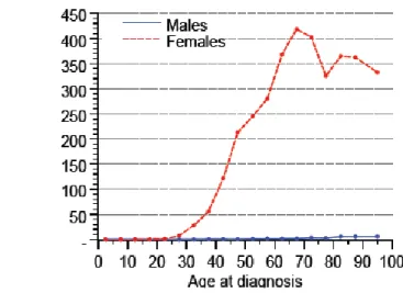
It has been suggested that breast cancer should be categorized at the time of diagnosis as either premenopausal or postmenopausal. Some studies divide women with breast cancer into those whose are 50 or younger and those who are older, which corresponds roughly these same two categories (146, 148, 149). Although breast cancer is more frequent among 60-70-year old women (Figure 6), the disease is more often aggressive among women younger than 50 (Table 2). Moreover, mortality declines with increasing age at the time of diagnosis.
The inverse association between age at the time of diagnosis and the risk of dying from breast cancer has been addressed in several publications (150-152). Han and Kang (143) observed that breast cancer mortality risk increased by 5% for each year of reducing in age among women younger than 35, but not for those between 35 and 50 years of age. In fact, they found that the pattern risk of mortality from breast cancer differs for women younger than 35 and those who are older and therefore suggested that 35 years is a suitable cutoff for young cases of breast cancer. Despite these findings, age at the time of diagnosis is not considered to be an independent prognostic factor (142) and the higher mortality risk among younger patients might reflect the more serious nature of their tumors (142, 152).
Table 2: Pathological features of breast cancer in premenopausal women Figure 6: Age distribution of women with breast cancer in Sweden in 2010
(Socialstyrelsen - The National Board of Health and Welfare, Official Statistics of Sweden, Statistics – Health and Medical Care, Cancer Incidence in Sweden 2010)
2.3.2.1.2 Age at first pregnancy
A younger age at first pregnancy in premenopausal women is associated with a higher breast cancer mortality rate (153-155). Although a study involving a population of more than 800,000 revealed that the mortality from premenopausal breast cancer was higher among women who had their first pregnancy after 30 years (153), other studies have reported that the risk of mortality from breast cancer is higher among women younger than 20 years at their first pregnancy than those between 20 and 24 (154, 155). Thus, although high age at first pregnancy is a risk factor for breast cancer, age at first pregnancy appears to be somewhat inversely associated with breast cancer mortality (155). It has been postulated that lower age at first pregnancy can prevent less aggressive forms of breast cancer. If so, the more aggressive forms that do develop might explain the higher risk of mortality associated with lower age at first pregnancy (154).
2.3.2.1.3 The time that elopes between pregnancy and the diagnosis of breast cancer: Pregnancy-associated breast cancer (PABC) is defined as disease diagnosed during pregnancy or within 2 years after delivery (156). Several previous examinations have found that the risk of breast cancer is increased during the two years immediately after delivery, and some have shown that survival among women whose breast cancer is diagnosed during this period is reduced (156-165). For example Philip and co-workers (163) found that the risk of mortality from breast cancer was 1.7 for women diagnosed during the two years after pregnancy and 0.9 for those diagnosed five or more years after. Although there is one report that this risk is higher among women diagnosed within the first year after pregnancy (164), a Swedish study found maximal risk 4-6 months after pregnancy (156).
The reduction in breast cancer mortality risk associated with a longer time interval between last pregnancy and diagnosis persists for more than 10 years among women who were younger than 20 at their first pregnancy(154). No association between the time between the last pregnancy and diagnosis of breast cancer and survival was seen among postmenopausal women (157). Although certain studies claim that the overall survival of cases with PABC and other is the same (166), Philips and colleagues (163) concluded that this survival increases by 8% for each year that elapses after pregnancy; while another report (167) presented a corresponding values of 15%, although the sample size was small in this latter case.
Although it is unclear why the risk of mortality from breast cancer falls as more time elapses after delivery, one possible mechanism could involve changes during pregnancy, particularly
more aggressive cancer cells (164). In addition, a longer delay before accurate diagnosis due to the increased density of the breasts of younger lactating women may play a role (163). It has also been proposed that the characteristics of the tumor are important in this connection.
Tumors diagnosed during first two years after pregnancy have been reported to be particularly aggressive (164, 165, 168) with a higher stage (160), an axillary node–positive nature (160, 163, 166), no expression of ER (159, 163, 166) or PR (159, 160, 166), a higher T class (159, 166), expression of p53 (160), a high mitotic count (160), with a fraction of cells in S phase (160), and a higher grade (160, 166). Thus, the association between diagnosis of PABC and risk of mortality from breast cancer has not been reported to be an independent prognostic factor (166) but, rather, to be confounded by the characteristics of the tumors (164). Certain other investigations suggest that additional, unknown characteristics of the tumors or pregnancy may exert an impact on this association (163).
2.3.2.1.4 Parity
Higher parity is associated with more extensive breast cancer mortality among premenopausal women (42, 157, 160, 161, 169, 170). For example, Olson and co-workers (42) found that the risk of such mortality is higher among women with three or more children than among nulliparous women. In contrast, certain reports indicate that parity either promotes (171) or has no effect on mortality from breast cancer, neither among premenopausal nor among postmenopausal women (155, 164). The tumors of parous women demonstrate a higher stage at the time of diagnosis, positive nodes and expression of p53 (160, 169).
2.3.2.2 The characteristics of pregnancy
2.3.2.2.1 Maternal weight, height and BMI
Some studies, including a literature review (48, 161, 172-174), show that most, but not all of the reports indicate that both overall and five-years survival from breast cancer is lower in pre- and postmenopausal women with higher BMI. Obesity is associated with cancer exhibiting less favorable features, including more nodule involvement (175) and a higher stage (176). In addition, a Norwegian study has shown that high body weight is associated with larger diameter, especially in the case of ER- and PR- negative tumors (177). Moreover, the risk of developing inflammatory breast cancer, the most lethal form, is higher among both pre- and postmenopausal women with higher BMI (48). In addition, all modalities of breast
cancer treatment, including surgery, radiotherapy, chemotherapy and hormone treatment, are reported to be adversely affected by obesity (48, 172).
2.3.2.2.2 Placental weight
The association between placental weight, suggested as an indicator of estrogen levels during pregnancy (58, 70, 178), and the risk of breast cancer has been examined by some investigators, but only one study has focused on the association between placental weight and breast cancer mortality risk. In this Swedish study, Larfors and colleagues (179) found that women whose placenta weighed 700 grams or more had an enhanced mortality risk, although they did not observe any significant association with other subgroups of placental weight. The association observed was more pronounced among uniparous women and those diagnosed with breast cancer during first two years after delivery.
2.3.2.3 Characteristics of the offspring
2.3.2.3.1 Birth size
Few investigations have examined the possible association between birth size and later offspring mortality from breast cancer. Sanderson and colleagues (180) reported that a birth weight of more than 4000 grams is associated with a higher subsequent breast cancer mortality risk. In addition, Norwegian researchers observed that a birth height of more than 52 cm was associated with a higher risk of mortality, but found no such associations with offspring birth weight or PI (181), which is in agreement with another study (155). Sovio et al. (182) have reported that the risk of mortality from breast cancer is increased 29% by an increase in birth weight by one standard deviation, with a weaker association for PI.
The possible association between offspring size and maternal breast cancer mortality risk has also been explored. Smith and co-workers (93) observed higher maternal mortality in association with higher PI, but no relationship to offspring birth weight or height. A meta-analysis revealed no association between birth weight and maternal mortality from breast cancer (183).
2.3.2.3.2 Gestational age
Little has been reported on the possible association between gestational age and mortality from breast cancer. Sanderson et al. (180) found no such association, whereas Sovio (182) reported 14-17% increased breast cancer mortality risk with an increase in gestational age by one standard deviation.
2.3.2.4 Tumor characteristics
2.3.2.4.1 Stage
Since 1953, solid tumors are categorized with staging system (184) (TNM, the latest edition of which was described in 2010 by the International Union Against Cancer (uicc) (185)), based on the size of the tumor (T), involvement of lymphatic system (N) and distribution throughout the body metastasis (M) as the primary prognostic factors. Based on this TNM, the American Joint Committee on Cancer (AJCC), has introduced a specific system for staging of breast cancer, (latest edition in 2009) (186), which is also used.
Various investigations have revealed that larger size, involvement of nodes and distant metastasis are poor prognostic factors for cancer. Higher tumor stage is associated with higher mortality (42, 187-189), but tumor size, nodule involvement and distant metastasis might valuable prognostic factors for individual breast cancers as well (42, 189). Previous findings indicate that axillary node involvement is the most independent indicator of overall survival from breast cancer (190), even if there is discordance between tumor staging and nodule involvement (191). Despite worldwide routine use of the TNM staging system for selecting the optimal therapeutic approach, there are suggestions that this system requires more refinement in order to become a really useful guideline for therapy (190, 192, 193).
It has been reported that the prognostic value of tumor size, nodule involvement and tumor grade decreases progressively with time (194, 195), although others suggest long-term effectiveness of these prognostic factors (196). One investigation including operable breast cancer cases found that the value of these prognostic factors tended to disappear after 10 years and that they were not related to mortality risk after 15 years (194).
2.3.2.4.2 Hormone receptors
The presence or absence of hormone receptors, including the ER and PR, individually or together has been suggested to be prognostic- and predictive factors for breast cancer. This expression increases with the age of the patient (197, 198) and the distribution of the receptors changes during the years following breast cancer diagnosis (199). Karlsson et al. (200) compared ductal carcinoma in situ with the subsequent nodule involvement found that 15% of the cases with ER expression and 30% of the cases with PR expression were changed in subsequent local relapse. Moreover, the prognostic value of hormone receptors appears to be short-time, and decreases with a long follow-up time (201-203). Bardou and colleagues
better survival than those with ER-/PR- tumors, this prognostic value disappears after 5 years, whereafter it becomes difficult to predict the future development of the tumor based on their receptor status. Notwithstanding, researchers still believe that breast cancer can be considered to be distinct diseases characterized by their hormone receptor status (204-208).
2.3.2.4.2.1 Estrogen receptors (ERs)
Estrogen receptors belong to a superfamily of nuclear receptors, including receptors for sex steroids, thyroid hormone and retinoid (99). The two different isoforms of ER, ERα and ERβ, are encoded on chromosomes 6 and 14, respectively (202, 209). A broad spectrum of tissues express ERα and ERβ; ERα is expressed at a higher level in the breast, prostate (stroma), ovary (theca cells), testis (Leydig cells), and liver, while ERβ is expressed more strongly in the prostate (epithelium), colon, testis, ovary (granulosa cells) and bone marrow (210).
In breast tissue, ERs are expressed by both normal and malignant cells. About 20% of the Terminal Duct Lobular Units (TDLU) in the breast of premenopausal women express the ER, a value that doubles during the follicular phase (211). The average extent of expression of ER by the TDLU cells of postmenopausal women is approximately 50%(211).
Expression of ERs increases dramatically in early hyper-proliferative premalignant lesions (211). Approximately 75% of breast tumors express ERs (211) with an elevated ratio of ER+ to ER- cells in comparison to normal breast tissue. Moreover, higher expression of ER is associated with higher breast cancer survival (212, 213). ER+ tumors tend to develop in older women (peaking of 70 years at age (214)), whereas ER- tumors tend to develop at an earlier age (peaking at 50) (49). Mortality from premenopausal ER+ breast cancer is higher in women younger than 35 than in older women (49).
2.3.2.4.2.2 Progesterone receptors (PRs)
Progesterone receptors belong to a nuclear or intracellular superfamily of ligand-dependent transcription factors (215, 216). After binding progesterone, PR changes conformation and is translocated to the nucleus, where it interacts with DNA to mediate the effects of progesterone (217, 218). In most target tissues, expression of PR is stimulated by estrogen (202) and reduced by progesterone.
The two isoforms of PR, PR-A and PR-B, are encoded by the same gene but their expression is initiated by different promoters (202, 215, 216, 219). Some studies suggest that expression
of PR is stimulated by atypia and increasing ratio of PR-A to PR-B, which is almost one in normal breast tissue, but varies extensively in malignant cells (220, 221). Approximately 60% of invasive breast tumors express PR-A or B (216).
2.3.2.4.3 Human epidermal growth factor receptor-2
Human epidermal growth factor receptor-2 (HER2) is a transmembrane tyrosine kinase receptor encoded on chromosome 17q21. HER2 is overexpressed in approximately 20-25% of invasive breast tumors as a result of gene amplification (222). This receptor has a role in regulating cell proliferation (50).
In newly diagnosed patients, HER2-positive breast cancer has a worse prognosis (50) and this factor may thus play a role in decision-making about treatment (223, 224). Such tumors are relatively resistant to endocrine therapy (222, 224, 225). Approximately 10% of primary HER2+ tumors becomes HER2- upon relapse (200). It has been reported that 6% of HER2- tumors becomes HER2+ during tumor progression and that 19% of HER2+ tumors becomes HER2-, although sample number in this case was small (226). The American Society of Clinical Oncology has suggested that HER2 expression should be used routinely as a prognostic- and predictive marker for breast cancer (222, 227)
2.3.2.4.4 Estrogen and progesterone receptors together
In light of the heterogeneity of breast cancer, it has been proposed that categorization should be based on expression of both ER and PR (207), which has a better and more independent prognostic value than their individual levels of expression (228, 229). ER+/PR+ tumors have a better prognosis than the other types of breast cancer (ER+/PR-, ER-/PR+ or ER-/PR-)(71, 228, 229), exhibiting smaller size, more favorable grade and better cancer-specific survival than ER-/PR- tumors (71). Among breast tumors, 60% are ER+/PR+, 20% ER-/PR-, 15-20% ER+/PR-, and less than 5% ER-/PR+ (71, 206-208).
Receptor status is associated with a number of factors, including maternal age, menopausal status, age at first pregnancy, nulliparity and age at menarche. ER+/PR+ tumors are much more common among older women, whereas ER-/PR- tumors are more frequent among younger patients (230). Thus, premenopausal women are diagnosed more after with ER-/PR- tumors, while ER+/PR+ tumors are more frequent after menopause (71). Some studies indicate that higher age at first pregnancy, nulliparity and later age at menarche exert stronger effects on the risk of developing ER+/PR+ than to ER-/PR- tumors in postmenopausal
women (207, 231). Others have shown that higher maternal BMI (232, 233) increases the risk of ER+/PR+ breast cancer. However, another investigation reported no influence of menstrual or reproductive characteristics and familial history on ER+/PR+ and ER-/PR- tumors (205).
2.3.2.5 Classification according to St
Gallen procedure
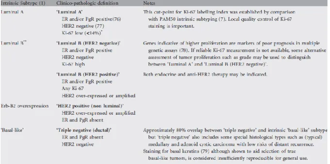
In 2011, an expert panel of the 12th St Gallen International Breast Cancer Conference introduced a new procedure for classification of breast cancer into 5 different subcategories on the basis of hormone receptor expression and epithelial cellular origin for therapeutic purposes (Table 3) (4). Tumors are categorized as follows:Luminal A: ER+ and/or PR+, low Ki56 (<14%), HER2-. Luminal B: ER+ and/or PR+, high Ki56 (>14%), HER2-; or HER2+ with any expression of Ki56. HER2+ (Erb-B2 or non-Luminal): ER- and PR- but HER2+. Triple Negative Breast Cancer: ER-, PR- and HER2-.
Table 3: The luminal classification of breast cancer
(Reprinted from Goldhirsch, A., et al., Strategies for subtypes—dealing with the diversity of breast cancer: highlights of the St Gallen International Expert Consensus on the Primary Therapy of Early Breast Cancer 2011. Annals of Oncology, 2011. 22:1736-1747. With permission from the publisher)
Luminal A tumor has a more favorable prognosis than the Luminal B subtype. TNBC tumors, which constitute 10-20% of breast cancer tumors, have a higher mortality rate and higher probability of distant metastasis than other subtypes of breast cancer (4, 51, 234). There is some discordance between the expression of factors in the original breast tumor and in lymph node metastases, as well as in the original tumor and relapse. For example, Falck and colleagues (234) showed 11% discordance between original tumors and with lymph node metastases and 16% of original Luminal A tumors were of a better subtype than lymph node metastases. Wilking and colleagues (226) found that in 15% of patients with HER2+ or HER2- original breast cancer, the recurrent tumor was of the opposite type. Moreover, Lindström and co-workers (235) found that 32.4% of ER+, 40.7% of PR+ and 14.5% of HER2+ original tumors change to the negative status upon relapse.
2.4 Mechanism(s) underlying the development of breast cancer
2.4.1 Normal maturation of the breast
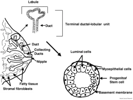
Mammary glands in women start to develop at the puberty, when the ductal structure begins to enlarge and branch by the beginning of the menstrual cycle. At onset of puberty, the epithelial cells in the ducts start to proliferate and form a tree-like pattern from the nipple to the end of the buds. Terminal Ductal Lobular Units (TDLU) develops and become more complex during subsequent menstrual cycles. The tree-like structures in the mammary gland are lined by epithelial cells and are surrounded by myoepithelial cells, which are in touch with basement cells. The TDLUs are embedded in fat tissue and surrounded by stromal cells (Figure 7) (216, 236, 237).
Development of the breast normally starts 3 years prior to menarche. Many hormones, and especially the female sex steroids estrogen and progesterone, are required for the proliferation and development of mammary cells. The minimal hormonal requirement in this connection includes estrogen, progesterone and prolactin or growth hormone (237). Estrogen produced by the ovary is primarily responsible for the development of breast stroma, growth of the ducts, and deposition of fat; while progesterone promotes lobular growth, alveolar budding and alveolar secretory changes. Both estrogen and progesterone are necessary for complete maturation of the ductal alveolar system (238).
Figure 7: Structure of the mammary gland and terminal ductal–lobular unit (TDLU).
At onset of pregnancy, the epithelial cells of the breast begin to divide again, a process which continues until delivery. The ductal trees expand and the number of ductules in the TDLUs increases during early pregnancy, and the ductules become mature to produce and secret milk during the last period of pregnancy and lactation. When lactation starts, epithelial DNA synthesis begins, but ends after lactation is completed, when the glands are switched off until onset of the next pregnancy. Thus, during pregnancy and during lactation, the breast tissue evolves from immature to fully developed (204, 237, 239).
2.4.2 Tumorogenesis
Histology reveals that the TDLU are the main origin of breast cancer in women. Moreover, since estrogen and progesterone receptors are expressed only in the luminal epithelial cells of these ducts, these cells could be in initial site of malignant transformation (204). Malignant cells may stay where they are, giving rise to Ductal Carcinoma In Situ (DCIS), the most common histological variant of the non-invasive stage of breast cancer. They may also penetrate the basal cells and extend to other parts of the body, resulting in, for example Invasive Ductal Cell (IDC), which accounts for 85-90% of all cases of invasive breast cancer (236).
The endogenous levels of steroid hormones fall sharply after menopause, due to the cessation of ovarian activity. At this point, adipose tissue becomes the main source of estrogen (102, 236). High levels of estrone (E1), the dominant form of the circulating hormone during postmenopausal period (102) is associated with a higher risk of postmenopausal breast cancer. Higher levels of estradiol and testosterone are also known to be associated with an enhanced risk of breast cancer (126, 240).
3. Aim of the present study
Main aim
To examine the independent association of indirect markers of pregnancy hormone exposure with breast cancer risk and survival, with special emphasis on young patients.
Aims of the individual studies
To investigate whether tumor characteristics modify the association between placental weight and maternal mortality from breast cancer.
To examine possible associations between offspring size at birth from the most recent pregnancy before diagnosis of premenopausal breast cancer and maternal risk of breast cancer mortality, taking tumor characteristics into consideration.
To explore the association between birth weight and offspring risk of breast cancer in females in opposite sexed twin pairs.
To investigate whether prenatal exposure to androgens from a twin brother influences the risk of breast cancer in the female twins, using male co-twin’s birth weight as an indicator of androgen exposure.
To examine whether associations between reproductive factors and risk of breast cancer are modified by genetic or early environmental factors.