http://create.canterbury.ac.uk
Please cite this publication as follows:
Vasu, V., Turner, K., George, S., Greenall, J., Slijepcevic, P. and Griffin, Darren K.
(2017) Preterm infants have significantly longer telomeres than their term born
counterparts. PLoS ONE, 12 (6). ISSN 1932-6203.
Link to official URL (if available):
https://doi.org/10.1371/journal.pone.0180082
This version is made available in accordance with publishers’ policies. All material
made available by CReaTE is protected by intellectual property law, including
copyright law. Any use made of the contents should comply with the relevant law.
Preterm infants have significantly longer
telomeres than their term born counterparts
Vimal Vasu1,2*, Kara J. Turner2, Shermi George1, John Greenall1, Predrag Slijepcevic3, Darren K. Griffin2
1Department of Child Health, East Kent Hospitals University Foundation NHS Trust, William Harvey Hospital, Ashford, Kent, United Kingdom,2University of Kent, School of Biosciences, Giles Lane, Canterbury, Kent, United Kingdom,3Brunel University London, Department of Life Sciences, College of Health and Life Sciences, Uxbridge, Middlesex, United Kingdom
*vimal.vasu@nhs.net
Abstract
There are well-established morbidities associated with preterm birth including respiratory, neurocognitive and developmental disorders. However several others have recently em-erged that characterise an ‘aged’ phenotype in the preterm infant by term-equivalent age. These include hypertension, insulin resistance and altered body fat distribution. Evidence shows that these morbidities persist into adult life, posing a significant public health concern. In this study, we measured relative telomere length in leukocytes as an indicator of biological ageing in 25 preterm infants at term equivalent age. Comparing our measurements with those from 22 preterm infants sampled at birth and from 31 term-born infants, we tested the hypothesis that by term equivalent age, preterm infants have significantly shorter telomeres (thus suggesting that they are prematurely aged). Our results demonstrate that relative telo-mere length is highly variable in newborn infants and is significantly negatively correlated with gestational age and birth weight in preterm infants. Further, longitudinal assessment in preterm infants who had telomere length measurements available at both birth and term age (n = 5) suggests that telomere attrition rate is negatively correlated with increasing gesta-tional age. Contrary to our initial hypothesis however, relative telomere length was signifi-cantlyshortestin the term born control group compared to both preterm groups and longest in the preterm at birth group. In addition, telomere lengths were not significantly different between preterm infants sampled at birth and those sampled at term equivalent age. These results indicate that other, as yet undetermined, factors may influence telomere length in the preterm born infant and raise the intriguing hypothesis that as preterm gestation declines, telomere attrition rate increases.
Introduction
Preterm birth, defined by the World Health Organisation as birth at less than 37 weeks com-pleted gestation, is estimated to account for 14.9 million or 11.1% of all births worldwide. Al-though survival rates are improving, recent data suggests that preterm birth is a risk factor in half of all neonatal deaths and that 1 million deaths each year are a direct result of complications a1111111111 a1111111111 a1111111111 a1111111111 a1111111111 OPEN ACCESS
Citation:Vasu V, Turner KJ, George S, Greenall J, Slijepcevic P, Griffin DK (2017) Preterm infants have significantly longer telomeres than their term born counterparts. PLoS ONE 12(6): e0180082. https://doi.org/10.1371/journal.pone.0180082
Editor:Gabriele Saretzki, University of Newcastle, UNITED KINGDOM
Received:March 21, 2016
Accepted:June 9, 2017
Published:June 28, 2017
Copyright:©2017 Vasu et al. This is an open access article distributed under the terms of the Creative Commons Attribution License, which permits unrestricted use, distribution, and reproduction in any medium, provided the original author and source are credited.
Data Availability Statement:All relevant data are within the paper and its Supporting Information files.
Funding:This work was supported from the Medical Research Council (GB) (case studentship sponsored by Digital Scientific UK) and via an East Kent Hospitals University NHS Foundation Trust internal project grant scheme award is acknowledged. The funders had no role in study design, data collection and analysis, decision to publish, or preparation of the manuscript.
associated with preterm birth [1,2]. Furthermore, immediate and long term morbidity rates in those that survive have remained high and largely correlate with the degree of prematurity [1, 3–6].
By term age, preterm babies display a phenotype that is different from that of the term born infant. Specifically, these babies have altered adipose tissue partitioning [7,8], ectopic fat depo-sition as intrahepatocellular lipid [9], hypertension [10–12] and insulin resistance [13,14]. Moreover, there is preliminary evidence indicating that many of these morbidities persist into early adult life and as such, may represent a significant public health issue [15–18]. As the mor-bidities described are associated with ageing in adults, the observed phenotype of the preterm infant may be indicative of premature ageing. Attenuation or early recognition of this pheno-type would therefore be desirable to reduce morbidity and to appropriately manage long-term health in these individuals.
Telomeres are repetitive DNA sequences at the end of chromosomes that become shorter with each cell cycle [19,20]. When telomeres have shortened beyond a critical length, a DNA damage response is initiated, committing the cell to apoptosis or senescence [21,22]. It is widely believed that an accumulation of senescent cells within a population leads to a loss of tissue function and ultimately organismal ageing [23]. Indeed, available data indicate a close negative correlation between chronological age and telomere length [24–28]. Furthermore, evidence supports a link between shortened telomeres and age-related morbidities in adults, many of which characterise the preterm infant phenotype described above [29–33].
Despite recent academic focus on telomere biology in newborns [34–49], little is known about telomere length and regulation in preterm infants. In summary the evidence to date demonstrates a reduction in telomere length with advancing gestational age, particularly in those born at less than 32 weeks completed gestation [50,51]. This effect appears to be specific to lifeex utero, since age matched foetuses do not conform to this trend [52]. Furthermore, other physiological events during labour, such as status of membrane rupture has also been shown to be relevant to telomere length in preterm infants [50,53].
Theex uteroenvironment of the neonatal intensive care unit differs fundamentally from both thein uteroenvironment and the postnatal environment experienced by the term infant. For example, exposure of preterm infants to elevated levels of oxygen may potentially lead to increased reactive oxygen species and induce DNA damage (to which the G-rich telomere sequence is par-ticularly susceptible) [54,55]. In addition, altered nutrition, sleep cycles and general routine care procedures may induce increased levels of stress. In adults, similar stresses are associated with telomere attrition [56–59]. Therefore these and otherex uterofactors might act to modulate telo-mere length in the preterm infant, resulting in a phenotype reminiscent of premature ageing.
Therefore, the aim of this study was to conduct a prospective observational study to com-pare telomere lengths of preterm infants sampled at birth and at term equivalent age with that of term infants. Although a small number of studies have compared telomere lengths in pre-term infants with that of those born at pre-term, none have assessed telomere length in prepre-term infants at term equivalent age (i.e. 37–42 weeks). Given that the evidence available demonstrate a reduction in telomere length with advancing gestational maturity in preterm infants and that theex uteroenvironment may be relevant, we sought to test the hypothesis that by term equiv-alent age, telomere length is shortened in preterm infants in comparison to term born infants.
Materials and methods
With institutional research ethics committee approval (10/H1109/51) and informed parental consent we conducted a prospective observational study over a four year period (June 2011 to
<
Competing interests:The authors have declared that no competing interests exist.
gestation were recruited from the level 3 (regional) neonatal unit at the William Harvey Hospi-tal in Ashford, Kent and the level 1 neonaHospi-tal unit at the Queen Elizabeth the Queen Mother Hospital in Margate, Kent. We chose to include preterm infants born<32 weeks completed gestation in light of previous data, which showed a rapid and significant decline in telomere length with advancing gestational age in infants born at<32 weeks [51]. In addition, this ges-tational age cut off is frequently utilised in other studies of preterm birth and allows for recruit-ment of a reasonable sample size within a reasonable time period. Of the 47 preterm infants sampled, 22 were sampled within 48 hours following birth and 25 were sampled at term equiv-alent age. In addition, a total of 31 term born infants who required blood sampling in the first 48 hours following birth for reasons such as evaluation of neonatal jaundice or suspected sepsis were recruited from the postnatal wards at these hospitals as a pragmatic comparator cohort. Babies were excluded from the study if they had an antenatal or postnatal diagnosis of a severe congenital malformation or were unlikely to survive. Once recruited, several demographic fac-tors were collected from each participant. These included infant gestational age at birth, infant weight at birth and maternal age in all three groups.
Telomere length measurement
1ml of additional blood was collected by venepuncture in a paediatric lithium heparin bottle taken at the time of routine blood sampling in study recruits. In preterm infants, we aimed to collect a sample within 48 hours of life or at term equivalent age (between 37–42 weeks post menstrual age). Term born infants underwent blood sampling at the time of routine blood sampling within the first 48 hours of life. Each blood sample was assigned a 2 digit numerical code and frozen at -80˚C. Samples were collected by a member of the research team who was blinded to the group allocation of the blood sample and transported to the School of Biosci-ences, University of Kent under dry ice. Samples were thawed and equilibrated to room tem-perature prior to genomic DNA extraction using a DNA isolation from mammalian blood kit (Roche). Isolated DNA was dissolved in Tris-EDTA pH8.0 and assessed for DNA concentra-tion and purity using a Nanodrop spectrophotometer. All samples were stored at -20˚C until telomere length analysis by a single observer, using multiplex quantitative real time polymerase chain reaction (qRT-PCR). Primer design for telomere and a single copy reference gene (hae-moglobin B) amplification was as previously described by Cawthonet al2009 [60]. Simulta-neous amplification of the telomere and single copy gene was achieved in a total reaction volume of 25μl using SensiMixTMSYBR No-ROX Kit (Bioline), 50nM each primer and 25ng of standard or unknown sample DNA. The reaction was carried out using a Rotor-gene Q 2 plex HRM platform under the following cycling conditions: 95˚C for 10 mins, two cycles of 94˚C for 15 secs and 49˚C for 15 secs, 37 cycles 94˚C for 15 secs, 62˚C for 10 secs, 74˚C for 15 secs, 84˚C for 10 secs and 88˚C for 10 secs. Each unknown and standard DNA sample was assayed in triplicate. Relative telomere length expressed as telomere to single copy gene ratio (T/S ratio) was calculated using a standard comparative method (delta delta Ct) [61] (Equation 1). Intra- and inter-assay variations were 1.05% and 0.41% respectively.
T=S¼2^ððSample telCt Sample scgCtÞ reference telCt ref erence scgCTÞÞ
Equation 1. Comparative method for the calculation of T/S ratio. ‘Sample telCt’ refers to the unknown sample telomere sequence amplification cycle threshold, ‘Sample scgCt’ refers to the unknown sample single copy reference gene amplification cycle threshold, ‘reference telCt’ refers to the reference DNA telomere amplification cycle threshold and ‘reference scgCt’ is the reference DNA single copy gene amplification cycle threshold.
Statistical analysis
Data were tested for normality using the Shapiro-Wilk test and analysed using SPSS version 21 (IBM Corp. Released 2012. IBM SPSS Statistics for Windows, Version 21.0. Armonk, NY: IBM Corp). The primary outcome, T/S, ratio was normally distributed and therefore parametric methods (univariate ANOVA) were used for analysis. Cohen’s d was used to determine effect size (small, medium, or large) for mean T/S ratio between the groups.
Results
During the study period we obtained blood samples for telomere length analysis in 31 term born infants and 47 preterm infants (22 sampled within 48 hours of preterm birth and 25 sampled at term equivalent age). The characteristics of recruited term and preterm infants recorded at the time of sampling are displayed inTable 1. Within the term cohort, no baby was identified as having either blood culture positive sepsis or severe neonatal jaundice requir-ing neonatal exchange blood transfusion. In addition, the characteristics recorded at birth of preterm infants sampled at term equivalent age are shown inTable 2. As would be expected, birth gestation and birth weight of preterm infants (both preterm infants sampled at birth and those sampled at term) were significantly different (p=<0.01) when compared to term infants (Table 1). Birth gestation and birth weight in preterm infants sampled at birth (Table 1) and preterm infants sampled at term equivalent age (Table 2) were not significantly different (p= 0.28 and 0.18 respectively). There were no significant differences between the groups with respect to maternal age (Table 1). In each of the 3 cohorts, there were a greater number of male infants than female infants, however the proportion of each sex was similar in each group (Fig 1.) The Preterm infants sampled at term age demonstrated satisfactory postnatal growth in comparison to published UK data [62].
T/S Ratio
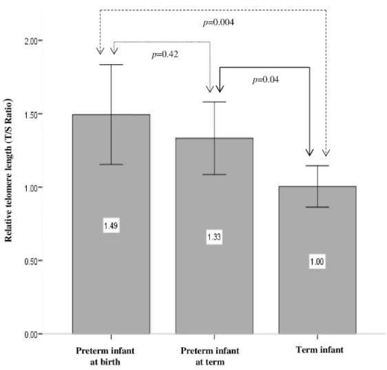
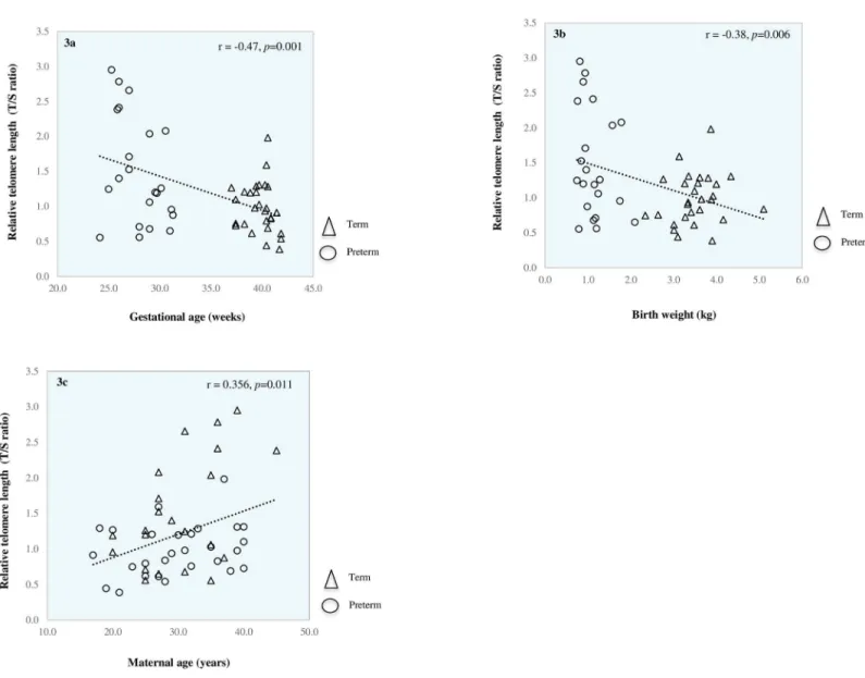
Mean T/S ratio and T/S variability for preterm infants sampled at birth, preterm infants sam-pled at term and term born infants are shown inFig 2. Mean T/S ratio and T/S variability of male and female infants overall and within each cohort are shown inTable 3. Finally,Fig 3A, 3B and 3Cshow correlation of T/S ratio with gestational age, birth weight and maternal age respectively in term infants and preterm infants sampled at birth.
Table 1. Baseline characteristics at the time of sampling in term infants, preterm infants sampled at birth and preterm infants sampled at term equivalent age.Data are presented as mean (95% CI).
Term (n = 31) Preterm at term age (n = 25) Preterm at birth (n = 22)
Gestational age (weeks) 39.85 (39.36–40.34) 38.11 (37.67–38.55) 27.99 (27.02–28.95)
Weight (kg) 3.58 (3.37–3.79) 2.27 (2.08–2.47) 1.14 (0.98–1.30)
Maternal age (years) 29.45 (26.80–32.11) 30.00 (27.76–32.24) 30.36 (27.56–33.17)
https://doi.org/10.1371/journal.pone.0180082.t001
Table 2. Baseline characteristics for gestational age and weight at the time of birth in preterm infants that were sampled at term equivalent age (n = 25).Data are presented as mean (95% CI).
Baseline characteristics Mean (95% CI)
Gestational age at birth (weeks) 27.30 (26.16–28.45)
Time between birth and sampling (weeks) 10.81 (9.56–12.06)
Birth weight (kg) 0.96 (0.84–1.09)
We did not observe any difference in T/S ratios in males compared to female infants either overall, or within each cohort. However, our results show that both gestational age and birth weight were negatively correlated with T/S ratio (p= 0.001,p= 0.006 respectively) and that maternal age was positively correlated with T/S ratio (p= 0.011). Moreover, our data demon-strate a gradient effect with T/S ratios being highest in preterm infants sampled at birth and lowest in term infants, with term infant T/S ratios being significantly shorter than those of both preterm infants sampled at term (p= 0.04) and preterm infants sampled at birth (p= 0.004) (Fig 2). Cohen’s d analyses revealed a large effect size when comparing T/S ratios between term and preterm infants sampled at birth (d = -0.85, r = -0.39) and a medium effect size when comparing T/S ratios in term infants versus preterm infants sampled at term equiva-lent age (d = -0.71, r = -0.33). Though there was a reduction in mean T/S ratio between pre-term infants sampled at birth and prepre-term infants sampled at pre-term equivalent age, this was not statistically significant (p= 0.42).
For reasons highlighted in the discussion section, we were only able to obtain longitudinal telomere length samples (at the time of birth and at term equivalent age) in five preterm infants. A post hoc analysis of this longitudinal data enabled calculation of telomere attrition rate (Equation 2) between birth and term age.
T=S ratioðbirthÞ T=S ratioðterm ageÞ
Number of weeks between birth sample and term age sample
Equation 2. Methodology for calculation of telomere attrition rate in five
preterm infants sampled at birth and at term equivalent age
Table 4shows that in agreement with the cross sectional data described above, a reduction in T/S ratio was observed between the time of birth and term equivalent age in all five babies. Fig 1. Proportion of males and females in term, preterm at birth and preterm at term infant cohorts. https://doi.org/10.1371/journal.pone.0180082.g001
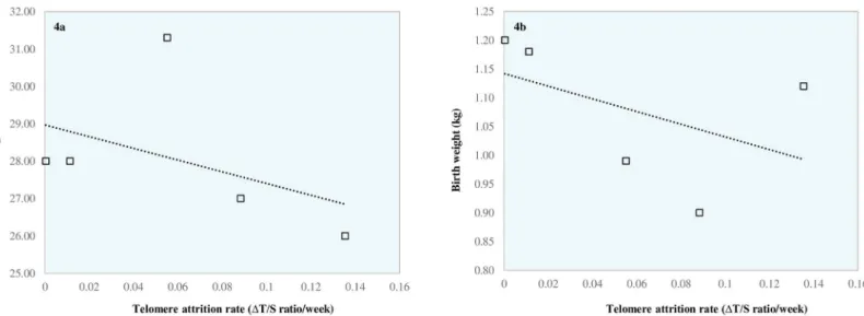
However, the magnitude of this reduction was highly variable between these five babies. Paired t-test analysis revealed no difference between birth sample T/S ratio and term sample T/S ratio (p= 0.07) for these 5 babies (in keeping with cross sectional data presented inFig 2. indicating no difference between birth sample T/S ratio and term sample T/S ratio in preterm infants). Intriguingly, our results also suggest an inverse relationship between telomere attrition rate and both birth weight (Pearson Correlation -0.47) and gestational age (Pearson Correlation -0.44). However, this was not of statistical significance (p= 0.42 andp =0.46 respectively). These data are shown inFig 4A and 4B.
Fig 2. Relative telomere length (T/S ratio) in preterm infants at birth, preterm infants at term age and term born infants.Data are Mean (95% CI).
https://doi.org/10.1371/journal.pone.0180082.g002
Table 3. Gender distribution and mean (95% CI) T/S ratios of male and female infants included in the study overall and within each study cohort. p value signifies results from univariate ANOVA analyses of T/S ratios in males compared to females overall and within each cohort. (*Gender data missing in 2/31 term infants).
Overall (n = 76) Term (n = 29)* Preterm at term (n = 25) Preterm at birth (n = 22)
Mean (95% CI) n Mean (95% CI) n Mean (95% CI) n Mean (95% CI) n
Male 1.29 (1.08–1.50) 49 1.13 (0.78–1.48) 18 1.25 (0.94–1.56) 17 1.22 (1.03–2.07) 14
Female 1.27 (1.05–1.48) 27 1.00 (0.68–1.30) 11 1.52 (1.03–2.00) 8 1.32 (0.98–1.82) 8
Fig 3. Relationship between relative telomere length (T/S ratio) and gestational age (a), birth weight (b) and maternal age (c). https://doi.org/10.1371/journal.pone.0180082.g003
Table 4. Longitudinal data and T/S ratios collected for five preterm infants.Data are Mean (95% CI).
Patient 1 2 3 4 5 Mean (95% CI)
Gestation at Birth (weeks) 27.00 26.00 31.30 28.00 28.00 28.06 (25.59–30.53)
Birth weight (kg) 0.90 1.12 0.99 1.20 1.18 1.08 (0.92–1.24)
T/S ratio (Birth) 2.66 2.41 0.87 0.56 0.71 1.44 (0.19–2.69)
Gestation at term sample (weeks) 37.00 37.43 38.00 36.86 36.86 37.23 (36.62–37.84)
Weight (kg) at term sample 1.93 3.06 1.95 3 2.72 2.53 (1.84–3.22)
T/S ratio (term) 1.77 0.86 0.50 0.56 0.59 0.86 (0.20–1.52)
Number of weeks between sample 1 and sample 2 10.00 11.43 6.71 8.86 8.86 9.17 (7.02–11.33)
Change in T/S ratio between sample 1 and sample 2 -0.884 -1.549 -0.37 -0.003 -0.123 -0.586 (-1.375–0.204)
Telomere attrition rate/week (ΔT/S ratio/week) -0.0884 -0.1355 -0.0552 -0.0004 -0.0139 -0.0587 (-0.127–0.010)
Discussion
In this study we assessed whole blood leukocyte telomere length in a cohort of preterm and term born infants shortly after birth, and for the first time, we also assessed telomere length in preterm infants at term equivalent age. Our data do not support our initial hypothesis that telomere length is significantly shortened by term equivalent age in preterm infants and in comparison to term infants. On the contrary, our results show that preterm infants at term equivalent age have significantlylongertelomere lengths than term born infants. However, in Fig 4. Relationship between telomere attrition rate (ΔT/S ratio per week) and birth gestation (a) and birth weight (b) in preterm infants with longitudinal samples.
https://doi.org/10.1371/journal.pone.0180082.g004
Table 5. A summary of studies to date that have assessed telomere length in preterm infants in comparison to term born infants or age matched fetus’.
Study Friedrichet al Holmeset al Menonet al Ferrariet al
Group assessed
Term (n = 11) versus preterm (n = 15)
Preterm (n = 5) versus age matched fetuses (n = 8)
Term (n = 35) versus preterm infants born with intact membrane (PTB, n = 69) and preterm infants born following preterm prelabour rupture of membranes (pPROM, n = 28)
Term (n = 43) versus PTB (n = 8), pPROM (n = 7) and stillborn (n = 42)
Sample type Leukocytes from cord blood Leukocyte from venous blood Leukocytes from cord blood and DNA from placental tissue (n = 5 PTB, n = 5 pPROM, n = 8 term)
Placental tissue
Methodology TRF analysis TRF analysis qRT-PCR qRT-PCR
Measure Absolute telomere length Absolute telomere length Absolute telomere length Relative telomere length Key Findings No difference in telomere
lengths of term (>37 weeks GA) versus preterm (<37 weeks GA). Significantly shorter telomeres in very low birth weight compared to low birth weight preterm infant. Significant decline in telomere length between 27 and 32 weeks gestation
Significant decline in telomere length in preterm infants sampled longitudinally between 23–35 weeks GA. No difference in telomere length observed in fetuses measured longitudinally over the same gestational period
Telomere length significantly longer in PTB (<37 weeks GA) compared to term born (>37 weeks GA) and pPROM (<37 weeks GA). No difference in telomere length between term and pPROM
Confirmation of findings by Menonet al. Significantly shorter telomere length in stillbirths (fetal death>22 weeks GA) compared to term (>37 weeks GA) and PTB (<37 weeks GA). No difference in telomere lengths of stillbirths compared to pPROM (<37 weeks GA)
keeping with other published data, our results indicate a significant decline in telomere length with advancing gestational age at birth [50–52]. We also observed a positive correlation between maternal age and T/S ratio, suggesting that older mothers deliver babies with longer relative telomere lengths. The strengths of this study are that it represents the only study to have directly compared telomere length measurement in preterm infants, assessed at term age and term born infants using a well established methodology. Potential limitations include the limited number of preterm infants in which we were able to obtain both birth and term age (longitudinal) telomere lengths and the observation that our ‘healthy’ comparator group of term infants cannot, by definition, be classified as entirely ‘healthy’ by the observation that they required blood testing. However, this latter point is merely reflective of the fact that obtaining research blood samples in completely healthy babies is not ethically sound.
Telomere length in preterm infants is a largely understudied area and to the best of our knowledge, only three other studies have assessed telomere length in preterm infantsin com-parison to term born babies: Friedrichet al. found no significant difference between the two groups when they assessed cord blood leukocyte telomere length [51], however Menonet al. and Ferrariet al. found significantly shortened leukocyte and placental telomere lengths respectively in term born infants compared to preterm infants born with intact membranes [50,51,53]. Interestingly, telomere length in preterm infants born following preterm pre-labour rupture of membranes was not different to term born infants and was also significantly shorter than preterm infants born with intact membranes. Furthermore, Ferrariet al. provided data to support the hypothesis that unexplained stillbirths are associated with placental telo-mere attrition by demonstrating a reduction in placental telotelo-mere length between stillbirths and term born infants [53]. Moreover, the placenta is known to contain sub-populations of karyotypically abnormal trophoblasts, which may have significant ramifications on telomere length measured in the Ferrari paper [63]. Indeed Ferrariet al. noted failed karyotype analysis in 15/42 stillborn cases.
While these findings (summarised inTable 5) offer interesting and novel insights into the physiological relevance of the events associated with telomere attrition that may lead to normal labour, preterm labour or unexplained stillbirth, none provide information on telomere length in the critical aberrant period of development that preterm born infants are exposed to in the neonatal intensive care unit during the weeks following preterm birth.
Our own data contradict the findings of Friedrich and Menon and are fundamentally dif-ferent to those presented by Ferrari, who examined placental telomere length. We propose that our finding that telomere length is longer in preterm infants sampled at birth compared to term born infants might be explained by a period of high cell turnover and replicative stress during a period of growth in the final weeks of pregnancy in the term infant that does not occur in those born preterm. As such, we suggest that the most prominent factors influencing telomere length in neonates are gestational maturity and birth weight (which are intrinsically linked except for where fetal growth restriction has occurred). Nonetheless, given the high var-iability in telomere lengths shown by our own and others’ data [26,27,48,49,64,65], we sug-gest that there may also be other as yet undiscovered determinants of telomere length in newborns, which might include both genetic and epigenetic factors. Indeed other studies indi-cate heritability of telomere length and significant correlations between offspring telomere length and parental age [66–73]. Our own results and those of others indicate that this pattern is present at birth [64].
In light of previous observations by Holmeset al. who found a significant shortening of telomeres in the weeks following preterm birth [52], the finding that telomere length is not shortened at the time of term equivalent age in the preterm infant was unexpected. However, Holmes’ study only examined 5 preterm infants and thus may have been underpowered. We
propose that there may be at least two possible explanations for this finding. Firstly, it is possi-ble that the results from our study reflect a slow telomere attrition rate in the preterm infant during the initial weeks after birth, arising from slow replication and cell turnover. Indeed, many preterm infants undergo an initial phase of slow early growth (in comparison to fetal growth rates) [74,75]. Alternatively, an opposing view arises from observations by others, who have noted that following birth, lymphocyte expansion rate occurs independently of gesta-tional age [76–78]. In order to sustain rapid expansion of immature lymphocytes in the first few weeks of life, whilst evading telomere loss and subsequent entry into cell cycle arrest, it is possible that telomeres may be lengthened. In support of this, previous work has demonstrated up-regulation of telomerase and lengthened telomeres in response to stimulated expansion of naïve B lymphocytes isolated from adults and young children [79]. Should telomerase expres-sion elicit a similar effect during the expanexpres-sion of immature lymphocytes in the neonatal period, one might expect a greater population of cells with longer telomeres in the preterm group that were sampled some weeks after birth (at term equivalent age) in comparison to term born controls sampled within 48 hours of birth. Therefore it is also possible that our results can be explained by differences in the actions of telomere maintenance mechanisms between the study cohorts.
To further investigate these areas of uncertainty, it would have been beneficial to assess pre-term infant telomere length longitudinally in all prepre-term babies recruited to our own study. This would additionally unmask any potential hidden effects attributable to a large spread in data as a result of variable genetic and epigenetic influences. Despite this being our original intention, obtaining samples for telomere length analysis at birth and at term equivalent age proved difficult as a number of preterm babies were either discharged from hospital or trans-ferred back to their local neonatal unit prior to term age. From the five preterm infants in whom we were able to collect longitudinal data (Table 4), we demonstrated a reduction in telo-mere length between birth and term age. Intriguingly our data additionally suggests an inverse relationship between telomere attrition rate and both birth weight and gestational age (Fig 4A and 4B). Though we acknowledge that the sample size is small and there is no statistical signifi-cance, these preliminary data raise, for the first time, the biologically plausible hypothesis that telomere attrition rate in preterm infants maybe associated with the degree of prematurity, with the most preterm infants manifesting higher telomere attrition rates. Naturally, these data require confirmation in larger studies evaluating longitudinal telomere measurements in pre-term infants. Our data also suggest that advanced maternal age is associated with increased rel-ative telomere length in newborn infants. Okudaet aldemonstrated a similar association, albeit using a telomere restriction fragment (TRF) methodology to measure newborn telomere length [64]. This finding is of interest and warrants replication in light of the increase in mater-nal age at time of first pregnancy that has been observed over recent years [80]. However, the relationship we describe may be a confounding variable given the relationship between mater-nal telomere length and newborn telomere length observed by others [81,82]. The increase in vitro fertilisation methods over the past two decades may also be a relevant factor but in our own cohort, the majority of preterm births (93%) were as a result of a natural pregnancy.
It may be that analysis of average telomere length in the preterm infant at term equivalent age cannot act as a suitable marker of the aged phenotype observed. Perhaps rather than mea-suring telomere length at term equivalent age in preterm infants, it may be more prudent to measure them further downstream e.g. during later childhood or adolescence. This has proved to be informative in previous studies where shortened telomere length was shown to be associ-ated with respiratory morbidity in the ex-preterm infant [34,83]. However, the disadvantage of doing this would be the introduction of known and unknown confounding variables (i.e. lifestyle and epigenetic changes) which may influence telomere length [84]. Alternatively,
there are a number of other putative markers of cellular senescence that may have more rele-vance to the detection of senescence associated with preterm birth that require further investi-gation. A plausible alternative candidate is SIRT 1, which is known to be down-regulated in association with insulin resistance, cardiovascular disease and metabolic disease. Moreover, SIRT 1 down-regulation is known to be associated with accelerated cord blood endothelial progenitor cell senescence in preterm infants [85]. Likewise, cell cycle regulators CDKN2A and CDK1A are known to be linked with ageing in adults and therefore warrant investigation in the newborn population [86–88].
In conclusion, our data indicate that preterm infants assessed at term equivalent age mani-fest longer telomere lengths than term born infants. In addition, our data and that of other groups [44,64,84,89] show considerable variability in telomere length in preterm and term infants, suggesting that other mechanisms may exist alongside gestational maturity that are as yet unexplored determinants of telomere length. This high level of variability leads to a degree of overlap between the data in each of the cohorts assessed here and as such, we suggest that our data should be replicated by other groups and that future work in this area should evolve to examine a panel of markers of cellular senescence longitudinally. Furthermore, our findings lead us to speculate whether, as a response to preterm birth, there are mechanisms as yet not understood that serve to up regulate telomerase. These should be the focus of future research.
Supporting information
S1 File. (PATENT data file for PLoS One_170316.xls).Anonymised patient data collected for the study.
(XLSX)
Acknowledgments
Support via an internal project grant scheme award from East Kent Hospitals University NHS Foundation Trust is acknowledged.
Author Contributions
Conceptualization:Vimal Vasu, Darren K. Griffin.
Data curation:Vimal Vasu, Shermi George, John Greenall.
Formal analysis:Vimal Vasu, Kara J. Turner, Shermi George, John Greenall.
Funding acquisition:Vimal Vasu, Kara J. Turner.
Investigation:Vimal Vasu, Kara J. Turner, Shermi George, John Greenall, Darren K. Griffin.
Methodology:Vimal Vasu, Kara J. Turner, Predrag Slijepcevic.
Project administration:Vimal Vasu, Kara J. Turner, Darren K. Griffin.
Supervision:Vimal Vasu, Darren K. Griffin.
Validation:Vimal Vasu, Kara J. Turner, Darren K. Griffin.
Writing – original draft:Vimal Vasu, Kara J. Turner, Predrag Slijepcevic, Darren K. Griffin.
Writing – review & editing:Vimal Vasu, Kara J. Turner, Shermi George, John Greenall, Pre-drag Slijepcevic, Darren K. Griffin.
References
1. Blencowe H, Cousens S, Chou D, Oestergaard M, Say L, Moller A-B, et al. Born too soon: the global epidemiology of 15 million preterm births. Reprod Health. 2013; 10(1):S2.
2. Howson CP, Kinney MV, McDougall L, Lawn JE. Born too soon: preterm birth matters. Reprod Health. 2013; 10(1):S1.
3. Beck S, Wojdyla D, Say L, Betran AP, Merialdi M, Requejo JH, et al. The worldwide incidence of pre-term birth: a systematic review of maternal mortality and morbidity. Bulletin of the World Health Organi-zation. 2010; 88(1):31–8.https://doi.org/10.2471/BLT.08.062554PMID:20428351
4. Costeloe KL, Hennessy EM, Haider S, Stacey F, Marlow N, Draper ES. Short term outcomes after extreme preterm birth in England: comparison of two birth cohorts in 1995 and 2006 (the EPICure stud-ies). BMJ. 2012; 345:e7976.https://doi.org/10.1136/bmj.e7976PMID:23212881
5. Moore T, Hennessy EM, Myles J, Johnson SJ, Draper ES, Costeloe KL, et al. Neurological and develop-mental outcome in extremely preterm children born in England in 1995 and 2006: the EPICure studies. BMJ. 2012; 345:e7961.https://doi.org/10.1136/bmj.e7961PMID:23212880
6. Blencowe H, Cousens S, Jassir FB, Say L, Chou D, Mathers C, et al. National, regional, and worldwide estimates of stillbirth rates in 2015, with trends from 2000: a systematic analysis. Lancet Glob Health. 2016; 4(2):e98–e108.https://doi.org/10.1016/S2214-109X(15)00275-2PMID:26795602
7. Vasu V TEL, Durighel G, Uthaya S, Bell J D, N M. Abstracts of the 50th Annual Meeting of the European Society for Paediatric Research. October 9–12, 2009. Hamburg, Germany. Acta Paediatr Suppl. 2009; 98(460):1–278.
8. Uthaya S, Thomas EL, Hamilton G, Dore CJ, Bell J, Modi N. Altered adiposity after extremely preterm birth. Pediatric Res. 2005; 57(2):211–5.
9. Thomas EL, Uthaya S, Vasu V, McCarthy JP, McEwan P, Hamilton G, et al. Neonatal intrahepatocellu-lar lipid. Arc Dis Child Fetal Neonatal Ed. 2008; 93(5):F382–F3.
10. Johansson S. Risk of High Blood Pressure Among Young Men Increases With the Degree of Immaturity at Birth. Circulation. 2005; 112(22):3430https://doi.org/10.1161/CIRCULATIONAHA.105.540906
PMID:16301344
11. Bhat R, Salas AA, Foster C, Carlo WA, Ambalavanan N. Prospective analysis of pulmonary hyperten-sion in extremely low birth weight infants. Pediatrics. 2012; 129(3):e682–e9.https://doi.org/10.1542/ peds.2011-1827PMID:22311993
12. VanDeVoorde RG, Mitsnefes MM. Neonatal Hypertension. Kidney and Urinary Tract Diseases in the Newborn: Springer; 2014. p. 349–61.
13. Hovi P, Andersson S, Eriksson JG, Ja¨rvenpa¨a¨ A-L, Strang-Karlsson S, Ma¨kitie O, et al. Glucose regula-tion in young adults with very low birth weight. NEJM. 2007; 356(20):2053–63.https://doi.org/10.1056/ NEJMoa067187PMID:17507704
14. Tinnion R, Gillone J, Cheetham T, Embleton N. Preterm birth and subsequent insulin sensitivity: a sys-tematic review. Arch Dis Child. 2013–304615.
15. Parkinson JR, Hyde MJ, Gale C, Santhakumaran S, Modi N. Preterm birth and the metabolic syndrome in adult life: a systematic review and meta-analysis. Pediatrics. 2013; 131(4):e1240–e63.https://doi. org/10.1542/peds.2012-2177PMID:23509172
16. Thomas EL, Parkinson JR, Hyde MJ, Yap IK, Holmes E, Dore´ CJ, et al. Aberrant adiposity and ectopic lipid deposition characterize the adult phenotype of the preterm infant. Pediatric Res. 2011; 70(5):507– 12.
17. de Jong F, Monuteaux MC, van Elburg RM, Gillman MW, Belfort MB. Systematic review and meta-anal-ysis of preterm birth and later systolic blood pressure. Hypertension. 2012; 59(2):226–34.https://doi. org/10.1161/HYPERTENSIONAHA.111.181784PMID:22158643
18. Liu J, Fox CS, Hickson DA, May WD, Hairston KG, Carr JJ, et al. Impact of abdominal visceral and sub-cutaneous adipose tissue on cardiometabolic risk factors: the Jackson Heart Study. J Clin Endocrinol Metab. 2010; 95(12):5419–26.https://doi.org/10.1210/jc.2010-1378PMID:20843952
19. Olovnikov AM. A theory of marginotomy. J Theor Biol. 1973; 41(1):181 PMID:4754905
20. Moyzis RK, Buckingham JM, Cram LS, Dani M, Deaven LL, Jones MD, et al. A highly conserved repeti-tive DNA sequence, (TTAGGG)n, present at the telomeres of human chromosomes. Proc Natl Acad Sci U S A. 1988; 85(18):6622–6. PMID:3413114
21. Herbig U, Jobling WA, Chen BP, Chen DJ, Sedivy JM. Telomere Shortening Triggers Senescence of Human Cells through a Pathway Involving ATM, p53, and p21, but not p16. Mol Cell. 2004; 14(4):501– 13. PMID:15149599
22. Shay JW. Telomerase therapeutics: Telomeres recognized as a DNA damage signal. Clin Cancer Res. 2003; 9(10):3521–5.
23. Faragher RG, Kipling D. How might replicative senescence contribute to human ageing? Bioessays. 1998; 20(12):985–91.https://doi.org/10.1002/(SICI)1521-1878(199812)20:12<985::AID-BIES4>3.0. CO;2-APMID:10048298
24. Aubert G, Lansdorp PM. Telomeres and aging. Physiol Rev. 2008; 88(2):557–79.https://doi.org/10. 1152/physrev.00026.2007PMID:18391173
25. Harley CB, Futcher AB, Greider CW. Telomeres shorten during ageing of human fibroblasts. Nature. 1990; 345(6274):458–60.https://doi.org/10.1038/345458a0PMID:2342578
26. Allsopp RC, Vaziri H, Patterson C, Goldstein S, Younglai EV, Futcher AB, et al. Telomere length pre-dicts replicative capacity of human fibroblasts. Proc Natl Acad Sci U S A. 1992; 89(21):10114–8. PMID:
1438199
27. Hastie ND, Dempster M, Dunlop MG, Thompson AM, Green DK, Allshire RC. Telomere reduction in human colorectal carcinoma and with ageing. Nature. 1990; 346(6287):866–8.https://doi.org/10.1038/ 346866a0PMID:2392154
28. Lindsey J, McGill NI, Lindsey LA, Green DK, Cooke HJ. In vivo loss of telomeric repeats with age in humans. Mutat Res. 1991; 256(1):45–8. PMID:1944386
29. Aviv A, Aviv H. Reflections on telomeres, growth, aging, and essential hypertension. Hypertension. 1997; 29(5):1067–72. PMID:9149667
30. Cawthon RM, Smith KR, O’Brien E, Sivatchenko A, Kerber RA. Association between telomere length in blood and mortality in people aged 60 years or older. The Lancet. 2003; 361(9355):393–5.
31. Salpea K, Talmud P, Cooper J, Maubaret C, Stephens J, Abelak K, et al. Association of telomere length with type 2 diabetes, oxidative stress and UCP2 gene variation. Atherosclerosis. 2010; 209(1):42–50.
https://doi.org/10.1016/j.atherosclerosis.2009.09.070PMID:19889414
32. von Zglinicki T, Martin-Ruiz CM. Telomeres as biomarkers for ageing and age-related diseases. Curr Mol Med. 2005; 5(2):197–203. PMID:15974873
33. Adaikalakoteswari A, Balasubramanyam M, Ravikumar R, Deepa R, Mohan V. Association of telomere shortening with impaired glucose tolerance and diabetic macroangiopathy. Atherosclerosis. 2007; 195 (1):83–9.https://doi.org/10.1016/j.atherosclerosis.2006.12.003PMID:17222848
34. Hadchouel A, Marchand-Martin L, Franco-Montoya M-L, Peaudecerf L, Ancel P-Y, Delacourt C, et al. Salivary Telomere Length and Lung Function in Adolescents Born Very Preterm: A Prospective Multi-center Study. PloS one. 2015; 10(9):e0136123.https://doi.org/10.1371/journal.pone.0136123PMID:
26355460
35. Entringer S, Epel ES, Lin J, Blackburn EH, Buss C, Simhan HN, et al. Maternal Estriol Concentrations in Early Gestation Predict Infant Telomere Length. J Clin Endocrinol Metab. 2014; 100(1):267–73. 36. Entringer S, Epel ES, Lin J, Blackburn EH, Buss C, Shahbaba B, et al. Maternal Folate Concentration in
Early Pregnancy and Newborn Telomere Length. Ann Nutr Metab. 2015; 66(4):202–8.https://doi.org/ 10.1159/000381925PMID:26067849
37. Drury SS, Esteves K, Hatch V, Woodbury M, Borne S, Adamski A, et al. Setting the trajectory: racial dis-parities in newborn telomere length. J Pediatr. 2015; 166(5):1181–6.https://doi.org/10.1016/j.jpeds. 2015.01.003PMID:25681203
38. Gielen M, Hageman G, Pachen D, Derom C, Vlietinck R, Zeegers M. Placental telomere length decreases with gestational age and is influenced by parity: A study of third trimester live-born twins. Pla-centa. 2014; 35(10):791–6.https://doi.org/10.1016/j.placenta.2014.05.010PMID:25096951
39. Salihu HM, King L, Patel P, Paothong A, Pradhan A, Louis J, et al. Association between maternal symp-toms of sleep disordered breathing and fetal telomere length. Sleep. 2015; 38(4):559.https://doi.org/10. 5665/sleep.4570PMID:25325479
40. Salihu HM, Pradhan A, King L, Paothong A, Nwoga C, Marty PJ, et al. Impact of intrauterine tobacco exposure on fetal telomere length. Am J Clin Exp Obstet Gynecol. 2015; 212(2):205. e1–. e8.
41. Tellechea M, Gianotti TF, Alvariñas J, Gonza´lez CD, Sookoian S, Pirola CJ. Telomere length in the two extremes of abnormal fetal growth and the programming effect of maternal arterial hypertension. Sci Rep. 2015; 5.
42. Bijnens E, Zeegers MP, Gielen M, Kicinski M, Hageman GJ, Pachen D, et al. Lower placental telomere length may be attributed to maternal residential traffic exposure; a twin study. Environ Int. 2015; 79:1–7.
https://doi.org/10.1016/j.envint.2015.02.008PMID:25756235
43. Xu J, Ye J, Wu Y, Zhang H, Luo Q, Han C, et al. Reduced fetal telomere length in gestational diabetes. PloS one. 2014; 9(1).
44. Factor-Litvak P, Susser E, Kezios K, Wapner R, Hoffman M, Bricca C, et al. Determinants of leukocyte telomere length in the newborn. Am J Clin Exp Obstet Gynecol. 2015; 212(1):S39.
45. Benetos A, Dalgård C, Labat C, Kark JD, Verhulst S, Christensen K, et al. Sex difference in leukocyte telomere length is ablated in opposite-sex co-twins. Int J Epidemiol. 2014; 43(6):1799–805.https://doi. org/10.1093/ije/dyu146PMID:25056338
46. Moreno-Palomo J, Creus A, Marcos R, Herna´ndez A. Genomic instability in newborn with short telo-meres. PloS one. 2014; 9(3):e91753.https://doi.org/10.1371/journal.pone.0091753PMID:24622247
47. Wenger SL, Hansroth J, Shackelford AL. Decreased telomere length in metaphase and interphase cells from newborns with trisomy 21. Gene. 2014.
48. Entringer S, Epel ES, Lin J, Buss C, Shahbaba B, Blackburn EH, et al. Maternal psychosocial stress during pregnancy is associated with newborn leukocyte telomere length. Am J Clin Exp Obstet Gynecol. 2013; 208(2):134. e1–. e7.
49. Marchetto NM, Glynn RA, Ferry ML, Ostojic M, Wolff SM, Yao R, et al. Prenatal stress and newborn telomere length. Am J Clin Exp Obstet Gynecol. 2016.
50. Menon R, Yu J, Basanta-Henry P, Brou L, Berga SL, Fortunato SJ, et al. Short fetal leukocyte telomere length and preterm prelabor rupture of the membranes. PloS one. 2012; 7(2):e31136.https://doi.org/ 10.1371/journal.pone.0031136PMID:22348044
51. Friedrich U, Schwab M, Griese EU, Fritz P, Klotz U. Telomeres in neonates: new insights in fetal hema-topoiesis. Pediatr Res. 2001; 49(2):252–6.https://doi.org/10.1203/00006450-200102000-00020PMID:
11158522
52. Holmes DK, Bellantuono I, Walkinshaw SA, Alfirevic Z, Johnston TA, Subhedar NV, et al. Telomere length dynamics differ in foetal and early post-natal human leukocytes in a longitudinal study. Biogeron-tology. 2009; 10(3):279–84.https://doi.org/10.1007/s10522-008-9194-yPMID:18989747
53. Ferrari F, Facchinetti F, Saade G, Menon R. Placental telomere shortening in stillbirth: a sign of prema-ture senescence? J Matern Fetal Med. 2016; 29(8):1283–8.
54. Kawanishi S, Oikawa S. Mechanism of telomere shortening by oxidative stress. Ann N Y Acad Sci. 2004; 1019:278.https://doi.org/10.1196/annals.1297.047PMID:15247029
55. Oikawa S, Kawanishi S. Site-specific DNA damage at GGG sequence by oxidative stress may acceler-ate telomere shortening. FEBS Lett. 1999; 453(3):365–8. PMID:10405177
56. Epel ES. Psychological and metabolic stress: a recipe for accelerated cellular aging. Hormones (Ath-ens). 2009; 8(1):7–22.
57. Epel ES, Blackburn EH, Lin J, Dhabhar FS, Adler NE, Morrow JD, et al. Accelerated telomere shorten-ing in response to life stress. Proc Natl Acad Sci U S A. 2004; 101(49):17312–5.https://doi.org/10. 1073/pnas.0407162101PMID:15574496
58. Prather AA, Puterman E, Lin J, O’Donovan A, Krauss J, Tomiyama AJ, et al. Shorter leukocyte telomere length in midlife women with poor sleep quality. J Aging Res. 2011; 2011.
59. Liang G, Schernhammer E, Qi L, Gao X, De Vivo I, Han J. Associations between rotating night shifts, sleep duration, and telomere length in women. PLoS One. 2011; 6(8):e23462.https://doi.org/10.1371/ journal.pone.0023462PMID:21853136
60. Cawthon RM. Telomere length measurement by a novel monochrome multiplex quantitative PCR method. Nucleic Acids Res. 2009; 37(3):e21–e.https://doi.org/10.1093/nar/gkn1027PMID:19129229
61. Schmittgen TD, Livak KJ. Analyzing real-time PCR data by the comparative CT method. Nature Protoc. 2008; 3(6):1101–8.
62. Cole TJ, Statnikov Y, Santhakumaran S, Pan H, Modi N, Unit NDA. Birth weight and longitudinal growth in infants born below 32 weeks’ gestation: a UK population study. Arch Dis Child Fetal Neonatal Ed. 2014; 99(1):F34–F40.https://doi.org/10.1136/archdischild-2012-303536PMID:23934365
63. Grati FR. Chromosomal mosaicism in human feto-placental development: implications for prenatal diagnosis. J Clin Med. 2014; 3(3):809–37.https://doi.org/10.3390/jcm3030809PMID:26237479
64. Okuda K, Bardeguez A, Gardner JP, Rodriguez P, Ganesh V, Kimura M, et al. Telomere length in the newborn. Pediatr Res. 2002; 52(3):377–81.https://doi.org/10.1203/00006450-200209000-00012
PMID:12193671
65. Rufer N, Bru¨mmendorf TH, Kolvraa S, Bischoff C, Christensen K, Wadsworth L, et al. Telomere fluores-cence measurements in granulocytes and T lymphocyte subsets point to a high turnover of hematopoi-etic stem cells and memory T cells in early childhood. J Exp Med. 1999; 190(2):157–68. PMID:
10432279
66. Njajou OT, Cawthon RM, Damcott CM, Wu S-H, Ott S, Garant MJ, et al. Telomere length is paternally inherited and is associated with parental lifespan. Proc Natl Acad Sci U S A. 2007; 104(29):12135–9.
https://doi.org/10.1073/pnas.0702703104PMID:17623782
67. Unryn BM, Cook LS, Riabowol KT. Paternal age is positively linked to telomere length of children. Aging Cell. 2005; 4(2):97–101.https://doi.org/10.1111/j.1474-9728.2005.00144.xPMID:15771613
68. Nordfja¨ll K, Svenson U, Norrback K-F, Adolfsson R, Lenner P, Roos G. The individual blood cell telo-mere attrition rate is telotelo-mere length dependent. PLoS Genet. 2009; 5(2):e1000375.https://doi.org/10. 1371/journal.pgen.1000375PMID:19214207
69. Asghar M, Bensch S, Tarka M, Hansson B, Hasselquist D. Maternal and genetic factors determine early life telomere length. Proceedings of the Royal Society of London B: Biological Sciences. 2015; 282 (1799):20142263.
70. Mangino M, Hwang S-J, Spector TD, Hunt SC, Kimura M, Fitzpatrick AL, et al. Genome-wide meta-analysis points to CTC1 and ZNF676 as genes regulating telomere homeostasis in humans. Hum Mol Genet. 2012; 21(24):5385–94.https://doi.org/10.1093/hmg/dds382PMID:23001564
71. Mangino M, Christiansen L, Stone R, Hunt SC, Horvath K, Eisenberg DT, et al. DCAF4, a novel gene associated with leucocyte telomere length. J Med Genet. 2015; 52(3):157–62.https://doi.org/10.1136/ jmedgenet-2014-102681PMID:25624462
72. Codd V, Nelson CP, Albrecht E, Mangino M, Deelen J, Buxton JL, et al. Identification of seven loci affecting mean telomere length and their association with disease. Nature Genet. 2013; 45(4):422–7.
https://doi.org/10.1038/ng.2528PMID:23535734
73. Levy D, Neuhausen SL, Hunt SC, Kimura M, Hwang S-J, Chen W, et al. Genome-wide association iden-tifies OBFC1 as a locus involved in human leukocyte telomere biology. Proc Natl Acad Sci U S A. 2010; 107(20):9293–8.https://doi.org/10.1073/pnas.0911494107PMID:20421499
74. Ehrenkranz RA, Younes N, Lemons JA, Fanaroff AA, Donovan EF, Wright LL, et al. Longitudinal growth of hospitalized very low birth weight infants. Pediatrics. 1999; 104(2):280–9.
75. Smeets CC, Codd V, Samani NJ, Hokken-Koelega AC. Leukocyte Telomere Length in Young Adults Born Preterm: Support for Accelerated Biological Ageing. PloS one. 2015; 10(11):e0143951.https:// doi.org/10.1371/journal.pone.0143951PMID:26619005
76. Berrington J, Barge D, Fenton A, Cant A, Spickett G. Lymphocyte subsets in term and significantly pre-term UK infants in the first year of life analysed by single platform flow cytometry. Clin Exp Immunol. 2005; 140(2):289–92.https://doi.org/10.1111/j.1365-2249.2005.02767.xPMID:15807853
77. Scheible KM, Emo J, Yang H, Holden-Wiltse J, Straw A, Huyck H, et al. Developmentally determined reduction in CD31 during gestation is associated with CD8+ T cell effector differentiation in preterm infants. Clin Immunol. 2015; 161(2):65–74.https://doi.org/10.1016/j.clim.2015.07.003PMID:26232733
78. Series I, Pichette J, Carrier C, Masson M, Bedard P, Beaudoin J, et al. Quantitative analysis of T and B cell subsets in healthy and sick premature infants. Early Hum Dev. 1991; 26(2):143–54. PMID:
1743119
79. Weng N-p, Granger L, Hodes RJ. Telomere lengthening and telomerase activation during human B cell differentiation. Proc Natl Acad Sci U S A 1997; 94(20):10827–32. PMID:9380719
80. McLaren E. Live Births in England and Wales by Characteristics of Mother: The age and living arrange-ments of mothers based on birth registration data Office for National Statistics2014. Available from:
https://www.ons.gov.uk/peoplepopulationandcommunity/birthsdeathsandmarriages/livebirths/bulletins/ livebirthsinenglandandwalesbycharacteristicsofmother1/2014-10-16.
81. Factor-Litvak P, Susser E, Kezios K, McKeague I, Kark JD, Hoffman M, et al. Leukocyte telomere length in newborns: implications for the role of telomeres in human disease. Pediatrics. 2016; 137(4):peds. 2015–3927.
82. Akkad A, Hastings R, Konje J, Bell S, Thurston H, Williams B. Telomere length in small-for-gestational-age babies. BJOG. 2006; 113(3):318–23.https://doi.org/10.1111/j.1471-0528.2005.00839.xPMID:
16487204
83. Henckel E, Brostro¨m EB, Hedlin G, Roos G, Bohlin K. Prematurity and Lung Function in Relation to Telomere Length and Inflammation in 10-Year Old Children. Pediatr Res. 2011; 70:136.https://doi.org/ 10.1203/PDR.0b013e3182207ce7PMID:21516056
84. Aviv A, Valdes AM, Spector TD. Human telomere biology: pitfalls of moving from the laboratory to epide-miology. Int J Epidemiol. 2006; 35(6):1424–9.https://doi.org/10.1093/ije/dyl169PMID:16997848
85. Vassallo PF, Simoncini S, Ligi I, Chateau A-L, Bachelier R, Robert S, et al. Accelerated senescence of cord blood endothelial progenitor cells in premature neonates is driven by SIRT1 decreased expression. Blood. 2014; 123(13):2116–26.https://doi.org/10.1182/blood-2013-02-484956PMID:24518759
86. Koppelstaetter C, Schratzberger G, Perco P, Hofer J, Mark W, O¨ llinger R, et al. Markers of cellular senescence in zero hour biopsies predict outcome in renal transplantation. Aging Cell. 2008; 7(4):491– 7.https://doi.org/10.1111/j.1474-9726.2008.00398.xPMID:18462273
87. Gingell-Littlejohn M, McGuinness D, McGlynn LM, Kingsmore D, Stevenson KS, Koppelstaetter C, et al. Pre-transplant CDKN2A expression in kidney biopsies predicts renal function and is a future com-ponent of donor scoring criteria. PloS one. 2013; 8(7):e68133.https://doi.org/10.1371/journal.pone. 0068133PMID:23861858
88. Pathai S, Gilbert CE, Lawn SD, Weiss HA, Peto T, Cook C, et al. Assessment of candidate ocular bio-markers of ageing in a South African adult population: relationship with chronological age and systemic biomarkers. Mech Ageing Dev. 2013.
89. Lansdorp PM. Telomeres, stem cells, and hematology. Blood. 2008; 111(4):1759–66.https://doi.org/ 10.1182/blood-2007-09-084913PMID:18263784