R E V I E W
Open Access
Gestational pemphigoid
Laura Huilaja
1*, Kaarin Mäkikallio
2and Kaisa Tasanen
1Abstract
Gestational pemphigoid (pemphigoid gestationis, PG) is a rare autoimmune skin disorder occurring characteristically during pregnancy. Autoantibodies against placental BP180 (also known as BPAG2 or collagen XVII) cause damage to the skin basement membrane, resulting in severe itching and blistering rash over the body and the extremities. The diagnosis of PG is confirmed by immunofluorescence analysis of a skin biopsy, while serum levels of pemphigoid antigen BP180 antibody can be used to assess disease activity. PG with mild symptoms can be treated with topical corticosteroids, while oral corticosteroids are the mainstay in treatment of severe PG. PG usually flares up at the time of delivery, and resolves spontaneously shortly after. However, relapses in subsequent pregnancies are common. As PG has been linked to the risk of prematurity and fetal growth restriction, prenatal monitoring jointly by a dermatologist and an obstetrician is recommended. Mothers should also be informed of the potential risk of re-activation of the disease in subsequent pregnancies and during hormonal contraception.
Introduction
Gestational pemphigoid (pemphigoid gestationis, PG) is a rare autoimmune skin disorder that occurs during preg-nancy. PG belongs to the pemphigoid group of auto-immune skin diseases that cause blistering of the skin and mucosal membranes [1]. The most common form is bul-lous pemphigoid (BP); other major forms include mucous membrane pemphigoid and linear IgA disease. In pem-phigoid diseases, autoantibodies target hemidesmosomal proteins that maintain adhesion between basal kerati-nocytes and the basement membrane, thereby breaking cell-matrix adhesion and typically causing subepidermal blisters. These proteins include bullous pemphigoid antigen 180 (BP180, i.e., BPAG1 or collagen XVII) and BP230 (i.e., BPAG1-e). The IgG autoantibodies to BP180 are pathogenic but the role of autoantibodies against BP230 in blister formation is unclear [1].
PG was previously called herpes gestationis, but this misnomer should be withdrawn, since there is no true connection to herpetic diseases [2]. Studies looking for the epidemiology of PG are rare. Population-based stud-ies have reported an annual incidence ranging between 0.5 and 2.0 cases per 1 million people in France, Kuwait and Germany [3-5]. In a retrospective study, PG was found in 4.2% of 505 pregnant patients evaluated in
university-based dermatologic pregnancy clinics [6]. Based on the current epidemiological data PG is estimated to occur in one out of about 40,000-50,000 pregnancies [7] with no difference in racial distribution [8,9]. Single cases have been described in association with molar pregnancies [10] and trophoblastic tumors [11].
Clinical features
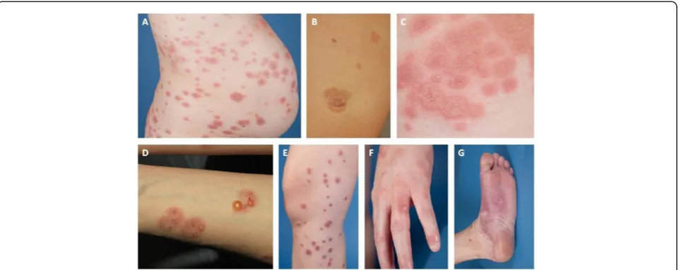
PG may appear at any time during pregnancy or puer-perium, but the most common time of symptom onset is during the second and third trimester. Intense abdom-inal itching usually begins around the navel, with varied red papules, urticarial plaques or annular target lesions
(erythema multiforme –like) appearing in the itchy
areas, followed by blistering after a few weeks (Figure 1). Bullous lesions vary from small vesicles to large blisters with a thick roof; however, some PG patients have no blisters at all (Figure 1). Typically, the skin symptoms first appear in the abdominal area, but according to an American study (n = 10) it is also common for cutaneous manifestations to appear first in the extremities [12]. In a Finnish study (n = 12) the symptoms started in the ab-dominal area in all patients, and 92% developed blisters as the disease progressed [13]. Facial and mucosal lesions are uncommon [12,14], but in some reports severe mucosal lesions were associated with more persistent disease [15].
The symptoms of PG usually alleviate a few weeks be-fore delivery, but the disease is re-activated in 75% of the patients at the time of delivery. The remitting, relapsing * Correspondence:[email protected]
1
Department of Dermatology, Medical Research Center, University of Oulu, Oulu University Hospital, Oulu, Finland
Full list of author information is available at the end of the article
course of the disease has been thought to be associated with progestin, which has immunosuppressive properties, and with changes in progestin levels: an increase in late pregnancy followed by a sharp fall during delivery [7,16]. According to a large PG study (n = 87), the average dur-ation of symptoms is 16 weeks and the majority of mothers are symptom-free 6 months after the delivery, the duration of postnatal manifestations varying between 2 weeks and 12 years [16].
Etiopathology
The pathogenesis of PG remains unknown. The pres-ence of MHC II-class HLA-antigens DR3 and DR4 or their combination has been shown to be clearly more common in women with PG compared to normal popu-lation [17]. Placental and fetal tissues contain paternal tissue antigens that are foreign to the maternal immune system. However, the maternal immune system does not normally react against these foreign antigens. In patients with PG, MHC II-class molecules that are normally not present in the placenta have been detected in tropho-blastic placental cells and amniochorionic stroma cells. As a result of partial breakdown of the syncytiotropho-blast cell layer of placental anchor villi, MHC II mole-cules are thought to get in contact with the maternal immune system, causing a (semi) allogeneic immune reac-tion against the BP180 molecule [18-20].
BP180 (also known as BPAG1 or collagen XVII) is a key structural protein of hemidesmosomes linking the epider-mis and derepider-mis. It consists of a short intracellular domain and a large extracellular domain [21]. Besides the skin basement membrane zone, BP180 is found in the placen-tal tissue and feplacen-tal membranes. Placenplacen-tal BP180 is detect-able in cytotrophoblastic cells as early as from the first
trimester [22]. In PG, antibodies are mainly directed against the same BP180 epitopes as in bullous pemphig-oid [23,24]: most commonly against the epitopes found in NC16A, the largest non-collagenous domain of BP180, but antibodies against intracellular BP180 domains and other extracellular domains of BP180 have also been ob-served [25]. In addition, antibodies against another struc-tural basement membrane protein, BP230, have been detected in about 10% of patients with PG, but this is con-sidered to be secondary and clinically insignificant [7,26]. The cross-reaction between placental antibodies and skin BP180 causes the typical skin symptoms of PG [7,27].
To the best of our knowledge, PG-related animal models do not exist. In BP, the pathogenicity of autoantibodies directed against the NC16A domain has been confirmed in several mouse models [1,28]. Severe blistering and high mortality prevents the use of experimental BP-mice for breeding to imitate a PG-like condition. The transfer of autoantibodies from mother with PG to newborn is to some extent simulated in a gene-targeted mice model in which the maternal transfer of NC16A antibodies results in blistering in the neonatal BP180-humanized mice [29].
Diagnosis
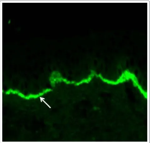
The diagnosis of PG is preferably made by a dermatologist, but all physicians treating pregnant women, i.e., general practitioners and obstetricians, should be able to consider PG. A biopsy for histopathology is not needed; the diagno-sis is based on clinical picture, direct immunofluorescence microscopy and serology [1,30,31]. Direct immunofluores-cence examination of a snap-frozen perilesional skin bi-opsy reveals the linear accumulation of complement C3 in the basement membrane zone at the interface of the epi-dermis and epi-dermis (Figure 2). Linear IgG positivity is also
detected in about 25-50% of the samples, but it is not a criterion for the diagnosis [27,32]. If PG is suspected, measurement of serum BP180 antibody level is recom-mended, as it correlates with the degree of disease severity and facilitates assessment of treatment response [33,34]. Since the BP180 NC16A ELISA is sensitive and specific to PG, it has even been proposed as a PG screening test or to be sufficient for the PG diagnosis in conjunction with typ-ical clintyp-ical symptoms [33-35]. Serum autoantibodies can also be detected with traditional indirect immunofluores-cence microscopy or the complement binding test on salt-split skin [1,30,31].
Differential diagnosis
Since PG is an extremely rare condition, other dermato-logic reasons for itchy cutaneous eruptions (Table 1) oc-curring during pregnancy should be ruled out. Pregnancy may influence the clinical picture of common skin dis-eases that either precede pregnancy or coincide with it by chance. Especially PG with atypical symptoms such as non-intense pruritus, mild erythematous papules and pla-ques or eczematous lesions represents a true challenge for clinical diagnostics.
The most important differential diagnosis alternatives for PG are the other specific dermatoses of pregnancy which
include atopic eruption of pregnancy (AEP), polymorphic eruption of pregnancy (PEP) and intrahepatic cholestasis of pregnancy (ICP) [6,36-40]. AEP is the most common pregnancy-specific skin disease, which typically appears in the first and second trimesters [40]. About 20% of the pa-tients with AEP have a pre-existing atopic dermatitis with a typical clinical picture, whereas the remaining 80% present widespread eczematous changes or papular lesions and have no previous history of atopic eczema or have been symptomless since childhood [31]. The greatest dif-ferential diagnostic challenge of PG is PEP, previously known as Pruritic Urticarial Papules and Plaques of Preg-nancy (PUPPP), with intensely pruritic urticarial papules and plaques during the last trimester. Despite rather simi-lar clinical features, negative immunofluorescence analysis of perilesional skin biopsy in PEP differentiates it explicitly
from PG [38,39]. Similar to PG, PEP symptoms usually start on the abdomen, but PEP lesions typically spare the umbilical region. ICP, which is associated with significant fetal risks, can present in the last trimester with pruritus, and thus it should be considered in differential diagnosis of PG [40]. Patients with ICP do not have primary skin le-sions, but due to severe pruritus and scratching may develop secondary excoriations or even prurigo nodularis-like changes, usually on the extremities [31].
Management
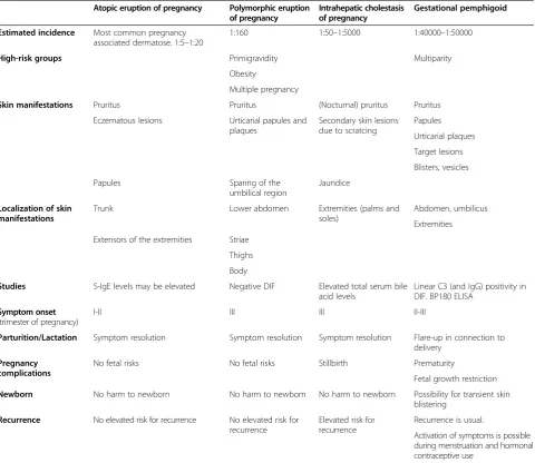
Due to the rarity of PG no randomized studies have been published and treatment recommendations are based on clinical experience and studies from treatment of other skin diseases. PG symptoms can be quite debili-tating, but the condition does not constitute a direct Table 1 Differential diagnostics of pregnancy associated pruritic dermatoses
Atopic eruption of pregnancy Polymorphic eruption of pregnancy
Intrahepatic cholestasis of pregnancy
Gestational pemphigoid
Estimated incidence Most common pregnancy associated dermatose. 1:5–1:20
1:160 1:50–1:5000 1:40000–1:50000
High-risk groups Primigravidity Multiparity
Obesity
Multiple pregnancy
Skin manifestations Pruritus Pruritus (Nocturnal) pruritus Pruritus
Eczematous lesions Urticarial papules and plaques
Secondary skin lesions due to scratcing
Papules
Urticarial plaques
Target lesions
Blisters, vesicles
Papules Sparing of the
umbilical region
Jaundice
Localization of skin manifestations
Trunk Lower abdomen Extremities (palms and
soles)
Abdomen, umbilicus
Extremities
Extensors of the extremities Striae
Thighs
Body
Studies S-IgE levels may be elevated Negative DIF Elevated total serum bile acid levels
Linear C3 (and IgG) positivity in DIF. BP180 ELISA
Symptom onset (trimester of pregnancy)
I-II III III II-III
Parturition/Lactation Symptom resolution Symptom resolution Symptom resolution Flare-up in connection to delivery
Pregnancy complications
No fetal risks No fetal risks Stillbirth Prematurity
Fetal growth restriction
Newborn No harm to newborn No harm to newborn No harm to newborn Possibility for transient skin blistering
Recurrence No elevated risk for recurrence No elevated risk for recurrence
Elevated risk for recurrence
Recurrence is usual.
Activation of symptoms is possible during menstruation and hormonal contraceptive use
health risk to the mother. When choosing a treatment, the benefit of the medication to the mother is critically weighed up against possible risks to the fetus. The aim of the treatment is to suppress the excessive itching and to prevent formation of new blisters [41].
According to current recommendations PG patients with mild symptoms (about 19% of the patients) should be treated with potent or very potent topical corticoste-roids (for example betamethasone valerate or clobetasol propionate) [1,30]. Controlled studies with BP patients have shown that topical treatment with highly potent corticosteroid is as effective and safe as oral prednisolone 0.5 mg/kg/day [42]. During pregnancy, mild or moderate topical corticosteroids are preferred to potent or very potent ones because of the risk of fetal growth restric-tion associated with the latter [43]. When necessary, po-tent or very popo-tent topical corticosteroids can be used for the therapy of PG for as short duration as possible, since their potential for fetotoxicity is less than that of systemic corticosteroids [43-45].
The combination of oral antihistamines with topical cor-ticosteroids, most commonly cetirizine, is usually employed to relieve the itching, despite the fact that clinical efficacy studies in PG are lacking [1,16,27,30]. In general, second-generation H1-antihistamines are currently preferred to first-generation antihistamines based on the potential ser-ious anticholinergic and central nervous system side ef-fects of old sedating antihistamines and the longer-lasting antipruritic effects of the modern antihistamines [46]. First-generation antihistamines have no definitive increased teratogenic risk, and the second-generation antihistamines cetirizine, levocetirizine and loratadine are also recom-mended for use in pregnancy [44,46].
Corticosteroid treatment has become the standard of care for first-line systemic therapy of severe PG thanks to its treatment response and tolerable safety profile. Most of prednisolone is inactivated by placental de-hydrogenase enzyme (11-hydroxysteroid dede-hydrogenase- dehydrogenase-2) before reaching the fetal circulation. As fluorinated corticosteroids (betamethasone and dexamethasone) are not metabolized by placental dehydrogenase enzyme, prednisolone is considered the primary treatment alterna-tive. [1,30,47]. The initial dose of prednisolone is usually 0.25-0.5 mg/kg/day, and the response is usually good. If formation of blisters does not decrease within a few days, the dose is increased until no new blisters appear. The
cortisone dose is gradually decreased about 1–2 weeks
after the symptoms have been brought under control, and discontinued altogether if possible.
The side effects of long-term systemic corticosteroid treatment are well-known. Previous studies have dem-onstrated that in the treatment of BP the use of oral prednisolone is associated with more frequent severe ad-verse events and increased mortality compared to topical
corticosteroids [1,30,42]. However, BP patients are much older and have more severe comorbidities than PG pa-tients. In addition, the duration of prednisolone treatment is shorter and the dosage is smaller in PG than in BP, which further decreases the risk of side effects. During pregnancy, the use of prednisolone in the first trimester causes an increased risk of malformations, especially orofa-cial clefts [44]. In the last trimester prednisolone may result in intrauterine growth retardation, gestational diabetes, eclampsia and premature delivery [44].
Plasmapheresis [48], immunoadsorption [49,50] and intravenous immunoglobulin G-infusion [51-54], which are not contraindicated during pregnancy, have in some cases been used to treat PG even prior to the delivery. Removal of antibodies with immunoadsorption gives quick symptom relief especially in PG cases with severe postnatal symp-toms, as there is no placenta to maintain an autoimmune reaction [50]. Prenatal treatment with cyclosporine com-bined to prednisolone has been reported in two cases with good treatment response [13,55], and in one case cyclo-sporine was used after intravenous immunoglobulin in persistent postnatal PG [56]. Case reports on the use of tetracycline, cyclophosphamide, azathioprine, dapsone and rituximab to treat PG with persisting postnatal symp-toms have been published, but these agents are avoided prenatally due to potential short- and long-term fetal effects. [7,41].
PG lesions usually disappear 12–16 weeks after the de-livery, with no scarring, and postnatal oral cortisone treat-ment can normally be discontinued fairly soon. However, sometimes treatment has to be resumed as the disease flares up again [16,27]. When systemic cortisone is given at the average doses used in the treatment of PG, it does not prevent breastfeeding, and breastfeeding has been shown to reduce the symptoms of PG [17,7,12].
Fetus and the newborn
The risk of preterm birth and fetal growth restriction is greater in PG pregnancies compared to normal popula-tion [57-60]. The pregnancy risks of PG are thought to be associated with mild placental failure caused by BP180 antibodies [13,27,60]. In addition to accumulation of C3 complement and IgG, mild villitis and collections of im-mature fibrotic villi have been observed in histologic ex-aminations of PG placentas [22]. Antibody concentrations do not as such correlate with the occurrence of pregnancy complications, and no association has been demonstrated between cortisone treatment and PG pregnancy complica-tions [60]. No follow-up guidelines for pregnancies com-plicated by PG have been published, most likely due to the rarity of the condition.
of miscarriages occurring in the first trimester [16]. How-ever, in a more recent British-Taiwanese study with 70 pa-tients late miscarriages and fetal deaths were observed in as many as 6% of the patients [60].
About 16-34% of PG patients are estimated to give birth prematurely [13,58-60]. Premature delivery is more likely if PG begins in the 1st or 2nd trimester or if the skin symptoms include blistering [60]. In a Finnish PG study, 25% of the deliveries were premature (the corre-sponding rate in the Finnish population during time of study was around 5%) [13,61]. The proportion of prema-ture deliveries among pregnant women with PG was similar to that in previously published studies, even though all patients, with one exception, had blistering PG. All pre-mature births occurred after the 35th gestational week, and PG had no effect on neonatal mortality [13]. Vaginal ultrasound is considered the gold standard in charting cer-vical dilation in women at risk of preterm delivery [62]. Although preterm delivery is difficult to predict, we rec-ommend obstetric follow-up with vaginal ultrasound due to the increased risk of preterm delivery.
In the British-Taiwanese study with 70 patients, fetal growth restriction was observed in 34% [60], the likeli-hood of its occurrence correlating with early onset of PG. In a Finnish study, only one mother developed pre-eclampsia combined with fetal growth restriction, which is in line with the general prevalence in Finnish popula-tion. However, 50% of the patients in our study had an abnormal placental weight/birth weight ratio [13]. The blood flow profile of the umbilical artery is used in clin-ical practice to diagnose placental failure [63]. In a PG case report where pregnancy was complicated by pre-eclampsia and fetal growth restriction, abnormal end-diastolic blood flow was reported in the umbilical artery [64]. Among 12 Finnish PG patients increased umbilical artery pulsatility was detected only in one pregnancy with pre-eclampsia and fetal growth restriction; all other PG pregnancies showed normal umbilical artery blood flow findings and biophysical scores [13], suggesting that clinically significant placental failure is rare in PG.
There is only little information available on the effect of PG on the newborn infant. No congenital abnormal-ities have been linked to PG [58,60]. According to the data from 12 Finnish PG patients, birth weight, umbilical artery pH, Apgar scores and neonatal morbidity did not differ from normal population [13]. The IgG antibodies of PG pass through the placenta, but PG blisters develop in only about 3% of newborn infants [14,16,57]. Skin symptoms in newborns usually resolve quickly without treatment as antibody levels decrease. According to a Japanese case report, antibody levels in newborn infants are comparable to those in mothers; the levels in both are reduced by half in about 15 days. Since the clinical status of the newborn often improves rapidly, it seems
that other factors besides autoantibodies contribute to the formation of blisters in newborns [65]. If PG in the mother was treated with large doses of cortisone, the pediatrician should be informed of the possibility of neo-natal adrenal insufficiency. There are no data on the long-term prognosis of children of PG mothers.
Prognosis
Recurrence of PG in subsequent pregnancies is likely, and symptoms are usually more severe, with earlier on-set. In patients with an earlier PG episode the likelihood of pregnancy with no symptoms is estimated to be 5-8%, but the reason for the lack of symptoms is unknown. [16,17]. In a Finnish study, PG recurred in two cases, while 67% of the subsequent pregnancies were symptom-free (n = 4/6). The large proportion of symptom-symptom-free preg-nancies is most likely due to the small number of patients, but ethnic factors may also play a role [13]. Perfect HLA-DR match between the mother and the fetus may explain some of the cases, but pregnancy may be free from PG symptoms even in the absence of identical HLA-DR type [16,17] and even if the symptoms of PG have been persistent [66].
At the postpartum examination, mothers with PG should be reminded of the possibility of relapse during menstruation and/or in connection with hormonal contra-ceptive use. Susceptibility for recurrence may persist for years [16,37]. In a large British study, about 11% of the pa-tients experienced a relapse during oral contraceptive use, but in smaller studies the incidence has been as high as 50% [14,16,17]. The low relapse rate in the British study was thought to be associated with the infrequent use of oral contraceptives after PG pregnancy [16]. Women who have had PG have also been described to be more suscep-tible to other autoimmune diseases; the prevalence of Graves’disease increases in particular, from 0.4% in nor-mal population to as high as 10.6% [16]. Proneness to
Hashimoto’s thyroiditis, autoimmune thrombocytopenia
and pernicious anemia has also been reported to be in-creased [16,67].
Conclusion
Abbreviations
PG:Pemphigoid gestationis, gestational pemphigoid; BP: Bullous pemphigoid; BP180: Bullous pemphigoid antigen 180; BP230: Bullous pemphigoid antigen 230; MHC: Major histocompatibility complex; HLA: Human leucocyte antigen; IgG: Immunoglobulin G; AEP: Atopic eruption of pregnancy; PEP: Polymorphic eruption of pregnancy; ICP: Intrahepatic cholestasis of pregnancy.
Competing interests
The authors declare that they have no competing interests.
Authors’contributions
Authors contributed equally to this review. All authors have read and approved the final version of the manuscript.
Author details
1Department of Dermatology, Medical Research Center, University of Oulu,
Oulu University Hospital, Oulu, Finland.2Department of Obstetrics and Gynecology, University of Oulu, Oulu University Hospital, Oulu, Finland.
Received: 20 May 2014 Accepted: 19 August 2014 Published: 2 September 2014
References
1. Schmidt E, Zillikens D:Pemphigoid diseases.Lancet2013,381(9863):320–332. 2. Holmes RC, Black MM:The specific dermatoses of pregnancy.J Am Acad
Dermatol1983,8(3):405–412.
3. Nanda A, Dvorak R, Al-Saeed K, Al-Sabah H, Alsaleh QA:Spectrum of autoimmune bullous diseases in Kuwait.Int J Dermatol2004,
43(12):876–881.
4. Bernard P, Vaillant L, Labeille B, Bedane C, Arbeille B, Denoeux JP, Lorette G, Bonnetblanc JM, Prost C:Incidence and distribution of subepidermal autoimmune bullous skin diseases in three French regions. Bullous Diseases French Study Group.Arch Dermatol1995,131(1):48–52. 5. Bertram F, Brocker E, Zillikens D, Schmidt E:Prospective analysis of the
incidence of autoimmune bullous disorders in Lower Franconia, Germany.J Dtsch Dermatol Ges2009,7(5):434–439.
6. Ambros-Rudolph CM, Mullegger RR, Vaughan-Jones SA, Kerl H, Black MM:
The specific dermatoses of pregnancy revisited and reclassified: results of a retrospective two-center study on 505 pregnant patients.J Am Acad Dermatol2006,54(3):395–404.
7. Semkova K, Black M:Pemphigoid gestationis: current insights into pathogenesis and treatment.Eur J Obstet Gynecol Reprod Biol2009,
145(2):138–144.
8. Shornick JK, Meek TJ, Nesbitt LT Jr, Gilliam JN:Herpes gestationis in blacks. Arch Dermatol1984,120(4):511–513.
9. Kneisel A, Hertl M:Autoimmune bullous skin diseases. Part 1: clinical manifestations.J Dtsch Dermatol Ges2011,9(10):844–857.
10. Takatsuka Y, Komine M, Ohtsuki M:Pemphigoid gestationis with a complete hydatidiform mole.J Dermatol2012,39(5):474–476. 11. Djahansouzi S, Nestle-Kraemling C, Dall P, Bender HG, Hanstein B:Herpes
gestationis may present itself as a paraneoplastic syndrome of choriocarcinoma-a case report.Gynecol Oncol2003,89(2):334–337. 12. Castro LA, Lundell RB, Krause PK, Gibson LE:Clinical experience in
pemphigoid gestationis: report of 10 cases.J Am Acad Dermatol2006,
55(5):823–828.
13. Huilaja L, Makikallio K, Sormunen R, Lohi J, Hurskainen T, Tasanen K:
Gestational pemphigoid: placental morphology and function.Acta Derm Venereol2013,93(1):33–38.
14. Shornick JK, Bangert JL, Freeman RG, Gilliam JN:Herpes gestationis: clinical and histologic features of twenty-eight cases.J Am Acad Dermatol1983,
8(2):214–224.
15. Boulinguez S, Bedane C, Prost C, Bernard P, Labbe L, Bonnetblanc JM:
Chronic pemphigoid gestationis: comparative clinical and immunopathological study of 10 patients.Dermatology2003,
206(2):113–119.
16. Jenkins RE, Hern S, Black MM:Clinical features and management of 87 patients with pemphigoid gestationis.Clin Exp Dermatol1999,
24(4):255–259.
17. Holmes RC, Black MM, Jurecka W, Dann J, James DC, Timlin D, Bhogal B:
Clues to the aetiology and pathogenesis of herpes gestationis. Br J Dermatol1983,109(2):131–139.
18. Kelly SE, Black MM, Fleming S:Pemphigoid gestationis: a unique mechanism of initiation of an autoimmune response by MHC class II molecules?J Pathol1989,158(1):81–82.
19. Kelly SE, Fleming S, Bhogal BS, Wojnarowska F, Black MM:
Immunopathology of the placenta in pemphigoid gestationis and linear IgA disease.Br J Dermatol1989,120(6):735–743.
20. Kelly SE, Black MM, Fleming S:Antigen-presenting cells in the skin and placenta in pemphigoid gestationis.Br J Dermatol1990,122(5):593–599. 21. Powell AM, Sakuma-Oyama Y, Oyama N, Black MM:Collagen XVII/BP180: a
collagenous transmembrane protein and component of the dermoepidermal anchoring complex.Clin Exp Dermatol2005,
30(6):682–687.
22. Huilaja L, Hurskainen T, Autio-Harmainen H, Hofmann SC, Sormunen R, Rasanen J, Ilves M, Franzke CW, Bruckner-Tuderman L, Tasanen K:
Pemphigoid gestationis autoantigen, transmembrane collagen XVII, promotes the migration of cytotrophoblastic cells of placenta and is a structural component of fetal membranes.Matrix Biol2008,27(3):190–200. 23. Giudice GJ, Emery DJ, Zelickson BD, Anhalt GJ, Liu Z, Diaz LA:Bullous
pemphigoid and herpes gestationis autoantibodies recognize a common non-collagenous site on the BP180 ectodomain.J Immunol 1993,151(10):5742–5750.
24. Herrero-Gonzalez JE, Brauns O, Egner R, Ronspeck W, Mascaro JM Jr, Jonkman MF, Zillikens D, Sitaru C:Immunoadsorption against two distinct epitopes on human type XVII collagen abolishes dermal-epidermal separation induced in vitro by autoantibodies from pemphigoid gestationis patients.Eur J Immunol2006,36(4):1039–1048.
25. Di Zenzo G, Calabresi V, Grosso F, Caproni M, Ruffelli M, Zambruno G:The intracellular and extracellular domains of BP180 antigen comprise novel epitopes targeted by pemphigoid gestationis autoantibodies.J Invest Dermatol2007,127(4):864–873.
26. Kelly SE, Bhogal BS, Wojnarowska F, Whitehead P, Leigh IM, Black MM:
Western blot analysis of the antigen in pemphigoid gestationis. Br J Dermatol1990,122(4):445–449.
27. Shimanovich I, Brocker EB, Zillikens D:Pemphigoid gestationis: new insights into the pathogenesis lead to novel diagnostic tools.BJOG2002,
109(9):970–976.
28. Nishie W:Update on the pathogenesis of bullous pemphigoid: an autoantibody-mediated blistering disease targeting collagen XVII. J Dermatol Sci2014,73(3):179–186.
29. Nishie W, Sawamura D, Natsuga K, Shinkuma S, Goto M, Shibaki A, Ujiie H, Olasz E, Yancey KB, Shimizu H:A novel humanized neonatal autoimmune blistering skin disease model induced by maternally transferred antibodies.J Immunol2009,183(6):4088–4093.
30. Kneisel A, Hertl M:Autoimmune bullous skin diseases. Part 2: diagnosis and therapy.J Dtsch Dermatol Ges2011,9(11):927–947.
31. AmbrosRudolph CM:Schwangerschaftsspezifische Dermatosen: S04/05. J Dtsch Dermatol Ges2011,9(Sup 1):45–46.
32. Holmes RC, Black MM, Dann J, James DC, Bhogal B:A comparative study of toxic erythema of pregnancy and herpes gestationis.Br J Dermatol1982,
106(5):499–510.
33. Sitaru C, Powell J, Messer G, Brocker EB, Wojnarowska F, Zillikens D:
Immunoblotting and enzyme-linked immunosorbent assay for the diagnosis of pemphigoid gestationis.Obstet Gynecol2004,103(4):757–763. 34. Sitaru C, Dahnrich C, Probst C, Komorowski L, Blocker I, Schmidt E,
Schlumberger W, Rose C, Stocker W, Zillikens D:Enzyme-linked immunosorbent assay using multimers of the 16th non-collagenous domain of the BP180 antigen for sensitive and specific detection of pemphigoid autoantibodies.Exp Dermatol2007,16(9):770–777. 35. Powell AM, Sakuma-Oyama Y, Oyama N, Albert S, Bhogal B, Kaneko F,
Nishikawa T, Black MM:Usefulness of BP180 NC16a enzyme-linked immunosorbent assay in the serodiagnosis of pemphigoid gestationis and in differentiating between pemphigoid gestationis and pruritic urticarial papules and plaques of pregnancy.Arch Dermatol2005,
141(6):705–710.
36. Al-Fares SI, Jones SV, Black MM:The specific dermatoses of pregnancy: a re-appraisal.J Eur Acad Dermatol Venereol2001,15(3):197–206. 37. Kroumpouzos G, Cohen LM:Specific dermatoses of pregnancy: an
38. Rudolph CM, Al-Fares S, Vaughan-Jones SA, Mullegger RR, Kerl H, Black MM:
Polymorphic eruption of pregnancy: clinicopathology and potential trigger factors in 181 patients.Br J Dermatol2006,154(1):54–60. 39. Roth MM:Pregnancy dermatoses: diagnosis, management, and
controversies.Am J Clin Dermatol2011,12(1):25–41. 40. Beard MP, Millington GW:Recent developments in the specific
dermatoses of pregnancy.Clin Exp Dermatol2012,37(1):1–4. 41. Intong LR, Murrell DF:Pemphigoid gestationis: current management.
Dermatol Clin2011,29(4):621–628.
42. Joly P, Roujeau J, Benichou J, Picard C, Dreno B, Delaporte E, Vaillant L, D’Incan M, Plantin P, Bedane C, Young P, Bernard P:A comparison of oral and topical corticosteroids in patients with Bullous Pemphigoid.N Engl J Med2002,346(5):321–327.
43. Chi CC, Kirtschig G, Aberer W, Gabbud JP, Lipozencic J, Karpati S, Haustein UF, Zuberbier T, Wojnarowska F:Evidence-based (S3) guideline on topical corticosteroids in pregnancy.Br J Dermatol2011,165(5):943–952. 44. Murase JE, Heller MM, Butler DC:Safety of dermatologic medications in
pregnancy and lactation.J Am Acad Dermatol2014,70(3):401e1–401e14. 45. Tyler KH, Zirwas MJ:Pregnancy and dermatologic therapy.J Am Acad
Dermatol2013,68(4):663–671.
46. Church D, Baiardini I, Staevska M, Popov T, Kralimarkova T, Dimitrov V, Church M:The effectiveness of antihistamines in up to four-times conventional doses on urticarial discomfort and quality of life in difficult-to-treat urticaria: 1501.Allergy2009,64(Supplement 90):571. 47. Jackson S, Gilchrist H, Nesbitt LT Jr:Update on the dermatologic use of
systemic glucocorticosteroids.Dermatol Ther2007,20(4):187–205. 48. Patsatsi A, Vavilis D, Tsikeloudi M, Kalabalikis D, Sotiriadis D:Refractory
pemphigoid gestationis postpartum.Acta Obstet Gynecol Scand2012,
91(5):636–637.
49. Marker M, Derfler K, Monshi B, Rappersberger K:Successful
immunoapheresis of bullous autoimmune diseases: pemphigus vulgaris and pemphigoid gestationis.J Dtsch Dermatol Ges2011,9(1):27–31. 50. Westermann L, Hugel R, Meier M, Weichenthal M, Zillikens D, Glaser R,
Schmidt E:Glucocorticosteroid-resistant pemphigoid gestationis: successful treatment with adjuvant immunoadsorption.J Dermatol2012,
39(2):168–171.
51. Gan DC, Welsh B, Webster M:Successful treatment of a severe persistent case of pemphigoid gestationis with antepartum and postpartum intravenous immunoglobulin followed by azathioprine.Australas J Dermatol2012,53(1):66–69.
52. Doiron P, Pratt M:Antepartum intravenous immunoglobulin therapy in refractory pemphigoid gestationis: case report and literature review. J Cutan Med Surg2010,14(4):189–192.
53. Rodrigues Cdos S, Filipe P, Solana Mdel M, Soares De Almeida L, Cirne De Castro J, Gomes MM:Persistent herpes gestationis treated with high-dose intravenous immunoglobulin.Acta Derm Venereol2007,87(2):184–186. 54. Kreuter A, Harati A, Breuckmann F, Appelhans C, Altmeyer P:Intravenous
immune globulin in the treatment of persistent pemphigoid gestationis. J Am Acad Dermatol2004,51(6):1027–1028.
55. Paternoster DM, Bruno G, Grella PV:New observations on herpes gestationis therapy.Int J Gynaecol Obstet1997,56(3):277–278. 56. Hern S, Harman K, Bhogal BS, Black MM:A severe persistent case of
pemphigoid gestationis treated with intravenous immunoglobulins and cyclosporin.Clin Exp Dermatol1998,23(4):185–188.
57. Holmes RC, Black MM:The fetal prognosis in pemphigoid gestationis (herpes gestationis).Br J Dermatol1984,110(1):67–72.
58. Shornick JK, Black MM:Fetal risks in herpes gestationis.J Am Acad Dermatol1992,26(1):63–68.
59. Mascaro JM Jr, Lecha M, Mascaro JM:Fetal morbidity in herpes gestationis.Arch Dermatol1995,131(10):1209–1210.
60. Chi CC, Wang SH, Charles-Holmes R, Ambros-Rudolph C, Powell J, Jenkins R, Black M, Wojnarowska F:Pemphigoid gestationis: early onset and blister formation are associated with adverse pregnancy outcomes. Br J Dermatol2009,160(6):1222–1228.
61. Finnish birth registry data.http://www.stakes.fi/tilastot/tilastotiedotteet/ 2010/Tr26_10.pdf.
62. Mella MT, Berghella V:Prediction of preterm birth: cervical sonography. Semin Perinatol2009,33(5):317–324.
63. Giles WB, Trudinger BJ, Baird PJ:Fetal umbilical artery flow velocity waveforms and placental resistance: pathological correlation.Br J Obstet Gynaecol1985,92(1):31–38.
64. Dolkart L, Harter M, Snyder M:Pemphigoid gestationis: report of a case with umbilical artery Doppler assessment.J Reprod Med2006,
51(7):591–594.
65. Aoyama Y, Asai K, Hioki K, Funato M, Kondo N, Kitajima Y:Herpes gestationis in a mother and newborn: immunoclinical perspectives based on a weekly follow-up of the enzyme-linked immunosorbent assay index of a bullous pemphigoid antigen noncollagenous domain. Arch Dermatol2007,143(9):1168–1172.
66. Black MM, Najem NM:Remarkable follow-up experiences of a severe persistent case of pemphigoid gestationis.Clin Exp Dermatol2005,
30(5):593–594.
67. Shornick JK, Black MM:Secondary autoimmune diseases in herpes gestationis (pemphigoid gestationis).J Am Acad Dermatol1992,
26(4):563–566.
doi:10.1186/s13023-014-0136-2
Cite this article as:Huilajaet al.:Gestational pemphigoid.Orphanet Journal of Rare Diseases20149:136.
Submit your next manuscript to BioMed Central and take full advantage of:
• Convenient online submission
• Thorough peer review
• No space constraints or color figure charges
• Immediate publication on acceptance
• Inclusion in PubMed, CAS, Scopus and Google Scholar
• Research which is freely available for redistribution