AMERICAN ACADEMY OF PEDIATRICS
C
LINICALP
RACTICEG
UIDELINEAmerican Academy of Family Physicians, American Academy of Otolaryngology-Head and Neck Surgery, and American Academy of Pediatrics Subcommittee on Otitis Media With Effusion
Otitis Media With Effusion
ABSTRACT. The clinical practice guideline on otitis media with effusion (OME) provides evidence-based rec-ommendations on diagnosing and managing OME in children. This is an update of the 1994 clinical practice guideline “Otitis Media With Effusion in Young Chil-dren,” which was developed by the Agency for Health-care Policy and Research (now the Agency for HealthHealth-care Research and Quality). In contrast to the earlier guide-line, which was limited to children 1 to 3 years old with no craniofacial or neurologic abnormalities or sen-sory deficits, the updated guideline applies to children aged 2 months through 12 years with or without devel-opmental disabilities or underlying conditions that pre-dispose to OME and its sequelae. The American Acad-emy of Pediatrics, American AcadAcad-emy of Family Physicians, and American Academy of Otolaryngology-Head and Neck Surgery selected a subcommittee com-posed of experts in the fields of primary care, otolaryn-gology, infectious diseases, epidemiology, hearing, speech and language, and advanced-practice nursing to revise the OME guideline.
The subcommittee made a strong recommendation that clinicians use pneumatic otoscopy as the primary diagnos-tic method and distinguish OME from acute otitis media.
The subcommittee made recommendations that clini-cians should 1) document the laterality, duration of effu-sion, and presence and severity of associated symptoms at each assessment of the child with OME, 2) distinguish the child with OME who is at risk for speech, language, or learning problems from other children with OME and more promptly evaluate hearing, speech, language, and need for intervention in children at risk, and 3) manage the child with OME who is not at risk with watchful waiting for 3 months from the date of effusion onset (if known) or diagnosis (if onset is unknown).
The subcommittee also made recommendations that 4) hearing testing be conducted when OME persists for 3 months or longer or at any time that language delay, learn-ing problems, or a significant hearlearn-ing loss is suspected in a child with OME, 5) children with persistent OME who are not at risk should be reexamined at 3- to 6-month intervals until the effusion is no longer present, significant hearing loss is identified, or structural abnormalities of the eardrum or middle ear are suspected, and 6) when a child becomes a surgical candidate (tympanostomy tube insertion is the preferred initial procedure). Adenoidectomy should not be performed unless a distinct indication exists (nasal
ob-struction, chronic adenoiditis); repeat surgery consists of adenoidectomy plus myringotomy with or without tubeinsertion. Tonsillectomy alone or myringotomy alone should not be used to treat OME.
The subcommittee made negative recommendations that 1) population-based screening programs for OME not be performed in healthy, asymptomatic children, and 2) because antihistamines and decongestants are ineffective for OME, they should not be used for treat-ment; antimicrobials and corticosteroids do not have long-term efficacy and should not be used for routine management.
The subcommittee gave as options that 1) tympanom-etry can be used to confirm the diagnosis of OME and 2) when children with OME are referred by the primary clinician for evaluation by an otolaryngologist, audiolo-gist, or speech-language patholoaudiolo-gist, the referring clini-cian should document the effusion duration and specific reason for referral (evaluation, surgery) and provide ad-ditional relevant information such as history of acute otitis media and developmental status of the child. The subcommittee made no recommendations for 1) comple-mentary and alternative medicine as a treatment for OME, based on a lack of scientific evidence documenting efficacy, or 2) allergy management as a treatment for OME, based on insufficient evidence of therapeutic effi-cacy or a causal relationship between allergy and OME. Last, the panel compiled a list of research needs based on limitations of the evidence reviewed.
The purpose of this guideline is to inform clinicians of evidence-based methods to identify, monitor, and manage OME in children aged 2 months through 12 years. The guideline may not apply to children more than 12 years old, because OME is uncommon and the natural history is likely to differ from younger children who experience rapid developmental change. The target population includes chil-dren with or without developmental disabilities or under-lying conditions that predispose to OME and its sequelae. The guideline is intended for use by providers of health care to children, including primary care and specialist phy-sicians, nurses and nurse practitioners, physician assistants, audiologists, speech-language pathologists, and child-de-velopment specialists. The guideline is applicable to any setting in which children with OME would be identified, monitored, or managed.
This guideline is not intended as a sole source of guidance in evaluating children with OME. Rather, it is designed to assist primary care and other clinicians by providing an evidence-based framework for decision-making strategies. It is not intended to replace clinical judgment or establish a protocol for all children with this condition and may not provide the only appropriate ap-proach to diagnosing and managing this problem. Pedi-atrics 2004;113:1412–1429; acute otitis media, antibacte-rial, antibiotic.
This document was approved by the American Academy of Otolaryn-gology–Head and Neck Surgery Foundation, Inc and the American Acad-emy of Pediatrics, and is published in the May 2004 issue of Otolaryngology-Head and Neck Surgeryand the May 2004 issue ofPediatrics.
ABBREVIATIONS. OME, otitis media with effusion; AOM, acute otitis media; AAP, American Academy of Pediatrics; AHRQ, Agency for Healthcare Research and Quality; EPC, Southern Cal-ifornia Evidence-Based Practice Center; CAM, complementary and alternative medicine; HL, hearing level.
O
titis media with effusion (OME) as discussed in this guideline is defined as the presence of fluid in the middle ear without signs or symptoms of acute ear infection.1,2 OME is consid-ered distinct from acute otitis media (AOM), which is defined as a history of acute onset of signs and symptoms, the presence of middle-ear effusion, and signs and symptoms of middle-ear inflammation. Persistent middle-ear fluid from OME results in de-creased mobility of the tympanic membrane and serves as a barrier to sound conduction.3 Approxi-mately 2.2 million diagnosed episodes of OME occur annually in the United States, yielding a combined direct and indirect annual cost estimate of $4.0 bil-lion.2OME may occur spontaneously because of poor eustachian tube function or as an inflammatory re-sponse following AOM. Approximately 90% of chil-dren (80% of individual ears) have OME at some time before school age,4 most often between ages 6 months and 4 years.5In the first year of life,⬎50% of children will experience OME, increasing to ⬎60% by 2 years.6 Many episodes resolve spontaneously within 3 months, but⬃30% to 40% of children have recurrent OME, and 5% to 10% of episodes last 1 year or longer.1,4,7
The primary outcomes considered in the guideline include hearing loss; effects on speech, language, and learning; physiologic sequelae; health care utilization (medical, surgical); and quality of life.1,2 The high prevalence of OME, difficulties in diagnosis and as-sessing duration, increased risk of conductive hear-ing loss, potential impact on language and cognition, and significant practice variations in management8 make OME an important condition for the use of up-to-date evidence-based practice guidelines.
METHODS General Methods and Literature Search
In developing an evidence-based clinical practice guideline on managing OME, the American Academy of Pediatrics (AAP), American Academy of Family Physicians, and American Acad-emy of Otolaryngology-Head and Neck Surgery worked with the Agency for Healthcare Research and Quality (AHRQ) and other organizations. This effort included representatives from each part-nering organization along with liaisons from audiology, speech-language pathology, informatics, and advanced-practice nursing. The most current literature on managing children with OME was reviewed, and research questions were developed to guide the evidence-review process.
The AHRQ report on OME from the Southern California Evi-dence-Based Practice Center (EPC) focused on key questions of natural history, diagnostic methods, and long-term speech, lan-guage, and hearing outcomes.2Searches were conducted through
January 2000 in Medline, Embase, and the Cochrane Library. Additional articles were identified by review of reference listings in proceedings, reports, and other guidelines. The EPC accepted 970 articles for full review after screening 3200 abstracts. The EPC reviewed articles by using established quality criteria9,10and
in-cluded randomized trials, prospective cohorts, and validations of diagnostic tests (validating cohort studies).
The AAP subcommittee on OME updated the AHRQ review with articles identified by an electronic Medline search through April 2003 and with additional material identified manually by subcommittee members. Copies of relevant articles were distrib-uted to the subcommittee for consideration. A specific search for articles relevant to complementary and alternative medicine (CAM) was performed by using Medline and the Allied and Complementary Medicine Database through April 2003. Articles relevant to allergy and OME were identified by using Medline through April 2003. The subcommittee met 3 times over a 1-year period, ending in May 2003, with interval electronic review and feedback on each guideline draft to ensure accuracy of content and consistency with standardized criteria for reporting clinical prac-tice guidelines.11
In May 2003, the Guidelines Review Group of the Yale Center for Medical Informatics used the Guideline Elements Model12to
categorize content of the present draft guideline. Policy statements were parsed into component decision variables and actions and then assessed for decidability and executability. Quality appraisal using established criteria13was performed with Guideline
Ele-ments Model-Q Online.14,15Implementation issues were predicted
by using the Implementability Rating Profile, an instrument under development by the Yale Guidelines Review Group (R. Shiffman, MD, written communication, May 2003). OME subcommittee members received summary results and modified an advanced draft of the guideline.
The final draft practice guideline underwent extensive peer review by numerous entities identified by the subcommittee. Comments were compiled and reviewed by the subcommittee cochairpersons. The recommendations contained in the practice guideline are based on the best available published data through April 2003. Where data are lacking, a combination of clinical experience and expert consensus was used. A scheduled review process will occur 5 years from publication or sooner if new compelling evidence warrants earlier consideration.
Classification of Evidence-Based Statements
Guidelines are intended to reduce inappropriate variations in clinical care, produce optimal health outcomes for patients, and minimize harm. The evidence-based approach to guideline devel-opment requires that the evidence supporting a policy be identi-fied, appraised, and summarized and that an explicit link between evidence and statements be defined. Evidence-based statements reflect the quality of evidence and the balance of benefit and harm that is anticipated when the statement is followed. The AAP definitions for evidence-based statements16are listed in Tables 1
and 2.
Guidelines are never intended to overrule professional judg-ment; rather, they may be viewed as a relative constraint on individual clinician discretion in a particular clinical circumstance. Less frequent variation in practice is expected for a strong recom-mendation than might be expected with a recomrecom-mendation. Op-tions offer the most opportunity for practice variability.17 All
clinicians should always act and decide in a way that they believe will best serve their patients’ interests and needs regardless of guideline recommendations. Guidelines represent the best judg-ment of a team of experienced clinicians and methodologists addressing the scientific evidence for a particular topic.16
Making recommendations about health practices involves value judgments on the desirability of various outcomes associ-ated with management options. Value judgments applied by the OME subcommittee were made in an effort to minimize harm and diminish unnecessary therapy. Emphasis was placed on promptly identifying and managing children at risk for speech, language, or learning problems to maximize opportunities for beneficial out-comes. Direct costs also were considered in the statements con-cerning diagnosis and screening and to a lesser extent in other statements.
1A. PNEUMATIC OTOSCOPY: CLINICIANS SHOULD USE PNEUMATIC OTOSCOPY AS THE PRIMARY DIAGNOSTIC METHOD FOR OME, AND
OME SHOULD BE DISTINGUISHED FROM AOM
1B. TYMPANOMETRY: TYMPANOMETRY CAN BE USED TO CONFIRM THE DIAGNOSIS OF OME
This option is based on cohort studies and a balance of benefit and harm.
Diagnosing OME correctly is fundamental to proper management. Moreover, OME must be dif-ferentiated from AOM to avoid unnecessary antimi-crobial use.18,19
OME is defined as fluid in the middle ear without signs or symptoms of acute ear infection.2The tym-panic membrane is often cloudy with distinctly im-paired mobility,20 and an air-fluid level or bubble may be visible in the middle ear. Conversely, diag-nosing AOM requires a history of acute onset of signs and symptoms, the presence of middle-ear ef-fusion, and signs and symptoms of middle-ear in-flammation. The critical distinguishing feature is that
only AOM has acute signs and symptoms. Distinct redness of the tympanic membrane should not be a criterion for prescribing antibiotics, because it has poor predictive value for AOM and is present in
⬃5% of ears with OME.20
The AHRQ evidence report2 systematically re-viewed the sensitivity, specificity, and predictive val-ues of 9 diagnostic methods for OME. Pneumatic otoscopy had the best balance of sensitivity and spec-ificity, consistent with the 1994 guideline.1 Meta-analysis revealed a pooled sensitivity of 94% (95% confidence interval: 91%–96%) and specificity of 80% (95% confidence interval: 75%– 86%) for validated observers using pneumatic otoscopy versus myrin-gotomy as the gold standard. Pneumatic otoscopy therefore should remain the primary method of OME diagnosis, because the instrument is readily available TABLE 1. Guideline Definitions for Evidence-Based Statements
Statement Definition Implication
Strong
Recommendation
A strong recommendation means that the subcommittee believes that the benefits of the recommended approach clearly exceed the harms (or that the harms clearly exceed the benefits in the case of a strong negative
recommendation) and that the quality of the supporting evidence is excellent (grade A or B).* In some clearly identified circumstances, strong recommendations may be made based on lesser evidence when high-quality evidence is impossible to obtain and the anticipated benefits strongly outweigh the harms.
Clinicians should follow a strong recommendation unless a clear and compelling rationale for an alternative approach is present.
Recommendation A recommendation means that the subcommittee believes that the benefits exceed the harms (or that the harms exceed the benefits in the case of a negative recommendation), but the quality of evidence is not as strong (grade B or C).* In some clearly identified circumstances, recommendations may be made based on lesser evidence when high-quality evidence is impossible to obtain and the anticipated benefits outweigh the harms.
Clinicians also should generally follow a recommendation but should remain alert to new information and sensitive to patient preferences.
Option An option means that either the quality of evidence that exists is suspect (grade D)* or that well-done studies (grade A, B, or C)* show little clear advantage to one approach versus another.
Clinicians should be flexible in their decision-making regarding appropriate practice, although they may set boundaries on alternatives; patient preference should have a substantial influencing role.
No Recommendation No recommendation means that there is both a lack of pertinent evidence (grade D)* and an unclear balance between benefits and harms.
Clinicians should feel little constraint in their decision-making and be alert to new published evidence that clarifies the balance of benefit versus harm; patient preference should have a substantial influencing role.
* See Table 2 for the definitions of evidence grades.
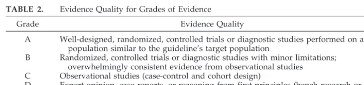
TABLE 2. Evidence Quality for Grades of Evidence
Grade Evidence Quality
A Well-designed, randomized, controlled trials or diagnostic studies performed on a population similar to the guideline’s target population
B Randomized, controlled trials or diagnostic studies with minor limitations; overwhelmingly consistent evidence from observational studies C Observational studies (case-control and cohort design)
in practice settings, cost-effective, and accurate in experienced hands. Non–pneumatic otoscopy is not advised for primary diagnosis.
The accuracy of pneumatic otoscopy in routine clinical practice may be less than that shown in pub-lished results, because clinicians have varying train-ing and experience.21,22When the diagnosis of OME is uncertain, tympanometry or acoustic reflectometry should be considered as an adjunct to pneumatic otoscopy. Tympanometry with a standard 226-Hz probe tone is reliable for infants 4 months old or older and has good interobserver agreement of curve patterns in routine clinical practice.23,24Younger in-fants require specialized equipment with a higher probe tone frequency. Tympanometry generates costs related to instrument purchase, annual calibra-tion, and test administration. Acoustic reflectometry with spectral gradient analysis is a low-cost alterna-tive to tympanometry that does not require an air-tight seal in the ear canal; however, validation stud-ies primarily have used children 2 years old or older with a high prevalence of OME.25–27
Although no research studies have examined whether pneumatic otoscopy causes discomfort, ex-pert consensus suggests that the procedure does not have to be painful, especially when symptoms of acute infection (AOM) are absent. A nontraumatic examination is facilitated by using a gentle touch, restraining the child properly when necessary, and inserting the speculum only into the outer one third (cartilaginous portion) of the ear canal.28The pneu-matic bulb should be compressed slightly before in-sertion, because OME often is associated with a neg-ative middle-ear pressure, which can be assessed more accurately by releasing the already compressed bulb. The otoscope must be fully charged, the bulb (halogen or xenon) bright and luminescent,29and the insufflator bulb attached tightly to the head to avoid the loss of an air seal. The window must also be sealed.
Evidence Profile: Pneumatic Otoscopy
• Aggregate evidence quality: A, diagnostic studies in relevant populations.
• Benefit: improved diagnostic accuracy; inexpen-sive equipment.
• Harm: cost of training clinicians in pneumatic oto-scopy.
• Benefits-harms assessment: preponderance of ben-efit over harm.
• Policy level: strong recommendation.
Evidence Profile: Tympanometry
• Aggregate evidence quality: B, diagnostic studies with minor limitations.
• Benefit: increased diagnostic accuracy beyond pneumatic otoscopy; documentation.
• Harm: acquisition cost, administrative burden, and recalibration.
• Benefits-harms assessment: balance of benefit and harm.
• Policy level: option.
1C. SCREENING: POPULATION-BASED SCREENING PROGRAMS FOR OME ARE NOT RECOMMENDED
IN HEALTHY, ASYMPTOMATIC CHILDREN
This recommendation is based on randomized, con-trolled trials and cohort studies, with a preponderance of harm over benefit.
This recommendation concerns population-based screening programs of all children in a community or a school without regard to any preexisting symp-toms or history of disease. This recommendation does not address hearing screening or monitoring of specific children with previous or recurrent OME.
OME is highly prevalent in young children. Screening surveys of healthy children ranging in age from infancy to 5 years old show a 15% to 40% point prevalence of middle-ear effusion.5,7,30–36 Among children examined at regular intervals for a year,
⬃50% to 60% of child care center attendees32 and 25% of school-aged children37were found to have a middle-ear effusion at some time during the exami-nation period, with peak incidence during the winter months.
Population-based screening has not been found to influence short-term language outcomes,33 and its long-term effects have not been evaluated in a ran-domized, clinical trial. Therefore, the recommenda-tion against screening is based not only on the ability to identify OME but more importantly on a lack of demonstrable benefits from treating children so identified that exceed the favorable natural history of the disease. The New Zealand Health Technology Assessment38 could not determine whether pschool screening for OME was effective. More re-cently, the Canadian Task Force on Preventive Health Care39reported that insufficient evidence was available to recommend including or excluding rou-tine early screening for OME. Although screening for OME is not inherently harmful, potential risks in-clude inaccurate diagnoses, overtreating self-limited disease, parental anxiety, and the costs of screening and unnecessary treatment.
Population-based screening is appropriate for condi-tions that are common, can be detected by a sensitive and specific test, and benefit from early detection and treatment.40 The first 2 requirements are fulfilled by OME, which affects up to 80% of children by school entry2,5,7and can be screened easily with tympanom-etry (see recommendation 1B). Early detection and treatment of OME identified by screening, however, have not been shown to improve intelligence, receptive language, or expressive language.2,39,41,42 Therefore, population-based screening for early detection of OME in asymptomatic children has not been shown to im-prove outcomes and is not recommended.
Evidence Profile: Screening
• Aggregate evidence quality: B, randomized, con-trolled trials with minor limitations and consistent evidence from observational studies.
• Harm: inaccurate diagnosis (positive or false-negative), overtreating self-limited disease, paren-tal anxiety, cost of screening, and/or unnecessary treatment.
• Benefits-harms assessment: preponderance of harm over benefit.
• Policy level: recommendation against.
2. DOCUMENTATION: CLINICIANS SHOULD DOCUMENT THE LATERALITY, DURATION OF EFFUSION, AND PRESENCE AND SEVERITY OF ASSOCIATED SYMPTOMS AT EACH ASSESSMENT
OF THE CHILD WITH OME
This recommendation is based on observational studies and strong preponderance of benefit over harm.
Documentation in the medical record facilitates diagnosis and treatment and communicates perti-nent information to other clinicians to ensure patient safety and reduce medical errors.43Management de-cisions in children with OME depend on effusion duration and laterality plus the nature and severity of associated symptoms. Therefore, these features should be documented at every medical encounter for OME. Although no studies have addressed doc-umentation for OME specifically, there is room for improvement in documentation of ambulatory care medical records.44
Ideally, the time of onset and laterality of OME can be defined through diagnosis of an antecedent AOM, a history of acute onset of signs or symptoms directly referable to fluid in the middle ear, or the presence of an abnormal audiogram or tympanogram closely af-ter a previously normal test. Unfortunately, these conditions are often lacking, and the clinician is forced to speculate on the onset and duration of fluid in the middle ear(s) in a child found to have OME at a routine office visit or school screening audiometry. In ⬃40% to 50% of cases of OME, neither the affected children nor their parents or caregivers de-scribe significant complaints referable to a middle-ear effusion.45,46 In some children, however, OME may have associated signs and symptoms caused by inflammation or the presence of effusion (not acute infection) that should be documented, such as
• Mild intermittent ear pain, fullness, or “popping” • Secondary manifestations of ear pain in infants, which may include ear rubbing, excessive irritabil-ity, and sleep disturbances
• Failure of infants to respond appropriately to voices or environmental sounds, such as not turn-ing accurately toward the sound source
• Hearing loss, even when not specifically described by the child, suggested by seeming lack of atten-tiveness, behavioral changes, failure to respond to normal conversational-level speech, or the need for excessively high sound levels when using au-dio equipment or viewing television
• Recurrent episodes of AOM with persistent OME between episodes
• Problems with school performance
• Balance problems, unexplained clumsiness, or de-layed gross motor development47–50
• Delayed speech or language development
The laterality (unilateral versus bilateral), duration of effusion, and presence and severity of associated symptoms should be documented in the medical record at each assessment of the child with OME. When OME duration is uncertain, the clinician must take whatever evidence is at hand and make a rea-sonable estimate.
Evidence Profile: Documentation
• Aggregate evidence quality: C, observational stud-ies.
• Benefits: defines severity, duration has prognostic value, facilitates future communication with other clinicians, supports appropriate timing of inter-vention, and, if consistently unilateral, may iden-tify a problem with specific ear other than OME (eg, retraction pocket or cholesteatoma).
• Harm: administrative burden.
• Benefits-harms assessment: preponderance of ben-efit over harm.
• Policy level: recommendation.
3. CHILD AT RISK: CLINICIANS SHOULD DISTINGUISH THE CHILD WITH OME WHO IS AT
RISK FOR SPEECH, LANGUAGE, OR LEARNING PROBLEMS FROM OTHER CHILDREN WITH OME
AND SHOULD EVALUATE HEARING, SPEECH, LANGUAGE, AND NEED FOR INTERVENTION
MORE PROMPTLY
This recommendation is based on case series, the pre-ponderance of benefit over harm, and ethical limitations in studying children with OME who are at risk.
The panel defines the child at risk as one who is at increased risk for developmental difficulties (delay or disorder) because of sensory, physical, cognitive, or behavioral factors listed in Table 3. These factors are not caused by OME but can make the child less tolerant of hearing loss or vestibular problems sec-ondary to middle-ear effusion. In contrast the child with OME who is not at risk is otherwise healthy and does not have any of the factors shown in Table 3.
Earlier guidelines for managing OME have ap-plied only to young children who are healthy and exhibit no developmental delays.1Studies of the re-lationship between OME and hearing loss or speech/ language development typically exclude children with craniofacial anomalies, genetic syndromes, and other developmental disorders. Therefore, the avail-able literature mainly applies to otherwise healthy children who meet inclusion criteria for randomized,
TABLE 3. Risk Factors for Developmental Difficulties*
Permanent hearing loss independent of OME
Suspected or diagnosed speech and language delay or disorder Autism-spectrum disorder and other pervasive developmental
disorders
Syndromes (eg, Down) or craniofacial disorders that include cognitive, speech, and language delays
Blindness or uncorrectable visual impairment Cleft palate with or without associated syndrome Developmental delay
controlled trials. Few, if any, existing studies dealing with developmental sequelae caused by hearing loss from OME can be generalized to children who are at risk.
Children who are at risk for speech or language delay would likely be affected additionally by hear-ing problems from OME,51although definitive ies are lacking. For example, small comparative stud-ies of children or adolescents with Down syndrome52 or cerebral palsy53 show poorer articulation and re-ceptive language associated with a history of early otitis media. Large studies are unlikely to be forth-coming because of methodologic and ethical difficul-ties inherent in studying children who are delayed or at risk for further delays. Therefore, clinicians who manage children with OME should determine whether other conditions coexist that put a child at risk for developmental delay (Table 3) and then take these conditions into consideration when planning assessment and management.
Children with craniofacial anomalies (eg, cleft pal-ate; Down syndrome; Robin sequence; coloboma, heart defect, choanal atresia, retarded growth and development, genital anomaly, and ear defect with deafness [CHARGE] association) have a higher prev-alence of chronic OME, hearing loss (conductive and sensorineural), and speech or language delay than do children without these anomalies.54–57Other chil-dren may not be more prone to OME but are likely to have speech and language disorders, such as those children with permanent hearing loss independent of OME,58,59specific language impairment,60 autism-spectrum disorders,61 or syndromes that adversely affect cognitive and linguistic development. Some retrospective studies52,62,63 have found that hearing loss caused by OME in children with cognitive de-lays, such as Down syndrome, has been associated with lower language levels. Children with language delays or disorders with OME histories perform more poorly on speech-perception tasks than do chil-dren with OME histories alone.64,65
Children with severe visual impairments may be more susceptible to the effects of OME, because they depend on hearing more than children with normal vision.51 Any decrease in their most important re-maining sensory input for language (hearing) may significantly compromise language development and their ability to interact and communicate with others. All children with severe visual impairments should be considered more vulnerable to OME se-quelae, especially in the areas of balance, sound lo-calization, and communication.
Management of the child with OME who is at increased risk for developmental delays should in-clude hearing testing and speech and language eval-uation and may include speech and language ther-apy concurrent with managing OME, hearing aids or other amplification devices for hearing loss indepen-dent of OME, tympanostomy tube insertion,54,63,66,67 and hearing testing after OME resolves to document improvement, because OME can mask a permanent underlying hearing loss and delay detection.59,68,69
Evidence Profile: Child at Risk
• Aggregate evidence quality: C, observational stud-ies of children at risk; D, expert opinion on the ability of prompt assessment and management to alter outcomes.
• Benefits: optimizing conditions for hearing, speech, and language; enabling children with spe-cial needs to reach their potential; avoiding limi-tations on the benefits of educational interventions because of hearing problems from OME.
• Harm: cost, time, and specific risks of medications or surgery.
• Benefits-harms assessment: exceptional prepon-derance of benefits over harm based on subcom-mittee consensus because of circumstances to date precluding randomized trials.
• Policy level: recommendation.
4. WATCHFUL WAITING: CLINICIANS SHOULD MANAGE THE CHILD WITH OME WHO IS NOT AT RISK WITH WATCHFUL WAITING FOR 3 MONTHS
FROM THE DATE OF EFFUSION ONSET (IF KNOWN) OR DIAGNOSIS (IF ONSET IS
UNKNOWN)
This recommendation is based on systematic review of cohort studies and the preponderance of benefit over harm. This recommendation is based on the self-limited nature of most OME, which has been well docu-mented in cohort studies and in control groups of randomized trials.2,70
The likelihood of spontaneous resolution of OME is determined by the cause and duration of effu-sion.70 For example,⬃75% to 90% of residual OME after an AOM episode resolves spontaneously by 3 months.71–73Similar outcomes of defined onset dur-ing a period of surveillance in a cohort study are observed for OME.32,37 Another favorable situation involves improvement (not resolution) of newly de-tected OME defined as change in tympanogram from type B (flat curve) to non-B (anything other than a flat curve). Approximately 55% of children so de-fined improve by 3 months,70but one third will have OME relapse within the next 3 months.4Although a type B tympanogram is an imperfect measure of OME (81% sensitivity and 74% specificity versus my-ringotomy), it is the most widely reported measure suitable for deriving pooled resolution rates.2,70
Any intervention for OME (medical or surgical) other than observation carries some inherent harm. There is little harm associated with a specified period of observation in the child who is not at risk for speech, language, or learning problems. When ob-serving children with OME, clinicians should inform the parent or caregiver that the child may experience reduced hearing until the effusion resolves, espe-cially if it is bilateral. Clinicians may discuss strate-gies for optimizing the listening and learning envi-ronment until the effusion resolves. These strategies include speaking in close proximity to the child, facing the child and speaking clearly, repeating phrases when misunderstood, and providing prefer-ential classroom seating.78,79
The recommendation for a 3-month period of ob-servation is based on a clear preponderance of ben-efit over harm and is consistent with the original OME guideline intent of avoiding unnecessary sur-gery.1At the discretion of the clinician, this 3-month period of watchful waiting may include interval vis-its at which OME is monitored by using pneumatic otoscopy, tympanometry, or both. Factors to con-sider in determining the optimal interval(s) for fol-low-up include clinical judgment, parental comfort level, unique characteristics of the child and/or his environment, access to a health care system, and hearing levels (HLs) if known.
After documented resolution of OME in all af-fected ears, additional follow-up is unnecessary.
Evidence Profile: Watchful Waiting
• Aggregate evidence quality: B, systematic review of cohort studies.
• Benefit: avoid unnecessary interventions, take ad-vantage of favorable natural history, and avoid unnecessary referrals and evaluations.
• Harm: delays in therapy for OME that will not resolve with observation; prolongation of hearing loss.
• Benefits-harms assessment: preponderance of ben-efit over harm.
• Policy level: recommendation.
5. MEDICATION: ANTIHISTAMINES AND DECONGESTANTS ARE INEFFECTIVE FOR OME
AND ARE NOT RECOMMENDED FOR TREATMENT; ANTIMICROBIALS AND CORTICOSTEROIDS DO NOT HAVE LONG-TERM
EFFICACY AND ARE NOT RECOMMENDED FOR ROUTINE MANAGEMENT
This recommendation is based on systematic review of randomized, controlled trials and the preponderance of harm over benefit.
Therapy for OME is appropriate only if persistent and clinically significant benefits can be achieved beyond spontaneous resolution. Although statisti-cally significant benefits have been demonstrated for some medications, they are short-term and relatively small in magnitude. Moreover, significant adverse events may occur with all medical therapies.
The prior OME guideline1found no data support-ing antihistamine-decongestant combinations in treating OME. Meta-analysis of 4 randomized trials showed no significant benefit for antihistamines or decongestants versus placebo. No additional studies have been published since 1994 to change this rec-ommendation. Adverse effects of antihistamines and decongestants include insomnia, hyperactivity, drowsiness, behavioral change, and blood-pressure variability.
Long-term benefits of antimicrobial therapy for OME are unproved despite a modest short-term ben-efit for 2 to 8 weeks in randomized trials.1,80,81Initial benefits, however, can become nonsignificant within 2 weeks of stopping the medication.82Moreover,⬃7 children would need to be treated with antimicrobi-als to achieve one short-term response.1 Adverse effects of antimicrobials are significant and may in-clude rashes, vomiting, diarrhea, allergic reactions, alteration of the child’s nasopharyngeal flora, devel-opment of bacterial resistance,83 and cost. Societal consequences include direct transmission of resistant bacterial pathogens in homes and child care cen-ters.84
The prior OME guideline1did not recommend oral steroids for treating OME in children. A later meta-analysis85 showed no benefit for oral steroid versus placebo within 2 weeks but did show a short-term benefit for oral steroid plus antimicrobial versus an-timicrobial alone in 1 of 3 children treated. This benefit became nonsignificant after several weeks in a prior meta-analysis1 and in a large, randomized trial.86Oral steroids can produce behavioral changes, increased appetite, and weight gain.1Additional ad-verse effects may include adrenal suppression, fatal varicella infection, and avascular necrosis of the fem-oral head.3Although intranasal steroids have fewer adverse effects, one randomized trial87 showed sta-tistically equivalent outcomes at 12 weeks for intra-nasal beclomethasone plus antimicrobials versus an-timicrobials alone for OME.
Antimicrobial therapy with or without steroids has not been demonstrated to be effective in long-term resolution of OME, but in some cases this ther-apy can be considered an option because of short-term benefit in randomized trials, when the parent or caregiver expresses a strong aversion to impending surgery. In this circumstance, a single course of ther-apy for 10 to 14 days may be used. The likelihood that the OME will resolve long-term with these reg-imens is small, and prolonged or repetitive courses of antimicrobials or steroids are strongly not recom-mended.
Other nonsurgical therapies that are discussed in the OME literature include autoinflation of the eu-stachian tube, oral or intratympanic use of mucolyt-ics, and systemic use of pharmacologic agents other than antimicrobials, steroids, and antihistamine-de-congestants. Insufficient data exist for any of these therapies to be recommended in treating OME.3
Evidence Profile: Medication
• Benefit: avoid side effects and reduce cost by not administering medications; avoid delays in defin-itive therapy caused by short-term improvement then relapse.
• Harm: adverse effects of specific medications as listed previously; societal impact of antimicrobial therapy on bacterial resistance and transmission of resistant pathogens.
• Benefits-harms assessment: preponderance of harm over benefit.
• Policy level: recommendation against.
6. HEARING AND LANGUAGE: HEARING TESTING IS RECOMMENDED WHEN OME PERSISTS FOR 3
MONTHS OR LONGER OR AT ANY TIME THAT LANGUAGE DELAY, LEARNING PROBLEMS, OR A SIGNIFICANT HEARING LOSS IS SUSPECTED IN A CHILD WITH OME; LANGUAGE TESTING SHOULD BE CONDUCTED FOR CHILDREN WITH HEARING
LOSS
This recommendation is based on cohort studies and the preponderance of benefit over risk.
Hearing Testing
Hearing testing is recommended when OME per-sists for 3 months or longer or at any time that language delay, learning problems, or a significant hearing loss is suspected. Conductive hearing loss often accompanies OME1,88and may adversely affect binaural processing,89 sound localization,90 and speech perception in noise.91–94Hearing loss caused by OME may impair early language acquisition,95–97 but the child’s home environment has a greater im-pact on outcomes98; recent randomized trials41,99,100 suggest no impact on children with OME who are not at risk as identified by screening or surveillance. Studies examining hearing sensitivity in children with OME report that average pure-tone hearing loss at 4 frequencies (500, 1000, 2000, and 4000 Hz) ranges from normal hearing to moderate hearing loss (0 –55 dB). The 50th percentile is an⬃25-dB HL, and⬃20% of ears exceed 35-dB HL.101,102Unilateral OME with hearing loss results in overall poorer binaural hear-ing than in infants with normal middle-ear function bilaterally.103,104 However, based on limited re-search, there is evidence that children experiencing the greatest conductive hearing loss for the longest periods may be more likely to exhibit developmental and academic sequelae.1,95,105
Initial hearing testing for children 4 years old or older can be done in the primary care setting.106 Testing should be performed in a quiet environment, preferably in a separate closed or sound-proofed area set aside specifically for that purpose. Conven-tional audiometry with earphones is performed with a fail criterion of more than 20-dB HL at 1 or more frequencies (500, 1000, 2000, and 4000 Hz) in either ear.106,107Methods not recommended as substitutes for primary care hearing testing include tympanom-etry and pneumatic otoscopy,102caregiver judgment regarding hearing loss,108,109speech audiometry, and tuning forks, acoustic reflectometry, and behavioral observation.1
Comprehensive audiologic evaluation is recom-mended for children who fail primary care testing, are less than 4 years old, or cannot be tested in the primary care setting. Audiologic assessment includes evaluating air-conduction and bone-conduction thresholds for pure tones, speech-detection or speech-recognition thresholds,102 and measuring speech understanding if possible.94 The method of assessment depends on the developmental age of the child and might include visual reinforcement or con-ditioned orienting-response audiometry for infants 6 to 24 months old, play audiometry for children 24 to 48 months old, or conventional screening audiome-try for children 4 years old and older.106The auditory brainstem response and otoacoustic emission are tests of auditory pathway structural integrity, not hearing, and should not substitute for behavioral pure-tone audiometry.106
Language Testing
Language testing should be conducted for children with hearing loss (pure-tone average more than 20-dB HL on comprehensive audiometric evalua-tion). Testing for language delays is important, be-cause communication is integral to all aspects of human functioning. Young children with speech and language delays during the preschool years are at risk for continued communication problems and later delays in reading and writing.110–112 In one study, 6% to 8% of children 3 years old and 2% to 13% of kindergartners had language impairment.113 Language intervention can improve communication and other functional outcomes for children with his-tories of OME.114
Children who experience repeated and persistent episodes of OME and associated hearing loss during early childhood may be at a disadvantage for learn-ing speech and language.79,115Although Shekelle et al2concluded that there was no evidence to support the concern that OME during the first 3 years of life was related to later receptive or expressive language, this meta-analysis should be interpreted cautiously, because it did not examine specific language do-mains such as vocabulary and the independent vari-able was OME and not hearing loss. Other meta-analyses79,115 have suggested at most a small negative association of OME and hearing loss on children’s receptive and expressive language through the elementary school years. The clinical significance of these effects for language and learn-ing is unclear for the child not at risk. For example, in one randomized trial,100prompt insertion of tympa-nostomy tubes for OME did not improve develop-mental outcomes at 3 years old regardless of baseline hearing. In another randomized trial,116 however, prompt tube insertion achieved small benefits for children with bilateral OME and hearing loss.
as the MacArthur Communicative Development In-ventory118 and the Language Development Survey.119 For older children, the Denver Developmental Screen-ing Test II120 can be used to screen general develop-ment including speech and language. Comprehensive speech and language evaluation is recommended for children who fail testing or whenever the child’s parent or caregiver expresses concern.121
Evidence Profile: Hearing and Language
• Aggregate evidence quality: B, diagnostic studies with minor limitations; C, observational studies. • Benefit: to detect hearing loss and language delay
and identify strategies or interventions to improve developmental outcomes.
• Harm: parental anxiety, direct and indirect costs of assessment, and/or false-positive results.
• Balance of benefit and harm: preponderance of benefit over harm.
• Policy level: recommendation.
7. SURVEILLANCE: CHILDREN WITH PERSISTENT OME WHO ARE NOT AT RISK SHOULD BE REEXAMINED AT 3- TO 6-MONTH INTERVALS UNTIL THE EFFUSION IS NO LONGER PRESENT, SIGNIFICANT HEARING LOSS IS IDENTIFIED, OR
STRUCTURAL ABNORMALITIES OF THE EARDRUM OR MIDDLE EAR ARE SUSPECTED
This recommendation is based on randomized, con-trolled trials and observational studies with a preponder-ance of benefit over harm.
If OME is asymptomatic and is likely to resolve spontaneously, intervention is unnecessary even if OME persists for more than 3 months. The clinician should determine whether risk factors exist that would predispose the child to undesirable sequelae or predict nonresolution of the effusion. As long as OME persists, the child is at risk for sequelae and must be reevaluated periodically for factors that would prompt intervention.
The 1994 OME guideline1 recommended surgery for OME persisting 4 to 6 months with hearing loss but requires reconsideration because of later data on tubes and developmental sequelae.122 For example, selecting surgical candidates using duration-based criteria (eg, OME⬎3 months or exceeding a cumu-lative threshold) does not improve developmental outcomes in infants and toddlers who are not at risk.41,42,99,100 Additionally, the 1994 OME guideline did not specifically address managing effusion with-out significant hearing loss persisting more than 6 months.
Asymptomatic OME usually resolves spontane-ously, but resolution rates decrease the longer the effusion has been present,36,76,77and relapse is com-mon.123Risk factors that make spontaneous resolu-tion less likely include124,125:
• Onset of OME in the summer or fall season • Hearing loss more than 30-dB HL in the
better-hearing ear
• History of prior tympanostomy tubes • Not having had an adenoidectomy
Children with chronic OME are at risk for struc-tural damage of the tympanic membrane126because the effusion contains leukotrienes, prostaglandins, and arachidonic acid metabolites that invoke a local inflammatory response.127Reactive changes may oc-cur in the adjacent tympanic membrane and mucosal linings. A relative underventilation of the middle ear produces a negative pressure that predisposes to focal retraction pockets, generalized atelectasis of the tympanic membrane, and cholesteatoma.
Structural integrity is assessed by carefully exam-ining the entire tympanic membrane, which, in many cases, can be accomplished by the primary care cli-nician using a handheld pneumatic otoscope. A search should be made for retraction pockets, ossic-ular erosion, and areas of atelectasis or atrophy. If there is any uncertainty that all observed structures are normal, the patient should be examined by using an otomicroscope. All children with these tympanic membrane conditions, regardless of OME duration, should have a comprehensive audiologic evaluation. Conditions of the tympanic membrane that gener-ally mandate inserting a tympanostomy tube are posterosuperior retraction pockets, ossicular erosion, adhesive atelectasis, and retraction pockets that ac-cumulate keratin debris. Ongoing surveillance is mandatory, because the incidence of structural dam-age increases with effusion duration.128
As noted in recommendation 6, children with per-sistent OME for 3 months or longer should have their hearing tested. Based on these results, clinicians can identify 3 levels of action based on HLs obtained for the better-hearing ear using earphones or in sound field using speakers if the child is too young for ear-specific testing.
1. HLs ofⱖ40 dB (at least a moderate hearing loss): A comprehensive audiologic evaluation is indi-cated if not previously performed. If moderate hearing loss is documented and persists at this level, surgery is recommended, because persistent hearing loss of this magnitude that is permanent in nature has been shown to impact speech, lan-guage, and academic performance.129–131
2. HLs of 21 to 39 dB (mild hearing loss): A compre-hensive audiologic evaluation is indicated if not previously performed. Mild sensorineural hearing loss has been associated with difficulties in speech, language, and academic performance in school,129,132and persistent mild conductive hear-ing loss from OME may have a similar impact. Further management should be individualized based on effusion duration, severity of hearing loss, and parent or caregiver preference and may include strategies to optimize the listening and learning environment (Table 4) or surgery. Repeat hearing testing should be performed in 3 to 6 months if OME persists at follow-up evaluation or tympanostomy tubes have not been placed. 3. HLs ofⱕ20 dB (normal hearing): A repeat hearing
In addition to hearing loss and speech or language delay, other factors may influence the decision to intervene for persistent OME. Roberts et al98,133 showed that the caregiving environment is more strongly related to school outcome than was OME or hearing loss. Risk factors for delays in speech and language development caused by a poor caregiving environment included low maternal educational level, unfavorable child care environment, and low socioeconomic status. In such cases, these factors may be additive to the hearing loss in affecting lower school performance and classroom behavior prob-lems.
Persistent OME may be associated with physical or behavioral symptoms including hyperactivity, poor attention, and behavioral problems in some stud-ies134–136 and reduced child quality of life.46 Con-versely, young children randomized to early versus late tube insertion for persistent OME showed no behavioral benefits from early surgery.41,100Children with chronic OME also have significantly poorer ves-tibular function and gross motor proficiency when compared with non-OME controls.48–50 Moreover, vestibular function, behavior, and quality of life can improve after tympanostomy tube insertion.47,137,138 Other physical symptoms of OME that, if present and persistent, may warrant surgery include otalgia, unexplained sleep disturbance, and coexisting recur-rent AOM. Tubes reduce the absolute incidence of recurrent AOM by⬃1 episode per child per year, but the relative risk reduction is 56%.139
The risks of continued observation of children with OME must be balanced against the risks of surgery. Children with persistent OME examined regularly at 3- to 6-month intervals, or sooner if OME-related symptoms develop, are most likely at low risk for physical, behavioral, or developmental sequelae of OME. Conversely, prolonged watchful waiting of OME is not appropriate when regular surveillance is impossible or when the child is at risk for developmental sequelae of OME because of co-morbidities (Table 3). For these children, the risks of anesthesia and surgery (see recommendation 9) may be less than those of continued observation.
Evidence Profile: Surveillance
• Aggregate evidence quality: C, observational stud-ies and some randomized trials.
• Benefit: avoiding interventions that do not im-prove outcomes.
• Harm: allowing structural abnormalities to de-velop in the tympanic membrane, underestimating the impact of hearing loss on a child, and/or fail-ing to detect significant signs or symptoms that require intervention.
• Balance of benefit and harm: preponderance of benefit over harm.
• Policy level: recommendation.
8. REFERRAL: WHEN CHILDREN WITH OME ARE REFERRED BY THE PRIMARY CARE CLINICIAN FOR EVALUATION BY AN OTOLARYNGOLOGIST,
AUDIOLOGIST, OR SPEECH-LANGUAGE PATHOLOGIST, THE REFERRING CLINICIAN SHOULD DOCUMENT THE EFFUSION DURATION
AND SPECIFIC REASON FOR REFERRAL (EVALUATION, SURGERY) AND PROVIDE ADDITIONAL RELEVANT INFORMATION SUCH
AS HISTORY OF AOM AND DEVELOPMENTAL STATUS OF THE CHILD
This option is based on panel consensus and a prepon-derance of benefit over harm.
This recommendation emphasizes the importance of communication between the referring primary care clinician and the otolaryngologist, audiologist, and speech-language pathologist. Parents and care-givers may be confused and frustrated when a rec-ommendation for surgery is made for their child because of conflicting information about alternative management strategies. Choosing among manage-ment options is facilitated when primary care physi-cians and advanced-practice nurses who best know the patient’s history of ear problems and general medical status provide the specialist with accurate information. Although there are no studies showing improved outcomes from better documentation of OME histories, there is a clear need for better mech-anisms to convey information and expectations from primary care clinicians to consultants and subspe-cialists.140–142
When referring a child for evaluation to an otolar-yngologist, the primary care physician should ex-plain the following to the parent or caregiver of the patient:
• Reason for referral: Explain that the child is seeing an otolaryngologist for evaluation, which is likely to include ear examination and audiologic testing, and not necessarily simply to be scheduled for surgery.
• What to expect: Explain that surgery may be rec-ommended, and let the parent know that the oto-laryngologist will explain the options, benefits, and risks further.
• Decision-making process: Explain that there are many alternatives for management and that surgi-cal decisions are elective; the parent or caregiver should be encouraged to express to the surgeo-nany concerns he or she may have about the rec-ommendations made.
When referring a child to an otolaryngologist, au-diologist, or speech-language pathologist, the mini-TABLE 4. Strategies for Optimizing the Listening-Learning
Environment for Children With OME and Hearing Loss*
Get within 3 feet of the child before speaking.
Turn off competing audio signals such as unnecessary music and television in the background.
Face the child and speak clearly, using visual clues (hands, pictures) in addition to speech.
Slow the rate, raise the level, and enunciate speech directed at the child.
Read to or with the child, explaining pictures and asking questions.
Repeat words, phrases, and questions when misunderstood. Assign preferential seating in the classroom near the teacher. Use a frequency-modulated personal- or
sound-field-amplification system in the classroom.
mum information that should be conveyed in writ-ing includes:
• Duration of OME: State how long fluid has been present.
• Laterality of OME: State whether one or both ears have been affected.
• Results of prior hearing testing or tympanometry. • Suspected speech or language problems: State whether there had been a delay in speech and language development or whether the parent or a caregiver has expressed concerns about the child’s communication abilities, school achievement, or attentiveness.
• Conditions that might exacerbate the deleterious effects of OME: State whether the child has condi-tions such as permanent hearing loss, impaired cognition, developmental delays, cleft lip or pal-ate, or an unstable or nonsupportive family or home environment.
• AOM history: State whether the child has a history of recurrent AOM.
Additional medical information that should be provided to the otolaryngologist by the primary care clinician includes:
• Parental attitude toward surgery: State whether the parents have expressed a strong preference for or against surgery as a management option. • Related conditions that might require concomitant
surgery: State whether there have been other condi-tions that might warrant surgery if the child is going to have general anesthesia (eg, nasal obstruction and snoring that might be an indication for adenoidec-tomy or obstructive breathing during sleep that might mean tonsillectomy is indicated).
• General health status: State whether there are any conditions that might present problems for sur-gery or administering general anesthesia, such as congenital heart abnormality, bleeding disorder, asthma or reactive airway disease, or family his-tory of malignant hyperthermia.
After evaluating the child, the otolaryngologist, audiologist, or speech-language pathologist should inform the referring physician regarding his or her diagnostic impression, plans for additional assess-ment, and recommendations for ongoing monitoring and management.
Evidence Profile: Referral
• Aggregate evidence quality: C, observational stud-ies.
• Benefit: better communication and improved deci-sion-making.
• Harm: confidentiality concerns, administrative burden, and/or increased parent or caregiver anx-iety.
• Benefits-harms assessment: balance of benefit and harm.
• Policy level: option.
9. SURGERY: WHEN A CHILD BECOMES A SURGICAL CANDIDATE, TYMPANOSTOMY TUBE
INSERTION IS THE PREFERRED INITIAL PROCEDURE; ADENOIDECTOMY SHOULD NOT BE
PERFORMED UNLESS A DISTINCT INDICATION EXISTS (NASAL OBSTRUCTION, CHRONIC ADENOIDITIS). REPEAT SURGERY CONSISTS OF ADENOIDECTOMY PLUS MYRINGOTOMY, WITH OR WITHOUT TUBE INSERTION. TONSILLECTOMY ALONE OR MYRINGOTOMY ALONE SHOULD NOT
BE USED TO TREAT OME
This recommendation is based on randomized, con-trolled trials with a preponderance of benefit over harm.
Surgical candidacy for OME largely depends on hearing status, associated symptoms, the child’s de-velopmental risk (Table 3), and the anticipated chance of timely spontaneous resolution of the effu-sion. Candidates for surgery include children with OME lasting 4 months or longer with persistent hear-ing loss or other signs and symptoms, recurrent or persistent OME in children at risk regardless of hear-ing status, and OME and structural damage to the tympanic membrane or middle ear. Ultimately, the recommendation for surgery must be individualized based on consensus between the primary care phy-sician, otolaryngologist, and parent or caregiver that a particular child would benefit from intervention. Children with OME of any duration who are at risk are candidates for earlier surgery.
Tympanostomy tubes are recommended for initial surgery because randomized trials show a mean 62% relative decrease in effusion prevalence and an ab-solute decrease of 128 effusion days per child during the next year.139,143–145HLs improve by a mean of 6 to 12 dB while the tubes remain patent.146,147 Ade-noidectomy plus myringotomy (without tube inser-tion) has comparable efficacy in children 4 years old or older143but is more invasive, with additional sur-gical and anesthetic risks. Similarly, the added risk of adenoidectomy outweighs the limited, short-term benefit for children 3 years old or older without prior tubes.148Consequently, adenoidectomy is not recom-mended for initial OME surgery unless a distinct indication exists, such as adenoiditis, postnasal ob-struction, or chronic sinusitis.
Tonsillectomy or myringotomy alone (without ade-noidectomy) is not recommended to treat OME. Al-though tonsillectomy is either ineffective152 or of lim-ited efficacy,148,150the risks of hemorrhage (⬃2%) and additional hospitalization outweigh any potential ben-efits unless a distinct indication for tonsillectomy exists. Myringotomy alone, without tube placement or ade-noidectomy, is ineffective for chronic OME,144,145 be-cause the incision closes within several days. Laser-assisted myringotomy extends the ventilation period several weeks,153 but randomized trials with concur-rent controls have not been conducted to establish ef-ficacy. In contrast, tympanostomy tubes ventilate the middle ear for an average of 12 to 14 months.144,145
Anesthesia mortality has been reported to be⬃1: 50 000 for ambulatory surgery,154 but the current fatality rate may be lower.155 Laryngospasm and bronchospasm occur more often in children receiv-ing anesthesia than adults. Tympanostomy tube se-quelae are common156 but are generally transient (otorrhea) or do not affect function (tympanosclero-sis, focal atrophy, or shallow retraction pocket). Tympanic membrane perforations, which may re-quire repair, are seen in 2% of children after place-ment of short-term (grommet-type) tubes and 17% after long-term tubes.156Adenoidectomy has a 0.2% to 0.5% incidence of hemorrhage150,157and 2% inci-dence of transient velopharyngeal insufficiency.148 Other potential risks of adenoidectomy, such as na-sopharyngeal stenosis and persistent velopharyngeal insufficiency, can be minimized with appropriate pa-tient selection and surgical technique.
There is a clear preponderance of benefit over harm when considering the impact of surgery for OME on effusion prevalence, HLs, subsequent inci-dence of AOM, and the need for reoperation after adenoidectomy. Information about adenoidectomy in children less than 4 years old, however, remains limited. Although the cost of surgery and anesthesia is nontrivial, it is offset by reduced OME and AOM after tube placement and by reduced need for reop-eration after adenoidectomy. Approximately 8 ade-noidectomies are needed to avoid a single instance of tube reinsertion; however, each avoided surgery probably represents a larger reduction in the number of AOM and OME episodes, including those in chil-dren who did not require additional surgery.150
Evidence Profile: Surgery
• Aggregate evidence quality: B, randomized, con-trolled trials with minor limitations.
• Benefit: improved hearing, reduced prevalence of OME, reduced incidence of AOM, and less need for additional tube insertion (after adenoidec-tomy).
• Harm: risks of anesthesia and specific surgical pro-cedures; sequelae of tympanostomy tubes. • Benefits-harms assessment: preponderance of
ben-efit over harm.
• Policy level: recommendation.
10. CAM: NO RECOMMENDATION IS MADE REGARDING CAM AS A TREATMENT FOR OME
There is no recommendation based on lack of scientific evidence documenting efficacy and an uncertain balance of harm and benefit.
The 1994 OME guideline1made no recommenda-tion regarding CAM as a treatment for OME, and no subsequent controlled studies have been published to change this conclusion. The current statement of “no recommendation” is based on the lack of scien-tific evidence documenting efficacy plus the balance of benefit and harm.
Evidence concerning CAM is insufficient to deter-mine whether the outcomes achieved for OME differ from those achieved by watchful waiting and spon-taneous resolution. There are no randomized, con-trolled trials with adequate sample sizes on the effi-cacy of CAM for OME. Although many case reports and subjective reviews on CAM treatment of AOM were found, little is published on OME treatment or prevention. Homeopathy158 and chiropractic treat-ments159 were assessed in pilot studies with small numbers of patients that failed to show clinically or statistically significant benefits. Consequently, there is no research base on which to develop a recommen-dation concerning CAM for OME.
The natural history of OME in childhood (dis-cussed previously) is such that almost any interven-tion can be “shown” to have helped in an anecdotal, uncontrolled report or case series. The efficacy of CAM or any other intervention for OME can only be shown with parallel-group, randomized, controlled trials with valid diagnostic methods and adequate sample sizes. Unproved modalities that have been claimed to provide benefit in middle-ear disease in-clude osteopathic and chiropractic manipulation, di-etary exclusions (such as dairy), herbal and other dietary supplements, acupuncture, traditional Chi-nese medicine, and homeopathy. None of these mo-dalities, however, have been subjected yet to a pub-lished, peer-reviewed, clinical trial.
side effects.165Quadriplegia has been reported, how-ever, after spinal manipulation in an infant with torticollis.166
Evidence Profile: CAM
• Aggregate evidence quality: D, case series without controls.
• Benefit: not established.
• Harm: potentially significant depending on the intervention.
• Benefits-harms assessment: uncertain balance of benefit and harm.
• Policy level: no recommendation.
11. ALLERGY MANAGEMENT: NO RECOMMENDATION IS MADE REGARDING ALLERGY MANAGEMENT AS A TREATMENT FOR
OME
There is no recommendation based on insufficient evi-dence of therapeutic efficacy or a causal relationship be-tween allergy and OME.
The 1994 OME guideline1made no recommenda-tion regarding allergy management as a treatment for OME, and no subsequent controlled studies have been published to change this conclusion. The cur-rent statement of “no recommendation” is based on insufficient evidence of therapeutic efficacy or a causal relationship between allergy and OME plus the balance of benefit and harm.
A linkage between allergy and OME has long been speculated but to date remains unquantified. The prevalence of allergy among OME patients has been reported to range from less than 10% to more than 80%.167 Allergy has long been postulated to cause OME through its contribution to eustachian tube dysfunction.168 The cellular response of respiratory mucosa to allergens has been well studied. There-fore, similar to other parts of respiratory mucosa, the mucosa lining the middle-ear cleft is capable of an allergic response.169,170Sensitivity to allergens varies among individuals, and atopy may involve neutro-phils in type I allergic reactions that enhance the inflammatory response.171
The correlation between OME and allergy has been widely reported, but no prospective studies have examined the effects of immunotherapy com-pared with observation alone or other management options. Reports of OME cure after immunotherapy or food-elimination diets172 are impossible to inter-pret without concurrent control groups because of the favorable natural history of most untreated OME. The documentation of allergy in published reports has been defined inconsistently (medical history, physical examination, skin-prick testing, nasal smears, serum immunoglobulin E and eosinophil counts, inflammatory mediators in effusions). Study groups have been drawn primarily from specialist offices, likely lack heterogeneity, and are not repre-sentative of general medical practice.
Evidence Profile: Allergy Management
• Aggregate evidence quality: D, case series without controls.
• Benefit: not established.
• Harm: adverse effects and cost of medication, phy-sician evaluation, elimination diets, and desensiti-zation.
• Benefits-harms assessment: balance of benefit and harm.
• Policy level: no recommendation.
RESEARCH NEEDS Diagnosis
• Further standardize the definition of OME. • Assess the performance characteristics of
pneu-matic otoscopy as a diagnostic test for OME when performed by primary care physicians and ad-vanced-practice nurses in the routine office set-ting.
• Determine the optimal methods for teaching pneu-matic otoscopy to residents and clinicians. • Develop a brief, reliable, objective method for
di-agnosing OME.
• Develop a classification method for identifying the presence of OME for practical use by clinicians that is based on quantifiable tympanometric char-acteristics.
• Assess the usefulness of algorithms combining pneumatic otoscopy and tympanometry for de-tecting OME in clinical practice.
• Conduct additional validating cohort studies of acoustic reflectometry as a diagnostic method for OME, particularly in children less than 2 years old.
Child At Risk
• Better define the child with OME who is at risk for speech, language, and learning problems.
• Conduct large, multicenter, observational cohort studies to identify the child at risk who is most susceptible to potential adverse sequelae of OME. • Conduct large, multicenter, observational cohort studies to analyze outcomes achieved with alter-native management strategies for OME in children at risk.
Watchful Waiting
• Define the spontaneous resolution of OME in in-fants and young children (existing data are limited primarily to children 2 years old or older). • Conduct large-scale, prospective cohort studies to
obtain current data on the spontaneous resolution of newly diagnosed OME of unknown prior dura-tion (existing data are primarily from the late 1970s and early 1980s).
• Develop prognostic indicators to identify the best candidates for watchful waiting.