ISSN: 2319-6505
EFFECT OF CULTIVATION MEDIA COMPONENTS ON PYOCYANIN PRODUCTION AND ITS
APPLICATION IN ANTIMICROBIAL PROPERTY
Ankita H Agrawal1 and Poonam B Chauhan*2
Department of Microbiology, KBS Commerce and Nataraj Professional Sciences College, Vapi. Dist. Valsad, Gujarat, India. PIN: 396195
A R T I C L E I N F O A B S T R A C T
A total of eight isolates belonging to the genus Pseudomonas were isolated from environmental samples and were screened for pyocyanin production. Isolate U4 was selected as highest pyocyanin producer. Pyocyanin was extracted by using chloroform solvent system. Pigment production began during the first 24 hrs of growth and maximal bioactive pigment yield was achieved at 13.65μg/ml after 72 hrs. Neutral pH at 30 C showed the optimum conditions for maximum pyocyanin production (17.56μg/ml). Maximum pyocyanin production was obtained using mannitol as carbon source (23.32μg/ml) and peptone as nitrogen source (26.12μg/ml). The antibacterial activity of pyocyanin was determined by well diffusion method against gram negative and gram positive bacteria.
INTRODUCTION
Pyocyanin is a water soluble blue green phenazine nitrogen-containing heterocyclic compound. It is redox active secondary metabolite and extra cellular pigment produced by
Pseudomonas aeruginosa (El-Fouly et al., 2015).
P. aeruginosa is a gram negative, rod-shaped bacteria found in nosocomial (hospital-acquired) infections. It inhabits the soil, the water and many other environments. It causes a variety of wound infections, urinary tract infections, pulmonary infections, meningitis, eye infections, septicaemia, endocardi tis and a variety of other infections (Streeter and Katouli, 2016).
A variety of redox-active phenazine compounds are produced by strains of P. aeruginosa, including pyocyanin, phenazine-1-carboxylic acid and phenazine-1-carboxamide (Budziki ewicz, 1993). Pyocyanin production is abundant in medium with low iron content and plays an important role in iron metabolism. The presence of pyocyanin is easy to detect due
to its blue color that turns stationary phase cultures of
P.aeruginosa green (Cox. 1986). It has various pharmacological effects on prokaryotic cells and also used to control phytopathogens (Sudhakar et al., 2013).
Pyocyanin increases intracellular oxidant stress and exhibits a redox cycle under aerobic condition. The redox-active properties also play an important part for the toxicity of the substance. Pyocyanin can exist in an oxidized, blue form or in a reduced, colorless form, and can easily cross cell membranes.
The reduced form can react with molecular oxygen and give rise to reactive oxygen imposing oxidative stress on host cells which cause cellular damage (Hassan and Fridovich., 1980). Pyocyanin has antimicrobial activity (Chakraborty, 1996). The phenazine compounds have various biotechnological applications especially as redox agents and antibiotics. Due to its redox-active properties, pyocyanin can cause reduction and release of iron from transferrin, a protein that transports iron in our bodies (Cox, 1986).
Pyocyanin has been shown to have numerous pathogenic effects such as increasing IL-8, depressing host response, and inducing apoptosis in neutrophils (Denning et al., 1998; Das and Manefield., 2012). In biosensors, pyocyanin is used as a redox compound for carrying out electron transfer between enzyme molecules and the electrode material.
It can be used as electron shuttle in microbial fuel cells (MFC) enabling bacterial electron transfer towards the cell anode (Pham et al., 2008). Pyocyanin could bind to organic compounds and forming new complexes those are used in organic light emitting devices (OLED), gaining importance due to their low voltage requirements, wide color range, and light weight (Chen and Chang, 2004).
Pyocyanin causes a wide spectrum of cellular damage such as the inhibition of cell respiration, ciliary function, epidermal cell growth, and also disruption of calcium homeostasis (Denning, 1998). It helps in oxidative damage to the lung epithelium through inhibition of catalase activity and induces apoptosis of neutrophils (Lau et al., 2004).
Available Online at http://journalijcar.org
International Journal
of Current Advanced
Research
International Journal of Current Advanced Research Vol 5, Issue 4, pp 829-833, April 2016
Article History:
Received 16th January, 2016
Received in revised form 24th February, 2016 Accepted 23rd March, 2016
Published online 28th April, 2016
RESEARCH ARTICLE
ISSN: 2319 - 6475
Key words:
Pyocyanin, Pseudomonas, Production, Antibacterial Activity.
830
MATERIAL AND METHODS
Isolation, Screening and Morphological Characterization of Pyocyanin Producing Pseudomonas Microorganisms
A total of twenty-five environmental samples were collected from various biotopes of Vapi, Gujarat, India. The samples were cultured on MacConkey's agar media and incubated overnight at 30 C to isolate non-lactose fermented colonies. The isolates were further purified by streaking on kings B plates and incubated at 30 C. After incubation blue green pigmented colonies were selected. The identification of
Pseudomonas species was done based on morphological, cultural, biochemical and physicochemical characteristics as suggested by Schaad et al., (2001).
Pyocyanin Pigment Production
Inoculum Preparation
An isolated colony from the preserved culture plate was transferred into 100 ml of Erlenmeyer flask containing 50 ml of nutrient broth. The flasks were incubated at 30 C for 24 h at 150 rpm. The freshly grown 24 h old culture with 1.0 OD at 600 nm is used as inoculum to inoculate the production medium.
Production Medium (Glutamic Acid Medium)
The glutamic acid medium composed of 0.2 g of KH2PO4, 0.01 g of MgSO4 and 0.5 g of NaC1 in 100 ml of distilled water. Sodium glutamate was added in a concentration of 1%, and the pH of the solution was adjusted to 7.2. Solid medium was also prepared by adding 1.5% agar (glutamic acid-agar medium) (Osawa et al., 1963).
Extraction and Estimation of Pyocyanin
After incubation, the culture broth was centrifuged at 5,000 rpm for 20 min. Two volumes of chloroform was added to one volume of cell free supernatant and shaken well. The pyocyanin was then extracted from the chloroform into 0.2 N HCl to give pinkish red solution. To this acidic solution 0.4 M sodium borate buffer (pH 10) was added until the colour changed to blue and the blue colored pyocyanin was again extracted into chloroform. This step was repeated 2 or 3 times, resulting in a clear blue solution of pyocyanin in chloroform (Saha et al., 2008).
Pyocyanin was quantitatively assayed by measuring the absorbance of pyocyanin at 520 nm. A standard pyocyanin graph was prepared with known concentrations of pigment. Concentration in the culture broth was estimated by measuring the OD at 520 nm and the obtained OD values were compared with the standard graph and the concentration of pyocyanin were expressed as µg/ml (Saha et al., 2008).
The cell pellet was washed with equal volume of distilled water and two times normal saline then mixed with equal volume of normal saline and O.D. was taken at 600nm.
Optimization of Culture Conditions
To enhance the production of pyocyanin pigment condition such as: incubation period, pH, temperature, carbon source and nitrogen source during the growth of Pseudomonas was studied.
Effect of Incubation Period on Pyocyanin Production
The effect of incubation period was determined by incubating production medium for different incubation periods with 1% inoculum. Samples were collected from fermentation flask at every 24 hours and centrifuge at 5000 rpm for 20 min. The supernatant was subjected to chloroform extraction and concentration of pyocyanin was measured.
Effect of PH on Pyocyanin Production
The pH of production medium was adjusted in the pH range of 5-10. The pH of the medium was adjusted using 1 N HCl or 1 N NaOH. The effect of pH on pyocyanin production was done by inoculating the respective production medium with 1% inoculum. The flasks were incubated at 30 C for 72 h at 150 rpm. After incubation, samples were withdrawn and centrifuge at 5000 rpm for 20 min. The supernatant was subjected to chloroform extraction and pyocyanin concentrations were determined.
Effect of Temperature on Pyocyanin Production
The effect of temperature on pyocyanin production was done by incubating 100 ml of inoculated production medium with 1% inoculum. The inoculated medium was incubated in the temperature range of 10 C - 60 C for 72 h at 150 rpm. After incubation period, samples were withdrawn and centrifuge at 5000 rpm for 20 min. The supernatant was subjected to chloroform extraction and concentrations of pyocyanin were determined.
Effect of Carbon Sources on Pyocyanin Production
In present study, effects of various carbon sources (glucose, fructose, sucrose, mannitol, citric acid and glycerol) on pyocyanin production were evaluated. The production medium were inoculated with 1% inoculums and incubated at 30 C for 72 h at 150 rpm. After incubation period, samples were withdrawn and centrifuge at 5000 rpm for 20 min. The supernatant was subjected to chloroform extraction and concentrations of pyocyanin were determined.
Effect of Nitrogen Sources on Pyocyanin Production
In order to determine the effect of nitrogen sources on pyocyanin production various organic and inorganic nitrogen sources such as peptone, ammonium chloride, ammonium nitrate, ammonium sulfate, potassium nitrate, sodium nitrate and urea were evaluated for their effect on pyocyanin production. The production medium were inoculated with 1% inoculum and incubated at 30 C for 72 h at 150 rpm. After incubation, samples were withdrawn and centrifuge at 5000 rpm for 20 min. The supernatant was subjected to chloroform extraction and concentrations of pyocyanin were determined.
Antibacterial Activity of Pyocyanin
831
Antibacterial activity of the pyocyanin pigment was determined by measuring the zone of inhibition. Tetracycline (100mg/ml) was kept as positive control.RESULTS AND DISCUSSION
Isolation of Pyocyanin Producing Pseudomonas Strains
Various environmental samples were cultured on Mac Conkey’s agar. Isolates which don’t ferment lactose and give grape-like odor and shows blue- green pigmentation were selected and re-cultured on King’s B medium. All the isolates were confirmed as Pseudomonas which was gram negative, oxidase positive, catalase positive, utilize citrate and liquefy gelatin in various biochemical tests. A total of 8 isolates were characterized as Pseudomonas and which are P4, P5, P7, P31, P37, U4, U7 and U16 and screened for pyocyanin production. During screening of the isolates, isolate U4, P4 and U16 showed significant pyocyanin production i.e. 13.65, 6.58 and 3.56µg/ml, respectively. The higher pyocyanin pigment producer isolate U4 isolated was selected for further study (table 3.1). The variation in pyocyanin production among different strains could be attributed to regulator mechanism. Liang et al., (2011) identified a novel regulator of the quorum sensing system in P. aeruginosa and called it QteE.
Effect of Incubation Period on Pyocyanin Production
The effect of incubation period was determined by inculating production medium with 1% inoculum. The inoculated flasks were incubated for 168 h and pyocyanin production was estimated at an interval of 24 h. A gradual increase in pyocyanin yield was observed from 24 to 72h. Maximum pyocyanin production with 13.65µg/ml was observed at 72 h (Fig. 3.1). Pyocyanin production decreases with further increase in time. Saha et al., (2008) reported that pyocyanin pigment showed a steady increase in concentration throughout the culture period of 72 h. Onbasli and Belma, (2008) showed that that highest pyocyanin production occurred after 72 h of incubation, when 5% (w/v) of molasses were used.
Effect of pH on Pyocyanin Production
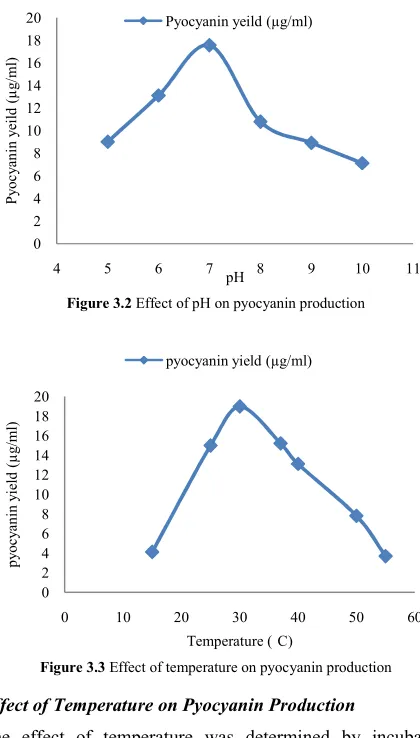
The effect of pH was determined by incubating production medium adjusted to different pH (5, 6, 7, 8, 9 and 10), inoculated with the 1% inoculum at 30 C at 150 rpm. Maximum pyocyanin production (17.56µg/ml) was observed at pH 7. Further increase in the pH resulted in decrease in pyocyanin production (Fig. 3.2). Culture conditions for
pyocyanin production by P. aeruginosa were studied at pH 7 and 30°C with a maximum yield of 10μg/ml (Liang et al., 2011). Similar results reported by Das et al., (2013) showed that P. aeruginosa PA14 strains produce pyocyanin at pH 7 at 30 °C.
Effect of Temperature on Pyocyanin Production
The effect of temperature was determined by incubating inoculated production medium at temperature range (15 C-60 C) with 1% inoculums at 130 rpm. Maximum pyocyanin production (18.98µg/ml) was obtained at temperature of 30 C. The pyocyanin production decreases with increase in temperature. Similar results reported by Das et al., (2013) showed that P. aeruginosa PA14 strains produce pyocyanin at pH 7 for at 30 °C. Kocka and Harris, (1974) showed that pyocyanin was generally formed after 2 days growth at 30 °C.
Effect of Carbon Source on Pyocyanin Production
To study the influence of carbon source on pyocyanin production, production medium was supplemented with different carbon source at 1.0% concentration. Isolate U4 showed efficient growth in all the substrate and maximum pyocyanin production was found in production media containing mannitol as carbon source with pyocyanin production, 23.32µg/ml. Whereas, minimum pyocyanin production was found in production media containing sucrose as carbon source with pyocyanin production, 10.88µg/ml as shown in the Fig. 3.4. In the present study, glucose, sucrose and fructose shows inhibitory effect on pyocyanin production by isolate U4. Inhibitory effect of glucose as an alternative
Figure 3.1 Effect of incubation period on pyocyanin production
0 0.2 0.4 0.6 0.8 1 1.2 1.4
0 2 4 6 8 10 12 14 16
0 24 48 72 96 120 144 168 192
B
io
m
as
s
(m
g
/m
l)
P
y
o
c
y
an
in
(
µ
g
/m
l)
Time (h)
Pyocyanin (µg/ml) Biomass (mg/ml)
Figure 3.2 Effect of pH on pyocyanin production
0 2 4 6 8 10 12 14 16 18 20
4 5 6 7 8 9 10 11
P
y
o
c
y
an
in
y
ei
ld
(
µ
g
/m
l)
pH
Pyocyanin yeild (µg/ml)
Figure 3.3 Effect of temperature on pyocyanin production
0 2 4 6 8 10 12 14 16 18 20
0 10 20 30 40 50 60
p
y
o
cy
an
in
y
ie
ld
(
µ
g
/m
l)
832
carbon source on pyocyanin production was previously reported by Kurachi, (1958a).Effect of Nitrogen Sources on Pyocyanin Production
To study the influence of organic and inorganic nitrogen source on pyocyanin production, production medium was supplemented with different nitrogen source at 1% w/v concentration (peptone, ammonium chloride, ammonium nitrate, ammonium sulfate, potassium nitrate, sodium nitrate and urea). Maximum pyocyanin production was found in production media containing peptone as nitrogen source with pyocyanin production of 26.12µg/ml. Whereas minimum pyocyanin production was found in production media containing sodium nitrate as nitrogen source with pyocyanin production of 11.03µg/ml as shown in the Fig. 3.5. Das and Das, (2015) suggested that peptone contains particular peptides or microelements or vitamins that could be essential for pigment synthesis.
Antibacterial Activity of Pyocyanin
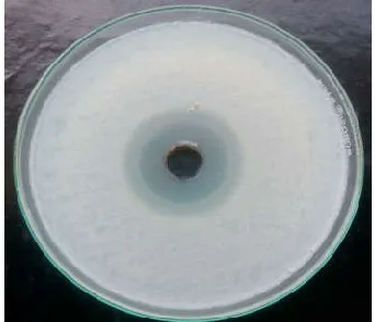
The antibacterial activity of pyocyanin was monitored against gram-positive bacteria (Staphylococcus aureus, Bacillus megaterium, Bacillus cereus) and gram-negative bacteria (Escherichia coli, Salmonella typhi A, Salmonella typhi B), results shown in Fig. 3.7 indicates that pyocyanin has antibacterial activity against all the tested microbial strains. The lowest zone of inhibition (2 mm) was exhibited by
Salmonella typhi B and highest zone of inhibition (14 mm) was observed against B. megaterium. Staphylococcus aureus
shows resistance against pyocyanin. El-Shouny., (2011) found
that the growth of all tested gram-positive bacteria and
Candida spp. were completely inhibited by pyocyanin; whereas Gram-negative bacteria, including S. typhi and
Pseudomonas mirabilis, were intermediately affected and K. pneumonia was resistant to pyocyanin.
Variation in the lipid content of cell wall of gram-positive and gram-negative bacteria may be responsible for the variation in the sensitivity of pyocyanin. Pyocyanin increases intracellular oxidant stress and exhibits a redox cycle under aerobic condition. This leads to reactive oxygen species (ROS) production such as superoxide and hydrogen peroxide; these ROS compounds are capable of inhibiting microbial growth (Denning et al., 1998; Das & Manefield., 2012). Another study based on denaturant gradient gel electrophoresis analysis revealed that the resistance or sensitivity to pyocyanin is related to level of catalase and superoxide dismutase enzymes (Norman et al., 2004). Yilmaz and Sidal., (2005) reported that the antimicrobial activity against gram positive bacteria was more efficient than against gram negative bacteria.
CONCLUSION
Pyocyanin is a water soluble blue green heterocyclic compound. Pyocyanin is redox active secondary metabolite. It is an extracellular pigment which is produced by
Pseudomonas aeruginosa. Pyocyanin have a variety of pharmacological activities. In present study a total of 8 bacterial strains were isolated and characterized as
Pseudomonas from different environmental samples. Isolate
Figure 3.4 Effect of carbon source on pyocyanin production
0 5 10 15 20 25
P
y
o
cy
an
in
y
ei
ld
(
µ
g
/m
l)
carbon source
Pyocyanin yeild (µg/ml)
Figure 3.5 Effect of nitrogen source on pyocyanin production
0 5 10 15 20 25 30
P
y
o
cy
an
in
y
ei
ld
(
µ
g
/m
l)
Nitrogen source Pyocyanin yeild (µg/ml)
Figure 3.6 Zone of inhibition of Pyocyanin against Bacillus
megaterium
Figure 3.7 Antibacterial activity of pyocyanin against bacteria
0 2 4 6 8 10 12 14 16
Z
o
n
e
o
f
in
h
ib
it
io
n
(
m
m
)
Bacterial strains Zone of inhibition (mm)
833
U4 was found to be most promising culture for production of pyocyanin in submerged fermentation amongst all the isolates tested. Maximum pyocyanin production was obtained at 72 h of incubation at pH 7.0. The pyocyanin production was maximum in production medium containing mannitol as carbon source and peptone as nitrogen source with pyocyanin production, 26.12µg/ml. The optimization process enhancing the pyocyanin production by factor of 2.17 fold.References
Baron, S. S and Rowe, J. J. (1981). Antibiotic action of pyocyanin. Antimicrobial Agents and Chemo therapy, 20, 814–820.
Budzikiewicz, H. (1993). Secondary metabolites from Pseudomnads. FEMS Mirobiology Reviews, 104, 209-228.
Chakraborty, P. (1996). A text book of microbiology.
Pseudomonas. Graphitech. India limited, 34, 304-309. Chen, J. and Xiao-Chang, C. (2004). Organic light-emitting
device having phenanthroline-fused phenazine. US patent 6713781.
Cox, C. D. (1986). Role of pyocyanin in the acquisition of iron from transferrin. Infection and immunity, 52, 263-270.
Das, S and Das, P. (2015). Effects of cultivation media components on biosurfactant and pigment production from Pseudomonas aeruginosa PAO1. Brazilian Journal of Chemical Engineering, 32, 317-324.
Das, T and Manefield, M. (2012). Pyocyanin promotes extracellular DNA release in Pseudomonas aeruginosa. PLoS ONE, 7. e46718.
Das, T., Kutty, S. K., Kumar, N., and Manefield, M. (2013). Pyocyanin Facilitates Extracellular DNA Binding to Pseudomonas aeruginosa Influencing Cell Surface Properties and Aggregation. PLoS ONE, 8, e58299.
Denning, G. M., Railsback, M. A., Rasmussen, G. T. Cox, C. D and Britigan, B. E. (1998). Pseudomonas pyocyanine alters calcium signaling in human airway epithelial cells. American Journal of Physiological lung cell and molecular physiology, 274, 893- 900.
El-Fouly, M. Z., Sharaf, A. M., Shahin, A. A. M., Heba, A. El-Bialy and Omara A. M. A. (2015). Biosynthesis of pyocyanin pigment by Pseudomonas aeruginosa. Journal of Radiation Research and Applied Sciences, 8, 36-48. El-Shouny, W. A., Al-Baidani, A. R and Hamza, W. T.
(2011). Antimicrobial activity of pyocyanin produced by
Pseudomonas aeruginosa isolated from surgical wound-infections. International Journal of Pharmacy and Medical Sciences, 1, 1-7.
Hassan, H. M and Fridovich, I. (1980). Mechanism of the antibiotic action ofpyocyanine. Journal of bacteriology, 141, 156-163.
Kocka, F. E and Harris, J. O. (1974). The influence of carbon sources on pigments produced by Pseudomonas aeruginosa. Transactions of the Illinois State Academy of Science, 67,345-351.
Kurachi, M. (1958a). Studies on the biosynthesis of pyocyanine. II. On the culture conditions for pyocyanine formation. Bulletin of the Institute for Chemical Research, 36, 163-173.
Lau, G., Hassett, H., and Kong, F. (2004). The role of pyocyanin in P. aeruginosa infection. Trends in Molecular Medicine, 10, 599-606.
Liang, H., Duan, J., Sibley, C. D., Surette, M. G., and Duan, K. (2011). Identification of mutants with altered phenazine production in Pseudomonas aeruginosa.
Journal of Medical Microbiology, 60, 22-34.
Norman, R. S., Moeller, P., Mc Donald, T. J., and Morris, P. J. (2004). Effect of pyocyanin on a crude-oil-degrading microbial community. Applied and Environmental Microbiology, 70, 4004-4011.
Onbasli, D and Belma, A. (2008). Determination of antimicrobial activity and production of some metabolites by Pseudomonas aeruginosa B1 B2 in sugar beet molasses. African Journal of Biotechnology, 7, 4614-4619.
Osawa, S., Yabuuchi, E., Narano, Y., Nakata, M., Kosono, Y., Takashina, K., and Tanabe, T. (1963). Pigment production by Pseudomonas aeruginosa on glutamic acid medium and gel filtration of the culture fluid filtrate.
Japanese Journal of Microbiology, 7, 87-95.
Ra’oof, W. M and Latif, I. A. R. (2010). In vitro study of the swarming phenomena and antimicrobial activity of different human infections, European Journal of Scientific Research, 47, 405-421.
Saha, S., Thavasi, R and Jayalakshmi, S. (2008). Phenazine pigments from Pseudomonas aeruginosa and their application as antibacterial agent and food colourants.
Research Journal of Microbiology, 3, 122-128.
Schaad, N. W., Jones, J. B and Chun, W. (2001). Laboratory Guide for Identication of Plant Pathogenic Bacteria. APS Press St. Paul M. N.
Streeter, K and Katouli, M. (2016). Pseudomonas aeruginosa: A review of their Pathogenesis and Prevalence in Clinical Settings and the Environment. Infection, Epidemiology and Medicine, 2, 25-32.
Sudhakar, T., Karpagam, S & Shiyama, S. (2013). Analysis of pyocyanin compound and its antagonistic activity against phytopathogens. International Journal of Chem Tech Research, 5, 1101-1106.
Yilmaz, E. S and Sidal, U. (2005). Investigation of antimicrobial effects of a P. aeruginosa-originated biosurfactant. Biologia, 60, 1-3.