ISSN: 2278-7461, www.ijeijournal.com
Volume 1, Issue 4 (September 2012) PP: 27-31
Classification of MRI Brain Images Using Neuro Fuzzy Model
Mr. Lalit P. Bhaiya
1, Ms. Suchita goswami
2, Mr. Vivek Pali
31Associate professor, HOD (ET&T), RCET,Bhilai(C.G.) 2,3M-tech scholar, RCET, Bhilai,
Abstract––It is difficult to identify the abnormalities in brain specially in case of Magnetic Resonance Image brain image processing. Artificial neural networks employed for brain image classification are being computationally heavy and also do not guarantee high accuracy. The major drawback of ANN is that it requires a large training set to achieve high accuracy. On the other hand fuzzy logic technique is more accurate but it fully depends on expert knowledge, which may not always available. Fuzzy logic technique needs less convergence time but it depends on trial and error method in selecting either the fuzzy membership functions or the fuzzy rules. These problems are overcome by the hybrid model namely, neuro-fuzzy model. This system removes essential requirements since it includes the advantages of both the ANN and the fuzzy logic systems. In this paper the classification of different brain images using Adaptive neuro-fuzzy inference systems (ANFIS technology). Experimental results illustrate promising results in terms of classification accuracy and convergence rate.
Keywords––Fuzzy logic, Neural network, ANFIS, Convergence rate
I.
INTRODUCTION
With the growing age, there is advancement in each and every field. As far as the medical field is concerned, it also has everyday progress. The medical imaging field in particular, has grown substantially in recent years, and has generated additional interest in methods and tools for the management, analysis, communication of medical image data. Medical imaging technology facilitates the doctors to see the interior portions of the body for easy diagnosis. It also helped doctors to make keyhole surgeries for reaching the interior parts without really opening too much of the body. CT scanners, ultra sound and magnetic resonance imaging took over X-ray imaging by making the doctors to look at the body’s elusive third dimension.MRI picks up signals from the body’s magnetic particles spilling to its magnetic tune and with the help of its powerful computer, convert scanner data into revealing pictures of internal organs. MRI differs from CT scan as it does not use radiations. MRI is a noninvasive medical test that helps physicians diagnose and treat medical conditions. It is a technique based on the measurement of magnetic field vectors generated after an appropriate excitation with strong magnetic fields and radio frequency pulses in the nuclei of hydrogen atoms present in water molecules of a patient’s tissues. We know that the content of water differ for each tissue, by using this fact one can quantify the differences of radiated magnetic energy and have elements to identify each tissue. When we measure the specific magnetic vector components under controlled conditions, different images can be taken and we can obtain the information related to tissue contrast which reveals the details that can be missed in other measurements[12]. Detailed MRI image allows the physicians to better evaluate various parts of the body and determine the presence of certain abnormalities that may not be accessed adequately with other imaging methods such as X-ray, CT scan, and ultra sound. Currently, MRI is the most sensitive imaging test of the head in routine clinical practice. MRI can detect a variety of conditions of the brain such as cysts, tumors, bleeding, swelling, developmental and structural abnormalities, infections, inflammatory conditions or problems with the blood vessels.MRI can provide clear images of parts of the brain that can not be seen as well with an X-ray, CAT scan, or ultrasound, making it particularly valuable for diagnosing problems with the pituitary gland and brain stem.
Applications of MRI segmentation include the diagnosis of brain trauma where a signature of brain injury, white matter lesions may be identified in moderate and mild cases. MRI segmentation methods are also useful in diagnosing multiple sclerosis, including the detection of lesions and the quantization of lesion volume using multispectral methods.[5]
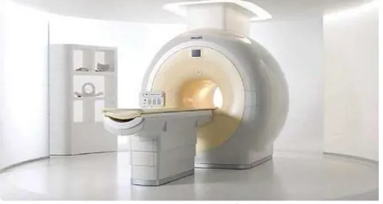
Figure 1.2: Schematic diagram of MRI machine
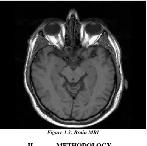
In MRI, water molecules give off radio signals which are converted into high resolution images that look like a picture shown in figure 1.3.
Figure 1.3: Brain MRI
II.
METHODOLOGY
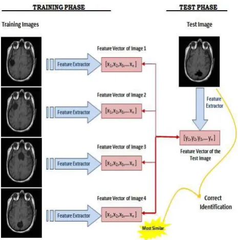
Figure 2.1: Proposed Methodology for Classification of MRI brain images
2.1 MRI image data set
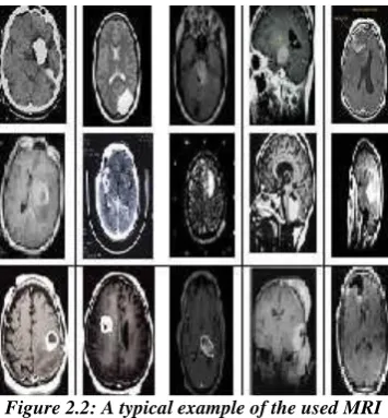
For the classification of normal and abnormal brain images a data set is collected from different sources one of the source is the Harvard medical school website. [http://www.med.harvard.edu/aanlib/home.html] The various types of brain images includes Axial, T2-weighted, 256-256 pixels MR brain images. Figure shows one of the database considered for the classification. The images are classified as normal and abnormal brain images.
Figure 2.2: A typical example of the used MRI
2.2 Feature Extraction
Figure 2.3 : Schematic diagram of a MR image recognizer.
2.3 Neuro-Fuzzy Classifier
A neuro-fuzzy classifier is used to detect the abnormalities in the MRI brain images. Generally the input layer consist of seven neurons corresponding to the seven features. The output layer consist of one neuron indicating whether the MRI is of a normal brain or abnormal and the hidden layer changes according to the number of rules that give best recognition rate for each group of features.[3] Here the neuro-fuzzy classifier used is based on the ANFIS technique. An ANFIS system is a combination of neural network and fuzzy systems in which that neural network is used to determine the parameters of fuzzy system. ANFIS largely removes the requirement for manual optimization of parameters of fuzzy system.The neuro-fuzzy system with the learning capabilities of neural network and with the advantages of the rule-base fuzzy system can improve the performance significantly and neuro-fuzzy system can also provide a mechanism to incorporate past observations into the classification process.In neural network the training essentially builds the system. However, using a neuro-fuzzy technique,the system is built by fuzzy logic definitions and and it is then refined with the help of neural network training algorithms.
Some advantages of ANFIS systems are:
It refines if-then rules to describe the behavior of a complex system.
It does not require prior human expertise
It uses membership functions plus desired dataset to approximate.
It provides greater choice of membership functions to use.
Very fast convergence time.[7]
2.3.1 ANFIS GUI
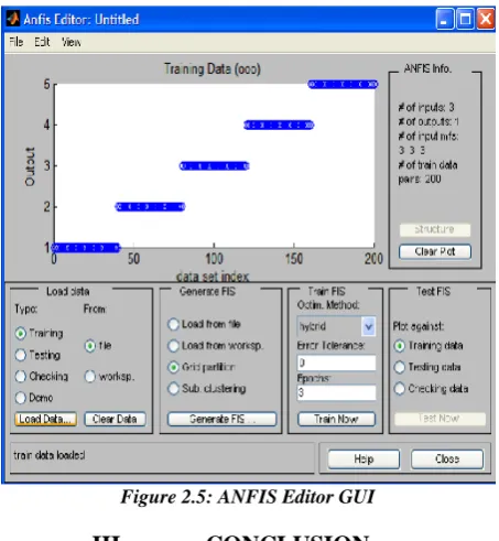
The ANFIS Editor GUI menu bar can be used to load a FIS training initialization, save the trained FIS, open a new Sugeno system or any of the other GUIs to interpret the trained FIS model. Any data set is loaded into the ANFIS Editor GUI, (or that is applied to the command-line function ANFIS) must be a matrix with the input data arranged as vectors in all but the last column. The output data must be in the last column. A sample of ANFIS Editor GUI with input is shown in Figure 2.5.
Figure 2.5: ANFIS Editor GUI
III.
CONCLUSION
Neural networks are performing successfully where other methods do not. There are many areas viz. medicine, weather forecasting, classification, resource allocation, and stock market predication; where such decision support system can help in setting priorities and making effective and productive decisions. Since the traditional connectionist systems do not provide explicit fuzzy interface, the proposed hybrid systems have wider scope/acceptability and presents dual advantages of a type-2 fuzzy logic based decision support using ANN techniques. this technique increases accuracy.
REFERENCES
1. Brain MRI Slices Classification Using Least Squares Support Vector Machine Vol. 1, No. 1, Issue 1, Page 21 of 33 ICMED 2007
2. MRI Fuzzy Segmentation of Brain Tissue Using IFCM Algorithm with Genetic Algorithm Optimization 1-4244-1031 2/07/$25.00©2007 IEEE
3. Brain Cancer Detection using Neuro Fuzzy Logic IJESS 2011
4. A Support Vector Machine Based Algorithm for Magnetic Resonance Image Segmentation 978-0-7695-3304-9/08 $25.00 © 2008 IEEE
5. An artificial neural network for detection of biological early brain cancer, 2010 Internation journal of computer applications(0975-8887) vol. I-no.6.
6. Tracking algorithm for De-noising of MR brain images IJCSNS,Vol.9 no. 11,Nov. 2009
7. Application of Neuro-Fuzzy Model for MR Brain Tumor Image Classification, International Journal of Biomedical Soft Computing and Human Sciences, Vol.16,No.1 (2010)
8. An Enhanced Implementation of Brain Tumor Detection Using Segmentation Based on Soft Computing International Journal of Computer Theory and Engineering, Vol. 2, No. 4, August, 2010 1793-8201
9. Segmentation of MR Brain Images Using FCM improved by Artificial Bee Colony (ABC) Algorithm 978-1-4244-6561-3/101$26.00 ©2010 IEEE
10. Biological Early Brain Cancer Detection Using Artificial Neural Network (IJCSE) Vol. 02, No. 08, 2010, 2721-2725
11. Improved Fuzzy C-Means Algorithm for MR Brain Image Segmentation (IJCSE) Vol. 02, No. 05, 2010, 1713-1715
12. Neural Network Based Brain Tumor Detection Using Mr Images IJCSC Vol. 2, No. 2, July-December 2011, pp. 325-331
13. A Hybrid Technique For automatic MRI brain Images Classification studia univ. babes_{bolyai, informatica, Volume LIV, 2009
14. Brain tumor detection based on multi-parameter MRI image analysis ICGST-GVIP journal,ISSN 1687-398x, vol. [9],issue [III],june 2009