R E S E A R C H A R T I C L E
Open Access
Combined anterior lumbar interbody fusion and
instrumented posterolateral fusion for
degenerative lumbar scoliosis: indication and
surgical outcomes
Ming-Kai Hsieh, Lih-Huei Chen, Chi-Chien Niu, Tsai-Sheng Fu, Po-Liang Lai and Wen-Jer Chen
*Abstract
Background:Traditional approaches to deformity correction of degenerative lumbar scoliosis include anterior-posterior approaches and posterior-only approaches. Most patients are treated with posterior-only approaches because the high complication rate of anterior approach. Our purpose is to compare and assess outcomes of combined anterior lumbar interbody fusion and instrumented posterolateral fusion with posterior alone approach for degenerative lumbar scoliosis with spinal stenosis.
Methods:Between November 2002 and November 2011, a total of 110 patients with degenerative spinal
deformity and curves measuring over 30°were included. Of the 110 patients who underwent surgery, 56 underwent the combined anterior and posterior approach and 54 underwent posterior surgery at our institution. The following were the indications of anterior lumbar interbody fusion: (1) rigid or frank lumbar kyphosis, (2) anterior or lateral bridged traction osteophytes, (3) gross coronal and sagittal deformity or imbalance, and (4) severe disc space narrowing that is not identifiable when performing posterior or transforaminal lumbar interbody fusion. The clinical outcomes were evaluated using the Oswestry disability index and the visual analog scale. The status of fusion were assessed according to the radiographic findings.
Results:All patients received clinical and radiographic follow-up for a minimum of 24 months, with an average follow-up of 53 months (range, 26–96 months). At the final follow-up, the mean ODI score improved from 28.8 to 6.4, and the mean back/leg VAS, from 8.2/5.5 to 2.1/0.9 in AP group and the mean ODI score improved from 29.1 to 6.2, and the mean back/leg VAS, from 9.0/6.5 to 2.3/0.5 in P group. The mean scoliotic angle changed from 41.3° preoperatively to 9.3°, and the lumbar lordotic angle, from 3.1° preoperatively to 35.7°in AP group and the mean scoliotic angle from 38.5 to 21.4 and the lumbar lordotic angle from 6 to 15.8 in P group. There were significant differences in sagittal (P = 0.009) and coronal (P = 0.02) plane correction between the two groups.
Conclusions:Our results demonstrate that combined anterior lumbar interbody fusion and instrumented
posterolateral fusion for adult degenerative lumbar scoliosis effectively improves sagittal and coronal plane alignment than posterior group and both group were effectively improves clinical scores.
Keywords:Degenerative lumbar scoliosis, De novo scoliosis, Anterior lumbar interbody fusion, Instrumented fusion
* Correspondence:chenwenj@adm.cgmh.org.tw
Department of Orthopedic Surgery, Chang Gung Memorial Hospital and Chang Gung University, 5, Fu-Hsin Street, Kweishan Shiang, Taoyuan 333, Taiwan
Background
Degenerative lumbar scoliosis is believed to develop sec-ondary to asymmetric collapse of the intervertebral disc spaces [1-5]. This leads to poor body posture, back pain, and neurological deterioration owing to decreased fo-raminal height with nerve root compression on the con-cave side of the deformity, and nerve stretching on the convex side [1,2,6]. The commonly presenting symptoms include chronic back pain and neurogenic claudication caused by concurrent stenosis with a structural degen-erative deformity [7]. Traditional approaches to deform-ity correction of degenerative lumbar scoliosis include anterior-posterior approaches and, more commonly, posterior-only approaches. Most patients are treated with posterior-only approaches because the anterior ap-proach has been shown to be associated with complica-tions such as vascular injury, ileus, and retrograde ejaculation and involves performing 2 large surgical pro-cedures and, hence, increases the operating time [8,9]. The use of posterior decompression with posterior spinal instrumentation and fusion may obviate the need for extensive abdominal surgery by enabling significant correction through a posterior-only approach. However, degenerative lumbar scoliosis secondary to an idiopathic curve tends to become rigid anteriorly, which gets more difficult to be corrected via a posterior-only approach [10]. Combined anterior lumbar interbody fusion and in-strumented posterolateral fusion provides several bene-fits over the posterior-only approach, in terms of improved stability, decreased stress on screws, improved fusion rates, and better lumbar lordosis [11-13]. To our knowledge, no study has yet mentioned the indications of combined anterior lumbar interbody fusion and in-strumented posterolateral fusion for degenerative lumbar scoliosis with spinal stenosis and compare and assess outcomes with posterior alone approach.
The goals of adult deformity surgery are to obtain sa-gittal and coronal balance, symptom relief, and solid fu-sion [14,15]. Various techniques have been reported for correcting degenerative lumbar deformities with instru-mentation and fusion using pedicle screw systems and various types of interbody cages. Interbody cages allow for the correction of the deformity, anterior column sup-port, increased foraminal height, circumferential arth-rodesis, and restoration of the anterior column height as well as lumbar lordosis. Interbody cages can be placed via either a posterior approach, as for the posterior lumbar interbody fusion (PLIF) and transforaminal lumbar inter-body fusion (TLIF), or an anterior approach, as for the an-terior lumbar interbody fusion (ALIF), by using either an autograft or allograft, metal cages, or poly-ether-ether-ke-tone (PEEK) cages [11,16-18]. Herein, we describe our ex-perience as well as indications for performing combined anterior lumbar interbody fusion and instrumented
posterolateral fusion for degenerative lumbar scoliosis with spinal stenosis.
Methods Patients
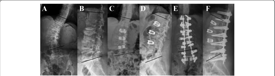
From November 2002 to November 2011, 1834 patients with degenerative lumbar scoliosis underwent surgery in our institution. The Chang Gung Medical Foundation In-stitutional Review Board approved this study (99-0771B) and waived the requirement for informed consent due to the retrospective nature of the study. All patients pre-sented with neurological claudication with mechanical back pain that was refractory to at least 6 months of con-servative management such as physical therapy, activity modification, chiropractic manipulation, administration of oral analgesics and nonsteroidal anti-inflammatory drugs, epidural steroids, and facet injections. The inclusion cri-teria of combined anterior and posterior approach were (1) rigid or frank lumbar kyphosis, (2) anterior or lateral bridged traction vertebral osteophytes, (3) gross coronal and sagittal deformity or imbalance, and (4) severe disc space narrowing that is not identifiable when performing PLIF or TLIF (Figure 1) and exclusion criteria were previ-ous abdominal or retroperitoneal surgery.
A total of 110 patients with degenerative spinal de-formity and curves measuring over 30°who underwent reconstructive spinal fusion surgery from 2002 to 2011 were included. Of the 110 patients who underwent sur-gery, 56 underwent the combined anterior and posterior approach and 54 underwent posterior surgery at our in-stitution. This posterior (P) group included 34 females and 20 males with an average age of 62 years. 56 patients underwent combined anterior release and fusion of mul-tiple lumbar levels followed by posterior instrumented fusion (AP group) included 35 females and 21 males with an average age of 61 years. Eighteen patients under-went ALIF followed by simultaneous instrumented pos-terolateral fusion. The remaining 38 patients underwent staged operations between 1 and 2 weeks.
Clinical assessment
The clinical outcome was evaluated using the Oswestry disability index (ODI) and the visual analog scale (VAS) preoperatively and at the final follow-up. All patients were scheduled for follow-up at 3 months, 6 months, and 1 year after the surgery and then annually. The fol-lowing comorbidities were preoperatively diagnosed in AP and P groups : diabetes mellitus (n = 6;8); hyperten-sion (n = 8:8); corticosteroid usage (n = 3;4); and valvular heart disease (n = 2;1).
Surgical technique
position with the concave side up, with the intention to directly correct the scoliosis. ALIF was performed using the flank retroperitoneal approach. After exposure of the anterior part of the disc, the anterior longitudinal ligament was transversely incised, and the disc was completely re-moved. Next, the vertebral endplates were cleared of car-tilage using sharp curettes, taking care that damage to the subchondral bone of the endplates is avoided. Maximum distraction of disc space was achieved by manual lordotic force. After a satisfactory trial implantation, the SynCage (Synthes Spine, West Chester, PA, USA), which was filled with a morselized cancellous allograft, was implanted.
Posterior surgery in the combined anterior-posterior group and the posterior group ,all posterior instrumen-tation was placed via an open posterior approach. After subperiosteal exposure of the dorsal spine using a stand-ard midline approach, an autologous bone graft was har-vested via a subcutaneous access to the posterior iliac crest. After adequate decompression, pedicle screw in-strumentation with TriFix G (Aspine, Oakland, CA, USA), with or without PLIF or TLIF was performed. Fi-nally, rod derotation maneuver and compression on the convex side with the rod carefully contoured to the lor-dosis was performed to restore lumbar lorlor-dosis and cor-rect lumbar scoliosis.
Radiographic evaluation
Plain radiographs in the standing posteroanterior (PA), lateral, and flexion-extension views were obtained for all patients preoperatively, postoperatively, 1 year after the surgery, and at the final follow-up. Preoperative and postoperative radiographs were compared to determine the degree of correction achieved following surgery. The coronal Cobb angle was determined from the standing PA radiograph by drawing a line parallel to the superior endplate of the most superior vertebra and a second line
parallel to the inferior endplate of the most inferior ver-tebra of the scoliotic curve. Lumbar lordosis was mea-sured using the Cobb method [18] between the superior endplate of L-1 and S-1. Hyperlordosis was defined as any Cobb angle >60°, and hypolordosis was defined as any angle <20°. We also measure the lordotic angle cor-rection for each level and SynCage position. End-plate fractures, cage malpositioning, and the status of the anter-ior and posterolateral fusion were also recorded. Anteranter-ior fusion was classified as solid, probable, or pseudoarthrosis. Solid fusion was defined as visible, continuous trabeculae of bridging fusion masses across the disc space and lack of instability in the flexion-extension radiographs. Probable fusion was defined as unclear bony trabecular continuity with no radiolucent interruption or motion seen in the stress radiographs. Pseudoarthrosis was defined as radio-lucent interruption of the cage, and as motion, in stress radiographs. Posterolateral fusion was also classified as solid, probable, and pseudoarthrosis. Solid fusion was de-fined as visible, continuous trabeculae of bridging fusion masses over the bilateral transverse processes and no mo-tion in the flexion and extension stress radiographs. Prob-able fusion was defined as unclear bony trabecular continuity with no radiolucent interruption or motion in stress radiographs. Radiolucent interruption of the fusion mass was labeled as pseudoarthrosis [19]. The fusion sta-tus was decided by the senior surgeons (W-J,Chen ).
Statistical analysis
Data were analyzed using the SPSS statistical software package (SPSS, Inc, Chicago, IL, USA). Means were cal-culated for different variables including the ODI score, VAS, and angles of lumbar lordosis and scoliosis. Pre-operative and postPre-operative measurements and values between the different subgroups were compared using the paired t-test with statistical significance set at a Figure 1A 64-year-old woman complained low back pain with bilateral sciatica and claudication for several years.Radiographs of
P value of <0.05. The position of SynCage and the lordo-tic angle correction was compared using the Student’s
t-test with statistical significance set at a P value of <0.05.
Results
All patients received clinical and radiographic follow-up for a minimum of 24 months, with an average follow-up of 53 months (range, 26–96 months). The operation time of anterior lumbar interbody fusion is 172.5 minutes (range,115-301 minutes) and instrumented posterolateral fusion is 262.5 minutes (range, 195–375 minutes) in AP group and 350.5 minutes (range, 210–452 minutes) in P group, estimated blood loss of anterior lumbar interbody fusion is 250 ml (range,150-1750 ml) and instrumented posterolateral fusion is 1650 ml (range,1000-4850 ml) in AP group and 3250 ml (range,2000-6500 ml) in P group, transfusion amount of anterior lumbar interbody fusion is 700 ml (range,500-2000 ml) and instrumented pos-terolateral fusion is 1500 ml (range,1000-5000 ml) in AP group and 4000 ml (range,2000-8000 ml) in P group, and length of stay is 16 days (range ,10-21 days ) in AP group and 10 days (range ,7-14 days ) in P group.
Clinical outcomes
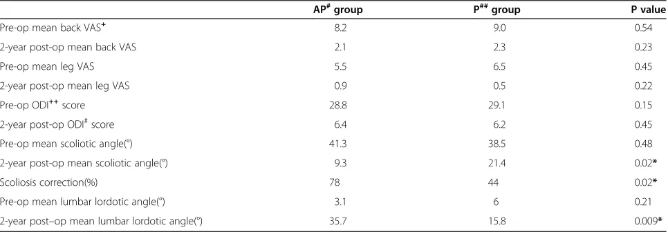
The VAS and ODI scores were evaluated preoperatively and at the final follow-up (Table 1). At final follow-up, the average ODI score was significantly lower than that determined preoperatively in both groups. The mean back and leg VAS scores also improved significantly in both groups.
Radiographic outcomes
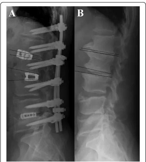
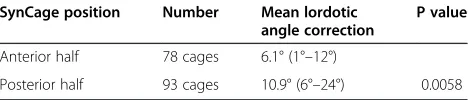
Preoperative and postoperative coronal Cobb angles and lumbar lordosis angles were compared (Table 1). The average preoperative coronal Cobb angle was 41.3° (range, 32°–85°), which decreased to 9.3° post-operatively in AP group, demonstrating a significant mean scoliosis correc-tion of 78% (P = 0.042). The mean preoperative lumbar lordosis angle increased from 3.1° (range, kyphosis 30° to lordosis 33°) to 35.7° (range, lordosis 9° to 60°), demon-strating a mean improvement of 32.6° (P = 0.009).In P group , the average preoperative coronal Cobb angle was 38.5° (range, 32°–55°), which decreased to 21.4° post-operatively, demonstrating a significant mean scoliosis correction of 44%. The mean preoperative lumbar lordosis angle increased from 6° (range, kyphosis 25° to lordosis 25°) to 15.8° (range, lordosis 10° to 40°), demonstrating a mean improvement of 9.8°. Both in coronal and sagittal plane ,angle improvement were better in AP group than P group.As shown in Table 2, ALIF was performed for a total of 171 disc levels in the 56 patients as follows: 1-level procedure (n = 3), 2-level (n = 15), 3-level (n = 18), 4-level (n = 16), and 5-level (n = 4). As seen in Figure 2, the ALIF procedures were correlated with a higher rate of scoliosis and lordosis correction. In Figure 3 and Table 3, an ALIF cage placed in the posterior half provides more lordosis at the instrumented level, whereas a cage placed in the anter-ior half may not provide better sagittal plane correction. (10.9° to 6.1°; P = 0.0058). Two patients exhibited tomatic SynCage subsidence, and 1 patient had asymp-tomatic S1 screw loosening. The fusion status was decided by the senior surgeons (W-J,Chen). At the final follow-up, 36 of the 56 patients (64.3%) exhibited solid anterior
Table 1 Clinical and radiographic outcomes
AP#group P##group P value
Pre-op mean back VAS+ 8.2 9.0 0.54
2-year post-op mean back VAS 2.1 2.3 0.23
Pre-op mean leg VAS 5.5 6.5 0.45
2-year post-op mean leg VAS 0.9 0.5 0.22
Pre-op ODI++score 28.8 29.1 0.15
2-year post-op ODI#score 6.4 6.2 0.45
Pre-op mean scoliotic angle(°) 41.3 38.5 0.48
2-year post-op mean scoliotic angle(°) 9.3 21.4 0.02*
Scoliosis correction(%) 78 44 0.02*
Pre-op mean lumbar lordotic angle(°) 3.1 6 0.21
2-year post–op mean lumbar lordotic angle(°) 35.7 15.8 0.009*
+
: VAS, Visual analog scale.
++
: ODI, Oswestry Disability Index.
#AP, Combined anterior and posterior approach. ##:P, Posterior approach.
fusion; 40 (71.4%), solid posterolateral fusion; 20 (35.7%), probable anterior fusion; and 16 (28.6%), probable pos-terolateral fusion in AP group and 39 of the 54 patients (72.2%) exhibited solid posterolateral fusion; 15 (27.8%), probable posterolateral fusion in P group. No anterior or posterolateral pseudoarthrosis was noted.
Complications
There were no major complications such as intraoperative cerebrospinal fluid (CSF) leak, postoperative weakness, deep venous thrombosis, ureteral trauma, or injury to the peritoneal or retroperitoneal structures. Perioperative complications, which presented as postoperative superfi-cial wound infections, occurred in 5 patients in the AP group and 7 in P group, but the symptoms subsided after debridement and antibiotic treatment. Six patients experi-enced transient postoperative anterior thigh numbness in the AP group, ipsilateral to the approach, in the distribu-tion of the anterior femoral cutaneous nerve.
Discussion
Adult degenerative scoliosis is believed to develop as a result of asymmetrical degeneration of the spine. It most commonly occurs in the lumbar spine and typically pre-sents as pain, which is the primary complaint in 90% of the patients [7]. This axial back pain occurs most com-monly due to a combination of muscle fatigue, trunk im-balance, facet arthropathy, and degenerative disc disease [20,21]. The flat-back deformity and forward sagittal im-balance have been shown to be a significant source of pain and disability in patients [22,23]. Several studies showed that radiographic parameters were correlated with clinical symptoms in adults [24-27]. Many radiographic parame-ters may affect functional scores in degenerative lumbar
Table 2 ALIF levels and angle correction
Patients Scoliotic angle (°) Lumbar lordotic angle (°)
ALIF* Pre-op 2-year F/U+ Correction Pre-op 2-year F/U Correction
1 3 22 2 20 2 20 18
2 15 39 6.8 32.2 4.1 32 27.9
3 18 41 9 32 2 33.7 31.7
4 16 44 11 33 7.8 46 38.2
5 4 62 22 40 0.5 56 55.5
*
ALIF: Anterior lumbar interbody fusion levels.
+
F/U: Follow up.
Figure 2Line chart of scoliotic angle correction compared with lordotic angle correction.
scoliosis, including the Cobb scoliosis angle in all major thoracolumbar or lumbar and lumbosacral curves, max-imal intervertebral lateral olisthesis, thoracic kyphosis, thoracolumbar kyphosis, lumbar lordosis, plumbline offset from C7 to the posterosuperior corner of the S1 vertebral body, and maximal intervertebral anteroposterior olisth-esis. Avraam et al. [26] reported that a loss of normal lor-dosis could affect health outcomes even when the sagittal balance is preserved in patients with degenerative lumbar scoliosis. Decreased lumbar lordosis and an increased lumbosacral hemicurve have led to poorer results of health status. The average curve correction in our series was 78% of maximal Cobb angle with maintenance of the correction seen at the 2-year follow-up. The mean scoli-otic and lumbar lordscoli-otic angle were improved from 41.3° and 3.1° pre-operatively to 9.3° and 35.7° at the 2-year follow-up in our combined group. However , the mean scoliotic and lumbar lordotic angle were improved from 38.5° and 6° pre-operatively to 21.4° and 15.8° in our pos-terior approach. There were significant differences in sa-gittal (P = 0.009) and coronal (P = 0.02) plane correction between the combined anterior-posterior group and the posterior group. The radiographic outcome in our com-bined approach was superior to that via a posterior ap-proach (Table 1).
Degenerative lumbar scoliosis with spinal stenosis has been traditionally treated with posterior decompression along with posterior spinal instrumentation and fusion. From 2002 to 2011, 1834 patients with degenerative lumbar scoliosis underwent surgery in our institution and 1778 patients were treated via a posterior-only ap-proach. Only 56 patients received combined anterior lumbar interbody fusion and instrumented posterolateral fusion. The obvious disadvantages of using an anterior ap-proach to the lumbar spine include retroperitoneal dissec-tion with associated vascular manipuladissec-tion, 2 major surgical procedures required, and increased operating time [8,9]. In our experience, the following are the indications for ALIF in addition to instrumented posterolateral fusion: (1) rigid or frank lumbar kyphosis; (2) anterior or lateral bridged traction vertebral osteophytes; (3) gross coronal and sagittal deformity or imbalance; and (4) severe disc space narrowing that is not identifiable when performing PLIF or TLIF. In de novo degenerative lumbar scoliosis, the curves tend to be more rigid posteriorly and, therefore, are amenable to correction via a posterior-only approach [10]. In our result ,rigid or frank lumbar kyphoscoliotic
deformity secondary to idiopathic curves were difficult to correct via a posterior-only approach with scoliosis correc-tion only 44% and lordotic angle correccorrec-tion only 9.8°, and better scoliosis and lordosis correction were achieved through the combined anterior and posterior approach with statistically significant. Anterior or lateral bridged traction vertebral osteophytes restrict not only graft place-ment but also lordosis restoration and can only be re-moved via an anterior approach. When performing PLIF or TLIF in osteoporotic endplate or severe disc narrowing cases, violation of the endplate always results in cage sub-sidence and poor angle correction.
The use of interbody grafts associated with posterior in-strumentation in deformity correction surgery has gained popularity for providing anterior column structural sta-bility, increased fusion rates, as well as enable restor-ation and preservrestor-ation of lumbar lordosis [11-13,28]. Graft placement has traditionally been achieved through either an anterior (ALIF) or a posterior (PLIF or TLIF) approach.
Anterior lumbar interbody fusion is superior to poster-ior lumbar fusion because of the larger surface area available between the vertebral bodies, which facilitates the use of wider cages that can rest on a strong periph-eral cortical bone on either side, thus minimizing the risk of cage subsidence, which is especially important in elderly patients with osteoporotic bones [11,17,29,30].
We found that the ALIF approach using the anterior-posterior wedge cages was effective for correcting sagittal plane deformities. An ALIF cage placed in the posterior half provides more lordosis at the instrumented level, whereas a cage placed in the anterior half may not provide better sagittal plane correction. (10.9° to 6.1°; P = 0.0058) (Figure 3, Table 3).
We have observed that the transpsoas approach leads to a high frequency of thigh numbness, pain, weakness, and dysesthesias, which are likely the result of retraction proximal to the lumbosacral plexus, and have been well described in previous anatomical studies [31]. Prior re-ports of lateral retroperitoneal approaches including mobilization of the psoas muscle from the lumbar spine have demonstrated high incidence (30%) of paresthesias in the thigh/groin region [32]. Knight et al. [33] reported 10% incidence of lateral femoral cutaneous nerve deficit and 3% incidence of L4 motor deficit using the lateral retroperitoneal transpsoas approach. In our study, we identified 6 patients (10.7%) with transient postoperative ipsilateral sensory deficits that had resolved before the last follow-up visit. Because these deficits were transient, we believe they were stretch or neuropraxic injuries.
Conclusions
ALIF with SynCages and supplemental instrumented pos-terolateral fusion resulted in better coronal and sagittal
Table 3 Position of SynCages and lordotic angle correction
SynCage position Number Mean lordotic angle correction
P value
Anterior half 78 cages 6.1° (1°–12°)
plane correction than posterior approach in all patients and was maintained in the 2-year follow-up. The VAS and ODI scores significantly improved after the operation, and no major complications occurred.
Competing interests
The authors declare that they have no competing interests.
Authors’contributions
Each author has made substantive intellectual contributions to this multicentre study: M–K H and W-J C participated in the study design, in collecting the data, the statistically analyses ,drafting and contributed equally to the manuscript. C-C N, T-S F , P-L L ,and L–H C, participated in the study design. W-J C advised and assisted drafting of the manuscript. All authors read and approved the final manuscript.
Acknowledgements
We thank the Department of Orthopaedic Surgery for their contribution to the study.
Received: 27 August 2014 Accepted: 10 February 2015
References
1. Ploumis A, Transfledt EE, Denis F. Degenerative lumbar scoliosis associated with spinal stenosis. Spine J. 2007;7:428–36.
2. Aebi M. The adult scoliosis. Eur Spine J. 2005;14:925–48.
3. Berven SH, Deviren V, Mitchell B, Wahba G, Hu SS, Bradford DS. Operative management of degenerative scoliosis:an evidence-based approach to surgical strategies based on clinical and radiographic outcomes. Neurosurg Clin N Am. 2007;18:261–72.
4. Oskouian Jr RJ, Shaffrey CI. Degenerative lumbar scoliosis. Neurosurg Clin N Am. 2006;17:299–315.
5. Daffner SD, Vaccaro AR. Adult degenerative lumbar scoliosis. Am J Orthop. 2003;32:77–82.
6. Anand N, Baron EM, Thaiyananthan G, Khalsa K, Goldstein TB. Minimally invasive multilevel percutaneous correction and fusion for adult lumbar degenerative scoliosis: a technique and feasibility study. J Spinal Disord Tech. 2008;21:459–67.
7. Winter RB, Lonstein JE, Denis F. Pain patterns in adult scoliosis. Orthop Clin North Am. 1988;19:339–45.
8. Crandall DG, Revella J. Transforaminal lumbar interbody fusion versus anterior lumbar interbody fusion as an adjunct to posterior instrumented correction of degenerative lumbar scoliosis: three year clinical and radiographic outcomes. Spine (Phila Pa 1976). 2009;34:2126–33. 9. Rajaraman V, Vingan R, Roth P, Heary RF, Conklin L, Jacobs GB. Visceral and
vascular complications resulting from anterior lumbar interbody fusion. J Neurosurg. 1999;91(1 Suppl):60–4.
10. Pateder DB, Kebaish KM, Cascio BM, Neubaeur P, Matusz DM, Kostuik JP. Posterior Only Versus Combined Anterior and Posterior Approaches to Lumbar Scoliosis in Adults. A Radiographic Analysis. Spine. 2007;32:1551–4. 11. Hsieh PC, Koski TR, O’Shaughnessy BA, Sugrue P, Salehi S, Ondra S, et al.
Anterior lumbar interbody fusion in comparison with transforaminal lumbar interbody fusion: implications for the restoration of foraminal height, local disc angle, lumbar lordosis, and sagittal balance. J Neurosurg Spine. 2007;7:379–86.
12. Ploumis A, Wu C, Fischer G, Mehbod AA, Wu W, Faundez A, et al. Biomechanical comparison of anterior lumbar interbody fusion and transforaminal lumbar interbody fusion. J Spinal Disord Tech. 2008;21:120–5. 13. Potter BK, Freedman BA, Verwiebe EG, Hall JM, Polly Jr DW, Kuklo TR.
Transforaminal lumbar interbody fusion: clinical and radiographic results and complications in 100 consecutive patients. J Spinal Disord Tech. 2005;18:337–46.
14. Birknes JK, White AP, Albert TJ, Shaffrey CI, Harrop JS. Adult degenerative scoliosis: a review. Neurosurgery. 2008;63(3 Suppl):94–103.
15. Heary RF, Kumar S, Bono CM. Decision making in adult deformity. Neurosurgery. 2008;63(3 Suppl):69–77.
16. Blumenthal SL, Ohnmeiss DD. Intervertebral cages for degenerative spinal diseases. Spine J. 2003;3:301–9.
17. Janssen ME, Lam C, Beckham R. Outcomes of allogenic cages in anterior and posterior lumbar interbody fusion. Eur Spine J. 2001;10(2 suppl):S158–68. 18. Groth AT, Kuklo TR, Klemme WR, Polly DW, Schroeder TM. Comparison of
sagittal contour and posterior disc height following interbody fusion: threaded cylindrical cages versus structural allograft versus vertical cages. J Spinal Disord Tech. 2005;18:332–6.
19. Wu CH, Wong CB, Chen LH, Niu CC, Tsai TT, Chen WJ. Instrumented Posterior Lumbar Interbody Fusion for Patients With Degenerative Lumbar Scoliosis. J Spinal Disord Tech. 2008;21:310–5.
20. Cobb JR. Outline for the study of scoliosis. Instructional course lectures. In: Bechtol CA, editor. American Academy of Orthopedic Surgeons, vol. 5. Edwards: Ann Arbor, MI; 1948. p. 261–75.
21. Bradford DS, Tay BK, Hu SS. Adult scoliosis: surgical indications, operative management, complications, and outcomes. Spine (Phila Pa 1976). 1999;24:2617–29.
22. Jang JS, Lee SH, Min JH, Maeng DH. Changes in sagittal alignment after restoration of lower lumbar lordosis in patients with degenerative flat back syndrome. J Neurosurg Spine. 2007;7:387–92.
23. Wiggins GC, Ondra SL, Shaffrey CI. Management of iatrogenic flat-back syndrome. Neurosurg Focus. 2003;15:E8.
24. Glassman SD, Berven S, Bridwell K, Horton W, Dimar JR. Correlation of radiographic parameters and clinical symptoms in adult scoliosis. Spine. 2005;30:682–8.
25. Jackson RP, Simmons EH, Stripinis D. Coronal and sagittal plane spinal deformities correlating with back pain and pulmonary function in adult idiopathic scoliosis. Spine. 1989;14:1391–7.
26. Ploumis A, Liu H, Mehbod AA, Transfeldt EE, Winter RB. A correlation of radiographic and functional measurements in adult degenerative scoliosis. Spine. 2009;34:1581–4.
27. Schwab FJ, Smith VA, Biserni M, Gamez L, Farcy JP, Pagala M. Adult scoliosis: a quantitative radiographic and clinical analysis. Spine. 2002;27:387–92. 28. Jagannathan J, Sansur CA, Oskouian Jr RJ, Fu KM, Shaffrey CI. Radiographic
restoration of lumbar alignment after transforaminal lumbar interbody fusion. Neurosurgery. 2009;64:955–64.
29. Gödde S, Fritsch E, Dienst M, Kohn D. Influence of cage geometry on sagittal alignment in instrumented posterior lumbar interbody fusion. Spine (Phila Pa 1976). 2003;28:1693–9.
30. Hart RA, Prendergast MA. Spine surgery for lumbar degenerative disease in elderly and osteoporotic patients. Instr Course Lect. 2007;56:257–72. 31. Benglis DM, Vanni S, Levi AD. An anatomical study of the lumbosacral
plexus as related to the minimally invasive transpsoas approach to the lumbar spine. J Neurosurg Spine. 2009;10:139–44.
32. Bergey DL, Villavicencio AT, Goldstein T, Regan JJ. Endoscopic lateral transpsoas approach to the lumbar spine. Spine (Phila Pa 1976). 2004;29:1681–8.
33. Knight RQ, Schwaegler P, Hanscom D, Roh J. Direct lateral lumbar interbody fusion for degenerative conditions: early complication profile. J Spinal Disord Tech. 2009;22:34–7.
Submit your next manuscript to BioMed Central and take full advantage of:
• Convenient online submission
• Thorough peer review
• No space constraints or color figure charges
• Immediate publication on acceptance
• Inclusion in PubMed, CAS, Scopus and Google Scholar
• Research which is freely available for redistribution