C A S E R E P O R T
Open Access
Diffuse alveolar haemorrhage associated
with subsequent development of ANCA
positivity and emphysema in three young
adults
Anna Stainer
1*, Alex Rice
2, Anand Devaraj
3, Joseph Luke Barnett
3, Jacqueline Donovan
4, Maria Kokosi
1,
Andrew Gordon Nicholson
2,5, Tom Cairns
6, Athol Umfrey Wells
1and Elisabetta Augusta Renzoni
1Abstract
Background:Diffuse alveolar haemorrhage (DAH) is characterized by the diffuse accumulation of red blood cells within the alveoli, presence of ground glass opacities and/or consolidation on computed tomography (CT). Aside from identifiable non-immune causes, DAH is classically subdivided into idiopathic (idiopathic pulmonary haemosiderosis, IPH) and autoimmune DAH. Here we describe three cases presenting with recurrent pulmonary haemorrhage, initially classified as IPH, who, several years after first presentation, develop anti myeloperoxidase antibodies (MPO) positivity, emphysema on CT and, in one case, renal involvement.
Case presentation:Patient 1 was diagnosed with IPH aged 14. Her disease remained poorly controlled despite immunosuppression, although ANCA remained negative over the years. Nineteen years from initial presentation, she developed MPO-ANCA positive antibodies and mild renal impairment. She was treated with Rituximab with good response. From first presentation, the chest CT was consistently characterized by diffuse ground-glass opacities and interlobular septal thickening. Ten years later, cystic opacities consistent with emphysema, with a striking peribronchovascular distribution, developed. Patient 2 was diagnosed with IPH aged 32. He was treated with corticosteroids and methotrexate, with fluctuating response. At 11 years from initial presentation, MPO-ANCA positivity was identified, and emphysema with a peribronchovascular distribution was observed on CT, with subsequent significant increase in extent. Patient 3 was diagnosed with IPH at the age of seven, and had recurrent episodes of haemoptysis of varying degree of severity, treated with intermittent courses of corticosteroids until age 11, when he was intubated due to severe DAH. Eight years after the diagnosis emphysematous changes were noted on CT and MPO-ANCA positivity developed for the first time 11 years after initial diagnosis.
Conclusions:We believe these three cases highlight: 1) the possibility of development of ANCA positivity several years down the line from first DAH presentation 2) the possibility that DAH may lead to cystic/emphysematous changes with peribronchovascular distribution on CT. Moreover, the need for ongoing immunosuppressive treatment and the development of emphysema, emphasize a possible role played by autoimmune phenomena, even when DAH is initially diagnosed as“idiopathic”. Further studies are required to better understand the relationship between DAH, ANCA positivity and development of emphysema.
Keywords:ANCA, Pulmonary haemosiderosis, Haemoptysis, Pulmonary haemorrage, Pulmonary vasculitis, Emphysema, AAV ANCA associated vasculitis.
© The Author(s). 2019Open AccessThis article is distributed under the terms of the Creative Commons Attribution 4.0 International License (http://creativecommons.org/licenses/by/4.0/), which permits unrestricted use, distribution, and reproduction in any medium, provided you give appropriate credit to the original author(s) and the source, provide a link to the Creative Commons license, and indicate if changes were made. The Creative Commons Public Domain Dedication waiver (http://creativecommons.org/publicdomain/zero/1.0/) applies to the data made available in this article, unless otherwise stated. * Correspondence:[email protected]
1Interstitial Lung Disease Unit, Royal Brompton Hospital, London, UK
Background
Diffuse alveolar haemorrhage (DAH) is characterized by intra-alveolar accumulation of red blood cells, with diffuse ground glass opacities and/or consolidation on chest high-resolution computed tomography (HRCT). The clinical spectrum of DAH ranges from incidental findings on im-aging and/or bronchoalveolar lavage (BAL) in asymptom-atic patients, to life-threatening acute respiratory failure. Histologically, DAH is characterized by the presence of hemosiderin-laden macrophages, fibrin deposition, type II pneumocyte hyperplasia, organizing pneumonia and acute inflammation. When present, capillaritis is associated with neutrophilic interstitial infiltration and disruption of the alveolar wall, although these changes can be subtle and difficult to detect [1].
DAH etiology is wide, including immunological and non-immunological causes. Among immunological causes of DAH, systemic vasculitides are one of the most fre-quent, particularly ANCA associated vasculitis (AAV). If no known cause or association can be found, DAH is clas-sified as idiopathic pulmonary hemosiderosis (IPH) [2].
We describe three cases presenting with recurrent pul-monary haemorrhage, who develop anti myeloperoxidase antibodies (MPO) positivity and radiologically, cystic areas resembling emphysema many years after their first presentation.
Case presentation
Patient 1
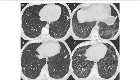
A 14 year-old young woman with lethargy, exertional dys-pnea, microcytic hypochromic anemia, persistent cough, and multiple episodes of haemoptysis was referred to the Royal Brompton Hospital (RBH) respiratory paediatric service. Lung function was characterized by a mild re-strictive pattern (forced vital capacity (FVC) 73%, forced expiratory volume in 1 s (FEV1) 77.5%, total lung capacity (TLC) 77%, carbon monoxide transfer factor (TLCO) 85% and transfer coefficient (KCO) 110%). HRCT revealed widespread ground glass opacification throughout both lungs (Fig.1-a). A bronchoalveolar lavage (BAL) revealed increasingly haemorrhagic returns and abundant haemosi-derin laden macrophages. A surgical lung biopsy showed findings consistent with DAH (Fig. 2). In the absence of identifiable associations, the patient was diagnosed with IPH and started on hydroxychloroquine. However, her disease remained inadequately controlled with frequent flares over the years, with poor compliance a possible contributor.
Aged 21, she was admitted to the RBH adult intersti-tial lung disease (ILD) unit with worsening breathless-ness, frequent“infectious”exacerbations, and daily small volume haemoptysis. At the time, she denied any con-nective tissue disease (CTD) symptoms. She only had an intermittent smoking history over the previous 3 years,
totaling one pack-year. Autoimmune screen was nega-tive. Although atypical non-specific anti-neutrophil cyto-plasmic antibody (ANCA) staining was found by indirect immunofluorescence [NOVA-lite, INOVA Diagnostics Inc., San Diego, USA]), both MPO and anti-proteinase 3 (PR3) were negative (fluorescence enzyme immunoassay on Immunocap 250 analyser [Thermo Scientific Immuno-Diagnostics, Milton Keynes, UK]). A chest HRCT again revealed diffuse ground glass opacification, in addition to interlobular septal thickening (Fig.1b). On lung function, she had developed mild obstruction and TLCO had signifi-cantly worsened (48%), without echocardiographic evi-dence of pulmonary hypertension. Over subsequent years, varying doses of oral and intravenous corticosteroids were used. Both Azathioprine and Methotrexate were not toler-ated. Mycophenolate mofetil achieved control of the haem-optysis for a number of years, although with the need of ongoing prednisolone associated with significant weight gain. In her late twenties, the intercurrent haemoptysis recurred. Mild renal impairment was identified with micro-scopic haematuria but no red blood cell casts.
At the age of 32, the patient developed p-ANCA, with MPO positivity of 3.5 u/ml, confirmed on a second sample (27 u/ml). On HRCT, in addition to the known ground-glass opacities, new emphysema was evident (Fig. 1 c-d), distributed alongside pulmonary vessels, in keeping with interstitital emphysema. She was started on intravenous cyclophosphamide with partial response followed by Rituximab which was continued at six monthly intervals. At her last follow up visit, 10 months after initiation of Rituximab, she reported improved breathlessness and no further episodes of haemoptysis. Renal function was stable although slightly abnormal and lung function remained stable with mild obstruction and disproportionate reduction in DLCO and KCO, sug-gestive of the development of emphysema.
Patient 2
antibodies. While positive transglutaminase IgA anti-bodies were found, gastroenterology review including duo-denal biopsies excluded coeliac disease. On BAL, Pearls stained pigmented macrophages were seen. Lung function tests were characterized by normal spirometry and mild gas transfer reduction (FEV1 127%, FVC 120.5%, TLC 106%, TLCO 84% and KCO 76%). He was started on low dose prednisone with resolution of the haemopty-sis. He remained functionally and clinically stable for several years, slowly tapering the prednisolone dose. However, 6 years later he started complaining of episodic small volume haemoptysis and arthralgia, responsive to short courses of increased dose prednisol one. A repeat HRCT revealed more obvious ground glass infiltrates (Fig.3
b). Eight years from first presentation p-ANCA with MPO positivity was identified (14 u/ml), confirmed on a second test (12 u/ml). In view of ongoing episodic small volume haemoptysis, methotrexate was added to background low
dose prednisolone. Despite relatively good control of haem-optysis, with only occasional episodes consistently respon-sive to increased short course of prednisolone, eight years after his initial presentation, minor areas of emphysema began to appear on HRCT (Fig. 3 c) which progressively worsened becoming more evident in HRCT 2 years later. Perivascular emphysema was again observed (Fig. 3 d). In parallel with CT changes, a progressive worsening in his gas transfer was observed, with essentially stable spirometry but TLCO of 41% and KCO of 42% and normal echocardio-graphic evaluation.
Patient 3
A 16-year old young man was transitioned to the Royal Brompton ILD service from the paediatric respiratory department with a diagnosis of IPH. He had the first epi-sode of haemoptysis aged 7 and since then experienced recurrent episodes of haemoptysis, shortness of breath
and cough. He was given a diagnosis of IPH based ini-tially on symptoms, recurrent shadowing on imaging (Fig.5a) and negative autoimmune screen. Interestingly, aged 6 he was diagnosed with Hashimoto thyroiditis. Exacerbations of his IPH were strongly associated with flare ups of his thyroiditis. Until the age of 10, his symp-toms and imaging findings responded to intermittent courses of oral corticosteroids of various doses.
Aged 11 he was admitted to the local hospital with haemoptysis and severe respiratory failure. He was intu-bated and transferred to the paediatric intensive care unit at RBH. He underwent BAL and surgical lung biopsy. The BAL revealed large numbers of haemosiderin-laden mac-rophages on Perl’s stain. The biopsy showed DAH without supporting evidence of vasculitis (Fig.6). His HRCT at the time of the biopsy demonstrated diffuse ground glass opa-city, perivascular emphysema and a small cavity in the right lower lobe (Fig. 5 b) which raised suspicion for vasculitis but repeat ANCA testing was negative. He was treated with antibiotics and intravenous corticosteroids
with good response and resolution of the cavity on subse-quent imaging. His spirometry prior to this acute episode showed FEV1 41% and FVC 37%.
Upon discharge, he was commenced on maintenance treatment with hydroxychloroquine, low dose prednisol-one and azithromycin 250 mg three times weekly. Short term courses of increased doses of prednisolone were used for flare ups. A repeat HRCT 4 years after the bi-opsy showed extensive emphysema with a perivascular distribution, and superimposed diffuse ground glass opacities (Fig. 5 c). The patient had never smoked nor had he ever been exposed to passive smoking.
When he transitioned to the adult ILD clinic and 8 years after the biopsy, aged 19, the radiological picture had further progressed and was characterized by exten-sive emphysema and pleuroparenchymal fibroelastosis (PPFE) of both upper lobes (Fig.6d). Lung function was severely impaired with FEV1 54%, FVC 46%, FEV1/FVC 99%, TLCO 33% and KCO 68% reflecting the combin-ation of severe emphysema and PPFE. A repeat
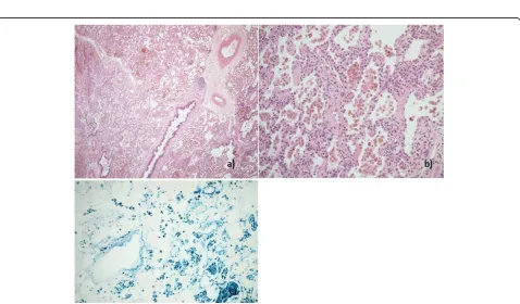
Fig. 2Patient 1. Histological sample, lung biopsy.aMarked accumulation of haemosiderin laden macrophages in alveolar spaces (H&E × 40).b
Mild chronic bronchiolitis with small lymphoid aggregates (H&E × 100)cFocal alveolar fibrin accumulation and mild chronic interstitial inflammation (H&E × 400)dMild chronic interstitial inflammation and reactive type 2 pneumocyte hyperplasia (H&E × 400)
autoimmune screen revealed a p-ANCA with MPO posi-tivity (4.6 u/mL). Following these results, he was started on low dose azathioprine, with ongoing hydroxycloro-quine and azithromycin, with current symptomatic and functional stability.
Discussion and conclusions
Here we present three cases of chronic DAH initially diag-nosed as IPH that subsequently developed ANCA-MPO positivity, and, on HRCT, progressive development of cys-tic areas in keeping with emphysema, all with a striking perivascular distribution. We believe these three cases are of interest in highlighting the 1) possibility of development of ANCA +/−renal involvement several years down the line from first IPH presentation; 2) the possibility that DAH changes may develop into emphysema.
Development of ANCA MPO antibodies
The development of specific antibodies +/−other typical autoimmune extrapulmonary manifestations after the initial pulmonary presentation have been described in a number of scenarios [3–6]. Specifically, to the develop-ment of ANCA MPO positivity, longitudinal assessdevelop-ments have revealed new onset of MPO+ or PR3+ and/or overt
vasculitis on follow up in patients initially presenting as idiopathic interstitial pneumonias [3].
However, there are only a few reports detailing subse-quent autoimmune serology (other than ANCA) conver-sions following an initial diagnosis of IPH [7, 8]. Le Clainche et al. [4] report on 15 children diagnosed with IPH, of which only one developed ANCA on follow up (specificity not reported). However, as the children were not screened for ANCA at presentation, it is impossible to conclude whether this was a real seroconversion.
ANCA MPO seroconversion occurring in adulthood in IPH after many years has been described, to our knowledge, only in another case [5]. However, the patient presented by Freitas et al. was a 21 year old girl diagnosed with IPH at the age of 4, who developed ANCA positivity 12 years after presentation, aged 16, and had a good response to immuno-suppression. Furthermore, no emphysematous changes on follow up were mentioned. By contrast, our cases developed ANCA positivity in adulthood, and all continue to require ongoing immunosuppressive treatment to control the DAH, suggesting the possible role played by autoimmune phe-nomena in this rare disease, even when initially deemed
“idiopathic”. In support of this, the second case had positive anti-transglutaminase IgA antibodies and autoimmune thy-roiditis and the third case had autoimmune thythy-roiditis.
Development of emphysema
During follow up, our cases developed cystic opacities consistent with emphysema, not seen at presentation. In the first patient, limited emphysema was first seen 10 years after presentation. The cystic areas became more prominent over the following 6 years, with concurrent isolated reduction in DLCO. The second patient was first noted to have developed emphysematous changes on HRCT 8 years after the initial presentation, and 20 years after having completely stopped smoking, with further significant worsening of radiological emphysema after another 2 years. In parallel, there was marked wors-ening in gas transfer with persistently normal lung volumes. The third patient was diagnosed with IPH in early childhood and was first noted to have emphysema on HRCT 4 years after the diagnosis, aged 11, with sub-sequent progression despite the lack of active or passive smoking exposure.
The development of emphysema in the context of interstitial processes even outside of a significant history
of smoking is being increasingly recognized. In a cohort of never-smoker patients with rheumatoid arthritis related ILD, 27% had associated emphysema with obstructive functional indices [6], while in systemic sclerosis related ILD patients with concomitant emphysema, the preva-lence of never smokers was 33% [9]. In a cohort of 233 hypersensitivity pneumonia (HP) patients evaluated for the presence of emphysema and PPFE, 23% of patients with emphysema were never-smokers [7].
In AAV, development of emphysema has been de-scribed in a few retrospective case series and case re-ports [8, 10–12]. In a cohort of AAV patients with 79 granulomatosis with polyangiitis (GPA) cases and 61 microscopic polyangiitis (MPA) cases, global emphyse-matous changes incidence was 13% with no significant differences between the two groups [12]. In the largest published series so far, Yagamata et al. reported em-physematous lesions in 37% of patients in a cohort of MPA patients, more frequently characterized by low attenuation areas rather than cystic lesions, although
Fig. 4Patient 2. Histological sample, lung biopsy.aMarked accumulation of haemosiderin laden macrophages in alveolar spaces (H&E × 40).b
Haemosiderin laden alveolar macrophages and focal perivascular lymphoid infiltrates (H&E × 200).cIron encrustation of elastin fibres of small artery (H&E × 200)dSmall perivascular non-necrotising granuloma (H&E × 200)
Fig. 6Patient 3. Histological sample, lung biopsyaSurgical lung biopsy showing marked accumulation of pigmented alveolar macrophages with scattered small lymphoid aggregates (H&E × 100).bAlveolar macrophages with coarse golden-brown cytoplasmic pigment. There is reactive type 2 hyperplasia and mild expansion of alveolar walls with scattered small lymphocytes and minimal fibrosis, but no features of capillaritis or vasculitis are seen (H&E × 200).cThe cytoplasmic pigment is positive with Perls Prussian Blue stain confirming it to be haemosiderin. Note also iron encrustation of elastin fibres in small vessels (Perls Prussian Blue stain × 200)
the Authors did not link the emphysematous lesions with alveolar haemorrhage per se [13].
The pathogenesis of emphysema on a background of DAH and ANCA positivity is unclear. One possibility is that the emphysema seen in our three cases could be secondary to the inflammatory process present within the capillaries/small vessels leading to the destruction of the alveolar walls though the release of noxious insults including proteolytic enzymes and free oxygen radicals. The observation of a striking peribronchovascular distri-bution to the emphysema supports this hypothesis. This has been suggested in the context of capillaritis [14] and in hypocomplementemic urticarial vasculitis, a rare im-mune complex related small vessel vasculitis, character-ized by low C1q and C4 complement levels. Serial histopathological analysis of one case described by Hunt et al. [15] showed that the initial changes of capillaritis evolved into emphysema, and obstructive airways dis-ease. Several other case reports have presented this asso-ciation in the context of hypocomplementemic urticarial vasculitis [16–22].
It is also possible that the ANCA antibodies themselves have pathogenic activity. Neutrophils can be activated by MPO-ANCA causing the release of MPO, a cationic pro-tein expressed by several immune cells including neutro-phils and macrophages, resulting in an inflammatory response [23]. In an animal model of smoke-induced em-physema, a myeloperoxidase inhibitor prevented progres-sion of the emphysematous changes [24] Further support for a role played by autoantibodies and specifically ANCA, comes from the finding of significantly increased propor-tion of combined pulmonary fibrosis an emphysema (CPFE) among patients with ANA positivity and a higher MPO-ANCA positivity compared to idiopathic pulmonary fibrosis (IPF) patients without emphysema. Furthermore, CPFE patients with ANA and/or ANCA positivity showed an increased number of CD20+ cells forming lymphoid follicles within the fibrotic interstitium in areas adjacent to fibroblastic foci [25].
In the first two cases, we cannot completely exclude that the development of cystic changes and/or emphy-sema was related to the previous history of smoking, and certainly cigarette smoking may have contributed to its development. However, the first case had a truly trivial smoking history (one pack year), and stopped by age 21, when her HRCT showed no cystic/emphysema-tous lesions. The second patient had a 5-pack year his-tory but he stopped and had been a non-smoker for 10 years by the time he had his initial HRCT, which did not reveal any emphysematous destruction. Further-more, the third patient developed extensive emphysema at a very young age with no exposure to cigarette smoke, supporting a direct link between DAH, ANCA positivity and emphysema.
Further studies are required to better understand the relationship between DAH, ANCA positivity and devel-opment of emphysema. ANCA positivity seems to be retrospectively associated with a worse outcome in chil-dren with IPH [26]. Also, the development of emphysema in the contest of CTDs and HP has been associated to a worse prognosis [6,7,9].
IPH is a rare disease and little is known about the mechanisms involved in its development. However, its known association with coeliac disease [27], its response to immunosuppressive therapy and its possible associ-ation with autoimmune features [4,28,29] suggests that a contribution of immunological overactivity, even if a clear cut autoimmune disease is not identified.
Immunosuppressive treatment should always be consid-ered in these patients even in the context of an idiopathic onset. Patients with IPH should have ANCA screening not only at presentation but also during follow up, espe-cially when the response to therapy is poor or in presence of suspected renal involvement. However, it is worth not-ing that ANCA positivity is not always identified in AAV, such that the presence of ANCA is not essential to con-firm the diagnosis or to start immunosuppressive therapy, if needed [30]. Full lung function tests including DLCO should be monitored frequently even in case of stable spi-rometric values, as subtle worsening due to development of emphysematous changes could develop even after many years. This could aid in identifying the cases which could benefit from a more aggressive therapy and perhaps pre-vent the development of complications such as emphyse-matous changes.
Abbreviations
ANA:Anti-nuclear antibodies; ANCA: Anti-neutrophil cytoplasmic antibody; anti-ds DNA: Anti-double-stranded DNA; anti-GBM : Anti–glomerular basement membrane; BAL: Bronchoalveolar lavage; CPFE: Combined pulmonary fibrosis and emphysema; CT: Computed tomography; CTD: Connective tissue disease; DAH: Diffuse alveolar haemorrhage; ECG: Electocardiogram; eGFR: Estimated glomerular filtration rate; ENA: Extractable nuclear antigens; FEV1: Forced expiratory volume in 1 s; FVC: Forced vital capacity; GPA: Granulomatosis with polyangiitis; HP: Hypersensitivity pneumonitis; HRCT: High resolution computed tomography; ILD: Interstitial lung disease; IPF: Idiopathic pulmonary fibrosis; IPH: Idiopathic pulmonary hemosiderosis; KCO: Transfer coefficient; MPA: Microscopic polyangiitis; MPO: Anti-myeloperoxidase antibodies; MRC: Medical Research Council; PPFE: Pleuroparenchymal fibroelastosis; PR3: Anti-proteinase 3 antibodies; RBH: Royal Brompton Hospital; RF: Rheumatoid factor; TLC: Total lung capacity; TLCO: Carbon monoxide transfer factor
Acknowledgements
Not applicable
Authors’contributions
the manuscript. Every Author contributed and approved the submitted version of the manuscript.
Funding
Not applicable
Availability of data and materials
Data sharing is not applicable to this article as no datasets were generated or analysed during the current study.
Ethics approval and consent to participate
Not applicable
Consent for publication
Written informed consent for the publication of patients’information has been obtained from all three patients. All information was obtained retrospectively at a time when the patients were over the age of 18. Parents/ guardians consent wasn’t required.
Competing interests
The Authors declare that they have no competing interests.
Author details
1Interstitial Lung Disease Unit, Royal Brompton Hospital, London, UK. 2
Department of Histopathology, Royal Brompton Hospital, London, UK. 3Department of Radiology, Royal Brompton Hospital, London, UK. 4Department of Clinical Biochemistry, Royal Brompton Hospital, London, UK. 5National Heart and Lung Institute, Imperial College London, London, UK. 6
Imperial College Healthcare NHS Trust, London, UK.
Received: 14 July 2019 Accepted: 23 September 2019
References
1. Scapa J, Fishbein G, Wallace W, Fishbein M. Diffuse alveolar hemorrhage and pulmonary Vasculitides: Histopathologic findings. Semin Respir Crit Care Med. 2018;39(04):425–33.
2. Nasser M, Cottin V. Alveolar hemorrhage in Vasculitis (primary and secondary). Semin Respir Crit Care Med. 2018;39(04):482–93.
3. Alba MA, Flores-Suárez LF, Henderson AG, Xiao H, Hu P, Nachman PH, et al. Interstital lung disease in ANCA vasculitis. Autoimmun Rev. 2017;16(7):722–9. 4. Le Clainche L, Le Bourgeois M, Fauroux B, Forenza N, Dommergues JP,
Desbois JC, et al. Long-term outcome of idiopathic pulmonary hemosiderosis in children. Medicine (Baltimore). 2000;79(5):318–26. 5. Freitas A, Senra V, Marinho A, Guedes M. Chronic alveolar haemorrhage in a
paediatric patient: a diagnostic and treatment challenge. BMJ Case Rep. 2015. (published online Apr21;2015). https://doi.org/10.1136/bcr-2014206856.
6. Jacob J, Song JW, Yoon H-Y, Cross G, Barnett J, Woo WL, et al. Prevalence and effects of emphysema in never-smokers with rheumatoid arthritis interstitial lung disease. EBioMedicine. 2018;28:303–10.
7. Jacob J, Odink A, Brun AL, Macaluso C, de Lauretis A, Kokosi M, et al. Functional associations of pleuroparenchymal fibroelastosis and emphysema with hypersensitivity pneumonitis. Respir Med. 2018;138:95–101.
8. Muray Cases S, Alcázar Fajardo C, Cabezuelo Romero JB. Enfisema pulmonar severo en un paciente joven con una vasculitis asociada a anticuerpos anticitoplasma de neutrófilo tipo proteinasa-3 (ANCA-PR3). Arch Bronconeumol. 2018;54(7):397–9.
9. Champtiaux N, Cottin V, Chassagnon G, Chaigne B, Valeyre D, Nunes H, et al. Combined pulmonary fibrosis and emphysema in systemic sclerosis: A syndrome associated with heavy morbidity and mortality. Semin Arthritis Rheum. 2018; Oct [cited 2019 Mar 12]; Available from:https://linkinghub. elsevier.com/retrieve/pii/S0049017217305115.
10. Gadre S, Mehta A, Stoller J. Granulomatosis with polyangiitis and associated pulmonary emphysema: breathtaking vasculitis. Lung India. 2015;32(4):367. 11. Mouly S, Brillet G, Stern M, Lesavre P, Guillevin L. Pulmonary giant bulla in
Wegener’s granulomatosis. Scand J Rheumatol. 2000;29(5):333–5. 12. Mohammad AJ, Mortensen KH, Babar J, Smith R, Jones RB, Nakagomi D,
et al. Pulmonary involvement in Antineutrophil cytoplasmic antibodies (ANCA)-associated Vasculitis: the influence of ANCA subtype. J Rheumatol. 2017;44(10):1458–67.
13. Yamagata M, Ikeda K, Tsushima K, Iesato K, Abe M, Ito T, et al. Prevalence and responsiveness to treatment of lung Abnormalities on chest computed tomography in patients with microscopic Polyangiitis: a multicenter, longitudinal, retrospective study of one hundred fifty consecutive hospital-based Japanese patients: CT LUNG ABNORMALITIES IN MICROSCOPIC POLYANGIITIS. Arthritis Rheum. 2016;68(3):713–23.
14. Schwarz MI, Mortenson RL, Colby TV, Waldron JA, Lynch DA, Hutt MP, et al. Pulmonary Capillaritis: the association with progressive irreversible airflow limitation and hyperinflation. Am Rev Respir Dis. 1993;148(2):507–11. 15. Hunt DPJ, Weil R, Nicholson AG, Burke MM, Du Bois RM, Wells AU.
Pulmonary capillaritis and its relationship to development of emphysema in hypocomplementaemic urticarial vasculitis syndrome. Sarcoidosis Vasc Diffuse Lung Dis. 2006;23(1):70–2.
16. Jamison SC, Brierre S, Sweet J, de Boisblanc B. A case of precocious emphysema and lung Cancer in a woman with a history of Hypocomplementemic urticarial Vasculitis. Chest. 2008;133(3):787–9. 17. Wisnieski JJ, Baer AN, Christensen J, Cupps TR, Flagg DN, Jones JV, et al.
Hypocomplementemic urticarial vasculitis syndrome. Clinical and serologic findings in 18 patients. Medicine (Baltimore). 1995;74(1):24–41.
18. Schwartz HR, McDuffie FC, Black LF, Schroeter AL, Conn DL. Hypocomplementemic urticarial vasculitis: association with chronic obstructive pulmonary disease. Mayo Clin Proc. 1982;57(4):231–8. 19. Ghamra Z, Stoller JK. Basilar hyperlucency in a patient with emphysema
due to hypocomplementemic urticarial vasculitis syndrome. Respir Care. 2003;48(7):697–9.
20. Pujara AC, Mohammed T-LH. Hypocomplementemic urticarial vasculitis syndrome: a rare cause of basilar panacinar emphysema. J Thorac Imaging. 2012;27(3):W50–1.
21. Breda L, Nozzi M, Harari S, Del Torto M, Lucantoni M, Scardapane A, et al. Hypocomplementemic urticarial Vasculitis (HUVS) with precocious emphysema responsive to azathioprine. J Clin Immunol. 2013;33(5):891–5. 22. Wisnieski JJ, Emancipator SN, Korman NJ, Lass JH, Zaim TM, Mcfadden ER.
Hypocomplementemic urticarial vasculitis syndrome in identical twins. Arthritis Rheum. 1994;37(7):1105–11.
23. Rutgers A, Heeringa P, Tervaert JW. The role of myeloperoxidase in the pathogenesis of systemic vasculitis. Clin Exp Rheumatol. 2003;21(6 Suppl 32):S55–63.
24. Churg A, Marshall CV, Sin DD, Bolton S, Zhou S, Thain K, et al. Late intervention with a myeloperoxidase inhibitor stops progression of experimental chronic obstructive pulmonary disease. Am J Respir Crit Care Med. 2012;185(1):34–43.
25. Tzouvelekis A, Zacharis G, Oikonomou A, Mikroulis D, Margaritopoulos G, Koutsopoulos A, et al. Increased incidence of autoimmune markers in patients with combined pulmonary fibrosis and emphysema. BMC Pulm Med. 2013 Dec [cited 2019 Mar 12];13(1). Available from: http:// bmcpulmmed.biomedcentral.com/articles/.https://doi.org/10.1186/ 1471-2466-13-31.
26. Blanco A, Solis P, Gomez S, Valbuena C, Telleria JJ. Antineutrophil cytoplasmic antibodies (ANCA) in idiopathic pulmonary hemosiderosis. Pediatr Allergy Immunol. 1994;5(4):235–9.
27. Nacaroglu HT, Sandal OS, Bag O, Erdem SB, Bekem Soylu O, Diniz G, et al. Association of Celiac Disease with Idiopathic Pulmonary Hemosiderosis. Lane Hamilton Syndrome Iran J Pediatr. 2015;25(5):e3312.
28. Taytard J, Nathan N, de Blic J, Fayon M, Epaud R, Deschildre A, et al. New insights into pediatric idiopathic pulmonary hemosiderosis: the French RespiRare® cohort. Orphanet J Rare Dis. 2013;8(1):161.
29. Zhang X, Wang L, Lu A, Zhang M. Clinical study of 28 cases of paediatric idiopathic pulmonary haemosiderosis. J Trop Pediatr. 2010;56(6):386–90. 30. Jennette JC, Falk RJ, Bacon PA, Basu N, Cid MC, Ferrario F, et al. 2012 revised
international Chapel Hill consensus conference nomenclature of Vasculitides. Arthritis Rheum. 2013;65(1):1–11.
Publisher’s Note