S H O R T R E P O R T
Open Access
Very low prevalence of epidermal growth factor
receptor (EGFR) protein expression and gene
amplification in Saudi breast cancer patients
Mohamed A Shawarby
*, Dalal M Al-Tamimi and Ayesha Ahmed
Abstract
Background:Breast cancers which demonstrate EGFR protein expression, gene amplification and/or gene mutations may benefit therapeutically from tyrosine kinase inhibitors. In Western studies, EGFR protein expression has been demonstrated in 7-36% of breast cancer patients, while gene amplification has been found in around 6% of cases and mutations were either absent or extremely rare. Studies addressing EGFR protein expression and gene amplification in Saudi breast cancer patients are extremely scanty and the results reported have been mostly non-conclusive. Herein we report the prevalence of EGFR protein expression and gene amplification in a cohort of Saudi breast cancer patients.
Findings:We noticed a remarkably low incidence of EGFR protein expression (1.3%) while analyzing the spectrum of molecular subtypes of breast cancer in a Saudi population by immunohistochemistry. Also,EGFRgene
amplification could not be demonstrated in any of 231 cases studied using silver enhancedin situ hybridization. Conclusions:The extremely low incidence of EGFR protein expression and gene amplification in Saudi breast cancer patients as compared to Western populations is most probably ethnically related as supported by our previous finding in the same cohort of a spectrum of molecular breast cancer types that is unique to the Saudi population and in stark contrast with Western and other regionally based studies. Further support to this view is provided by earlier studies from Saudi Arabia that have similarly shown variability in molecular breast cancer subtype distribution between Saudi and Caucasian populations as well as a predominance of the high-grade pathway in breast cancer development in Middle East women. More studies on EGFR in breast cancer are needed from different regions of Saudi Arabia before our assumption can be confirmed, however.
Virtual Slides:The virtual slide(s) for this article can be found here: http://www.diagnosticpathology.diagnomx.eu/ vs/1171467253537062.
Findings
Background and research hypothesis
EGFR is a tyrosine kinase receptor in the HER family which is widely expressed in a number of epithelial tumors and is believed to play a key role in cell prolif-eration. It is now well established that non-small-cell lung cancers which demonstrate EGFR protein expres-sion, gene amplification and/or gene mutations at exons 18 - 21 show a dramatic therapeutic response to tyro-sine kinase inhibitors such as gefitinib and erlotinib
[1-3]. Although the same may be true for other cancers including breast cancer, data regarding the presence or absence of EGFR abnormalities in tumors other than lung cancer and the response of such tumors to anti EGFR therapy are still limited and rather conflicting. EGFR protein expression as assessed by immunohisto-chemistry has been demonstrated in 7-36% of breast cancer patients, while gene amplification as assessed by CISH or FISH has been found in around 6% of cases [4-8]. Mutations in exons 18 - 21 of the EGFR gene investigated by PCR were either absent [1,7] or present in only rare breast cancer patients [9], such mutations being much frequent in lung cancer [10]. Differences in the prevalence of EGFR over-expression reported by * Correspondence: melshawarby46@hotmail.com
Department of Pathology, College of Medicine, University of Dammam, Dammam, Saudi Arabia
variability [7].
In a recent study that analyzed the spectrum of molecu-lar subtypes of breast cancer in a Saudi population [11], we noticed (but have not reported) a remarkably low inci-dence of EGFR protein expression in our patients. Also,
EGFRgene amplification could not be demonstrated in any of 231 cases studied using silver enhanced in situ hybridization (assessed after the study was published). In this article we aim to explore whether this extremely low incidence of protein expression and gene amplification reflects a truly low prevalence ofEGFRgene abnormalities in the Saudi population which may be ethnically related or is, alternatively, due to possible suboptimal sensitivity of the immunohistochemistry technique/antibodies or thein
situhybridization method used. Patients, methods and results
We have recently published a study that analyzed the spectrum of molecular subtypes of breast cancer in 231 Saudi patients [11]. The age of the patients ranged between 25 and 97 years with a mean of 49.5 years (SD ± 11). Representative cancerous tissues obtained from paraffin blocks of mastectomy and lumpectomy speci-mens were incorporated into 5 tissue microarray recep-tion blocks, from which 4 micron thick secrecep-tions were cut for immunohistochemical and in situ hybridization studies. For tru-cut biopsies, conventional paraffin blocks were utilized. The cases were randomly selected from the archives of our pathology department based on the availability of representative blocks and sufficient tis-sue material to perform the required procedures. An immunohistochemical panel including ER, PR, HER2, Ck5/6 and EGFR antibodies was used as a surrogate for gene expression profiling to classify the 231 breast can-cer specimens. Moreover, each class was correlated with its Ki-67 proliferation index and p53 gene over-expres-sion, as revealed by IHC, and also with the histologic type and grade of the tumor. The histopathological and molecular charcteristics of breast cancer in these patients are shown in table 1.
valence of EGFR protein expression among the studied cohort. A revisit to the study revealed that only three out of 231 cases were positive for EGFR (1.3%). Positiv-ity was defined as membrane staining (Figure 1A) and was scored according to the criteria originally developed for HER2/neu into 0, 1+, 2+ and 3+ [7]. Only 2+ and 3 + membrane staining of 10% or more of the tumor cells was considered positive. Cytoplasmic staining alone was interpreted as negative. We used a primary antibody manufactured by Dako (clone H11 at a dilution of 1:200). The staining was performed in a Ventana Bench-mark automated immunostainer according to the manu-facturer’s instructions (Ventana Medical Systems Inc., Tucson, Arizona). All three EGFR positive cases were negative for ER, PR and HER2 and two were also posi-tive for CK5/6. We classified the three cases as“basal” based on Ck5/6 and/or EGFR positivity coupled with ER, PR and HER2 negativity. Table 2 shows the immu-nohistochemical findings in the EGFR positive cases including the Ki67 proliferation index which was high (70-100%). The patients were aged 35, 61 and 78 years. All had a high grade (grade III) invasive carcinoma but only one had an advanced (stage IV) disease (table 3). Although the number of the EGFR positive breast can-cer cases is too small to allow for any correlation with clinical, pathologic or molecular variables, the presence of two“metaplastic” carcinomas out of three EGFR posi-tive cases is in keeping with what has already been reported in the literature that approximately 70-80% of metaplastic breast carcinomas overexpress EGFR [12]. On the other hand, EGFR gene amplification - assessed after the study was published using the newly intro-duced silver enhanced in situhybridization“SISH” tech-nique (Ventana Medical Systems Inc., Tucson, Arizona) - could not be demonstrated in any of the 231 cases. The SISH detection kit utuilizes an enzyme labeled anti-body that blocks the bound primary antianti-body. The com-plex is then visualized by silver acetate chromagen which produces a black precipitate. During the ISH pro-cess, labeled probes are bound to specific DNA or RNA
Table 1 Histolopathogical and molecular characteristics of cancer in a cohort of 231 Saudi breast cancer patients
Histologic type LUMA No.(%) LUMB No(%) HER2 No(%) Basal No(%) Hybrid No(%) UC N0(%) Total No(%)
IDC 6 (3.3) 27 (14.8) 29 (15.8) 20 (10.9) 16 (8.7) 85 (46.4) 183 (79.2)
ILC 3 (33.3) 5 (55.6) 0 (0) 0 (0) 0 (0) 1 (11.1) 9 (3.89)
ISC 0 (0) 3 (20.) 7 (46.7) 0 (0) 3 (20) 2 (13.3) 15 (6.49)
other* 0 (0) 2 (8.3) 4 (16.7) 3 (12.5) 4 (16.7) 11(45.8) 24 (10.38)
IDC = Invasive ductal carcinoma-NOS, ILC = Invasive lobular carcinoma, ISC = In situ carcinoma, LUMA = luminal A, LUMB = luminal B, HER2 = human epidermal growth factor receptor 2, UC = unclassified
target sequences in cells or tissues. Visualization of the bound linker antibody is accomplished through enzyme catalyzed deposition of silver. Silver ions are reduced by hydroquinone to metallic silver ions. The substrate for the enzyme catalyzed deposition of silver is hydrogen peroxide. The test was performed on 4 micron thick paraffin sections prepared from TMA and conventional blocks in a Ventana Benchmark IHC/ISH instrument (Ventana Medical Systems Inc., Tucson, Arizona) using an EGFR DNA probe (Ventana Medical Systems Inc., Tucson, Arizona) according to the manufacturer’s instructions. The results were evaluated by light micro-scopy under a 40× objective. The SISH signals (black) were counted in at least 20 nuclei (Figure 1B). Gene amplification was defined as copy number greater than 5/nucleus.
Comment, conclusions and recommendation
The remarkably lower incidence of protein expression and gene amplification in our breast cancer cases as compared to that reported in Western studies (table 4) may reflect a truly low prevalence of EGFR gene abnormalities in the Saudi population which may be ethnically related. Alternatively, it may be due to possi-ble suboptimal sensitivity of the immunohistochemistry technique/antibodies or the in situ hybridization
method used. However, a much greater likelihood of some ethnic variation in EGFRgene abnormalities in breast cancer is supported by our previous finding in the same cohort of a spectrum of molecular breast cancer types that is unique to our population with luminal tumors comprising 19.9% and unclassified (penta negative) tumors 42.9% [11]. This distribution is in stark contrast with Western and other regionally based studies that have reported a prevalence of 44.5 -80.2% for luminal cases [13-18] and 4.87 - 15.9% for the unclassified category [13-17]. Further support to this view is provided by earlier studies from Saudi Ara-bia that have similarly shown variability in molecular breast cancer subtype distribution between Saudi and Caucasian populations [19-21] as well as a predomi-nance of the high-grade pathway in breast cancer development in Middle East women [19]. There are also other features that distinguish breast cancer in Saudi women from what is seen in Western popula-tions. Breast cancers in Saudi women are generally locally advanced at the time of diagnosis, and affect predominantly females between 46-50 years of age, which is noticeably different from the median of 60-65 years seen in industrialized Western nations [22,23], where locally advanced disease is much less common.
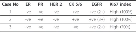
A better approach to verify our assumption, however, would be to attempt confirming an extremely low preva-lence ofEGFRgene amplification in Saudi patients using PCR which is a more sensitive method than all in situ hybridization techniques. Moreover, by PCR, we can explore mutations at various exons of theEGFR gene, which may not necessarily be reflected as gene amplifi-cation or protein expression but are still effective in determining prognosis and response to anti EGFR Table 2 Immunohistochemical findings and Ki67 index in
EGFR positive breast cancer cases
Case No ER PR HER 2 CK 5/6 EGFR Ki67 index
1 -ve -ve -ve +ve +ve (2+) High (100%)
2 -ve -ve -ve +ve +ve (3+) High (100%)
3 -ve -ve -ve -ve +ve (2+) High (70%)
A
B
therapy. Studies addressing EGFR protein expression and gene amplification in Saudi breast cancer patients are extremely scanty and the results reported have been mostly non-conclusive [19]. More studies in this direc-tion are encouraged from different regions of Saudi Arabia.
List of abbreviations
CK: Cytokeratin; CISH: Chromagenicin situhybridization; EGFR: Epidermal growth factor receptor; ER: Estrogen receptor; FISH: Fluorescentin situ hybridization; HER: Human epidermal growth factor receptor; IHC: Immunohistochemistry; ISH:In situhybridization; PCR: Polymerase chain reaction; PR: Progesterone receptor; SISH: Silver enhancedin situ hybridization;
Acknowledgements
We acknowledge the services of Mrs Khalda Al Johy, Mr Shakir Ahmad and Mrs Maria Rosario Lazaro for conducting the laboratory work.
Authors’contributions
MAS and DMT were responsible for interpretation of IHC and SISH results. MS, DT and AA were involved in the writing up of the manuscript. All authors have read and approved the final manuscript.
Competing interests
The authors declare that they have no competing interests.
Received: 8 April 2011 Accepted: 24 June 2011 Published: 24 June 2011
References
1. Lynch TJ, Bell DW, Sordella R,et al:Activating mutations in the epidermal growth factor receptor underlying responsiveness of non-small-cell lung cancer to gefitinib.N Engl J Med2004,350:2129-2139.
2. Paez JG, Janne PA, Lee JC,et al:EGFR mutations in lung cancer: correlation with clinical response to gefitinib therapy.Science2004, 304:1497-1500.
3. Pao W, Miller V, Zakowski M,et al:EGF receptor gene mutations are common in lung cancers from‘never smokers’and are associated with sensitivity of tumors to gefitinib and erlotinib.Proc Natl Acad Sci USA 2004,101:13306-13311.
4. Harris AL, Nicholson S, Sainsbury JR,et al:Epidermal growth factor receptors in breast cancer: association with early relapse and death, poor response to hormones and interactions with neu.J Steroid Biochem 1989,34:123-131.
5. Tsutsui S, Kataoka A, Ohno S,et al:Prognostic and predictive value of epidermal growth factor receptor in recurrent breast cancer.Clin Cancer Res2002,8:3454-3460.
6. Walker RA, Dearing SJ:Expression of epidermal growth factor receptor mRNA and protein in primary breast carcinomas.Breast Cancer Res Treat 1999,53:167-176.
7. Bhargava R, Gerald WL, Li AR,et al:EGFR gene amplification in breast cancer: correlation with epidermal growth factor mRNA and protein expression and Her-2 status and absence of EGFR-activating mutations.
Modern Pathology2005,18:1027-1033.
8. Press MF, Finn RS, Di Leo A,et al:Correlation of HER2 gene amplification, HER2 and EGFR expression (protein and mRNA) with lapatinib efficacy in women with metastatic breast cancer.J Clin Oncol2008,26, (May 20 suppl; abstr 1007).
9. Generali D, Leek R, Fox SB,et al:EGFR mutations in exons 18-21 in sporadic breast cancer.Ann Oncol2007,18:203-205.
10. Liang Z, Zhang J, Zeng X,et al:Relationship between EGFR expression, copy number and mutation in lung adenocarcinomas.BMC cancer2010, 10:376.
11. Al Tamimi DM, Shawarby MA, Ahmed A,et al:Protein expression profile and prevalence pattern of the molecular classes of breast cancer - a Saudi population based study.BMC Cancer2010,10:223.
12. Reis-Filho JS, Milanezi F, Carvalho S,et al:Metaplastic breast carcinomas exhibit EGFR, but not HER2, gene amplification and overexpression: immunohistochemical and chromagenicin situhybridization analysis.
Breast Cancer Research2005,7:R1028-R1035.
13. Kim MJ, Ro JY, Ahn SH,et al:Clinicopathologic significance of the basal-like subtype of breast cancer: a comparison with hormone receptor and HER2/neu-overexpressing phenotypes.Hum Pathol2006,37:1217-1226. 14. Carey LA, Perou CM, Livasy CA,et al:Race, breast cancer subtypes, and survival in the Carolina Breast Cancer Study.JAMA2006,295:2492-2502. 15. Cheang MCU, Chia SK, Voduc D,et al:Ki67 Index, HER2 Status, and
Prognosis of Patients With Luminal B Breast Cancer.J Natl Cancer Inst 2009,101:736-750.
16. Bhargava R, Striebel J, Beriwal S,et al:Prevalence, Morphologic Features and Proliferation Indices of Breast Carcinoma Molecular Classes Using Immunohistochemical Surrogate Markers.Int J clin Exp Pathol2009, 2:444-455.
17. Tamimi RM, Baer HJ, Marotti J,et al:Comparison of molecular phenotypes of ductal carcinoma in situ and invasive breast cancer.Breast Cancer Research2008,10:R67.
18. Adebamowo CA, Famooto A, Ogundiran TO,et al:Immunohistochemical and molecular subtypes of breast cancer in Nigeria.Breast cancer research and treatment2007,110:183-188.
19. Al-Kuraya K, Schraml P, Sheikh S,et al:Predominance of high-grade pathway in breast cancer development of Middle East women.Modern Pathology2005,18:891-897.
20. Bin Amer SN, Magbool Z, Nirmal MS,et al:Gene expression profiling in women with breast cancer in a Saudi population.Saudi Med J2008, 29:507-513.
21. Al-Tamimi DM, Bernard PS, Shawarby MA,et al:Distribution of molecular breast cancer subtypes in Middle Eastern-Saudi Arabian women: A pilot study.Ultrastructural Pathology2009,33:141-150.
22. Ibrahim EM, al-Mulhim FA, al-Amri A, al-Muhanna FA, Ezzat AA, Stuart RK, Ajarim D:Breast cancer in the Eastern province of Saudi Arabia.Med Oncol1998,15(4):241-247.
Table 4 Prevalence of EGFR protein expression and gene amplification in present study compared to Western studies
EGFR protein expression %
EGFRgene amplification %
Present study 1.3 0
Harris et al [4] 16 Not done
Tsutsui et al [5] 36 Not done
Walker & Dearing [6] 36 Not done
Bhargava et al [7] 7 6
Press et al [8] 27.9 Not done
2 Invasive ductal carcinoma, NOS 61 III pT2N0M0 (IIA)
23. Ezzat AA, Ibrahim EM, Raja MA, Al-Sobhi S, Rostom A, Stuart RK:Locally advanced breast cancer in Saudi Arabia: high frequency of stage III in a young population.Med Oncology1999,16(2):95-103.
doi:10.1186/1746-1596-6-57
Cite this article as:Shawarbyet al.:Very low prevalence of epidermal growth factor receptor (EGFR) protein expression and gene
amplification in Saudi breast cancer patients.Diagnostic Pathology2011
6:57.
Submit your next manuscript to BioMed Central and take full advantage of:
• Convenient online submission
• Thorough peer review
• No space constraints or color figure charges
• Immediate publication on acceptance
• Inclusion in PubMed, CAS, Scopus and Google Scholar
• Research which is freely available for redistribution