R E S E A R C H
Open Access
Pharmacological targeting of the ephrin
receptor kinase signalling by GLPG1790 in
vitro and in vivo reverts oncophenotype,
induces myogenic differentiation and
radiosensitizes embryonal
rhabdomyosarcoma cells
Francesca Megiorni
1*†, Giovanni Luca Gravina
2†, Simona Camero
1,3, Simona Ceccarelli
3, Andrea Del Fattore
4,
Vincenzo Desiderio
5, Federica Papaccio
6, Heather P. McDowell
1,7, Rajeev Shukla
8, Antonio Pizzuti
3, Filip Beirinckx
9,
Philippe Pujuguet
10, Laurent Saniere
10, Ellen Van der Aar
9, Roberto Maggio
11, Francesca De Felice
12,
Cinzia Marchese
3, Carlo Dominici
1, Vincenzo Tombolini
12, Claudio Festuccia
2†and Francesco Marampon
2*†Abstract
Background:EPH (erythropoietin-producing hepatocellular) receptors are clinically relevant targets in several malignancies. This report describes the effects of GLPG1790, a new potent pan-EPH inhibitor, in human embryonal rhabdomyosarcoma (ERMS) cell lines.
Methods:EPH-A2 and Ephrin-A1 mRNA expression was quantified by real-time PCR in 14 ERMS tumour samples and in normal skeletal muscle (NSM). GLPG1790 effects were tested in RD and TE671 cell lines, two in vitro models of ERMS, by performing flow cytometry analysis, Western blotting and immunofluorescence experiments. RNA interfering experiments were performed to assess the role of specific EPH receptors. Radiations were delivered using an x-6 MV photon linear accelerator. GLPG1790 (30 mg/kg) in vivo activity alone or in combination with irradiation (2 Gy) was determined in murine xenografts.
(Continued on next page)
* Correspondence:francesca.megiorni@uniroma1.it;f.marampon@gmail.com
Francesca Megiorni and Giovanni Luca Gravina are co-first authors. Claudio Festuccia and Francesco Marampon are co-last authors.
†Equal contributors
1
Department of Paediatrics and Infantile Neuropsychiatry,“Sapienza” University of Rome, Rome, Italy
2Department of Biotechnological and Applied Clinical Sciences, Division of
Radiation Oncology, University of L’Aquila, L’Aquila, Italy Full list of author information is available at the end of the article
(Continued from previous page)
Results:Our study showed, for the first time, a significant upregulation of EPH-A2 receptor and Ephrin-A1 ligand in ERMS primary biopsies in comparison to NSM. GLPG1790 in vitro induced G1-growth arrest as demonstrated by Rb, Cyclin A and Cyclin B1 decrease, as well as by p21 and p27 increment. GLPG1790 reduced migratory capacity and clonogenic potential of ERMS cells, prevented rhabdosphere formation and downregulated CD133, CXCR4 and Nanog stem cell markers. Drug treatment committed ERMS cells towards skeletal muscle differentiation by inducing a myogenic-like phenotype and increasing MYOD1, Myogenin and MyHC levels. Furthermore, GLPG1790 significantly radiosensitized ERMS cells by impairing the DNA double-strand break repair pathway. Silencing of both EPH-A2 and EPH-B2, two receptors preferentially targeted by GLPG1790, closely matched the effects of the EPH pharmacological inhibition. GLPG1790 and radiation combined treatments reduced tumour mass by 83% in mouse TE671 xenografts. Conclusions:Taken together, our data suggest that altered EPH signalling plays a key role in ERMS development and that its pharmacological inhibition might represent a potential therapeutic strategy to impair stemness and to rescue myogenic program in ERMS cells.
Keywords:Rhabdomyosarcoma, GLPG1790, Ephrin, Cancer stem cell, Radiation therapy, Tumour xenografts
Background
Rhabdomyosarcoma (RMS), the most common soft tissue sarcoma in childhood and adolescence, arises from undif-ferentiated mesenchymal cells with developing skeletal muscle features [1]. In this age range, RMS includes two main histological varieties, the embryonal (ERMS) and the alveolar (ARMS) subtypes, characterized by specific gen-etic alterations [2–4]. Despite refinements in dose and schedule of multimodality treatment, the 5-year overall survival of patients with high-risk RMS remains < 30%, thereby requiring the identification of novel targets for a more effective therapeutic intervention [5].
EPH (erythropoietin-producing hepatocellular) pro-teins are a large family of receptor tyrosine kinases (RTKs), comprehending EPH-A and EPH-B receptors, which respectively bind the cell/surface-associated pro-tein ligands, Ephrin-A and Ephrin-B [6–8]. EPH/Ephrin signalling represents a complex network of cell commu-nications. “Forward” signalling, corresponding to the prototypical RTK mode of signal transduction, triggers the activation of signal transduction effectors, such as Rho, Ras family GTPases and AKT/mTORC1.“Reverse” signalling transduces through the Src family kinase acti-vation [8, 9]. EPH/Ephrin network plays a key role in a wide array of developmental processes, such as cardio-vascular and skeletal development, axon guidance and tissue patterning [10]. EPH-A/Ephrin-A signalling has been shown to be essential in inducing myogenic differ-entiation of myoblasts through the suppression of the Ras/ERK1/2 cascade [11]. Due to their physiological im-portance, alterations in EPH/Ephrin cascade play a cen-tral role in the pathogenesis of several diseases [12], such as cancer [13, 14], in which EPH expression levels are frequently upregulated [13–15], with EPH-A2 and EPH-B4 being the most widely over-expressed EPH re-ceptors [13]. EPH/Ephrin role in cancer is complex by
interfering in tumour initiation, progression, neovascu-larization, invasion and metastatization [16]. Further-more, increasing evidence also indicates that EPH/ Ephrin signalling is involved in cancer stem cell self-renewal, facilitating tumour heterogeneity, metastasis and therapeutic resistance [17]. The role of EPH/Ephrin signalling in RMS cells is still largely unknown. Although a global upregulation of EPH-B receptors and Ephrin-B ligands was previously found in RMS tumours [18], the expression levels of EPH-A receptors and Ephrin-A ligand as well as the effects of EPH/Ephrin inhibition in RMS cell biology remain unclear.
Methods
Patient clinical features
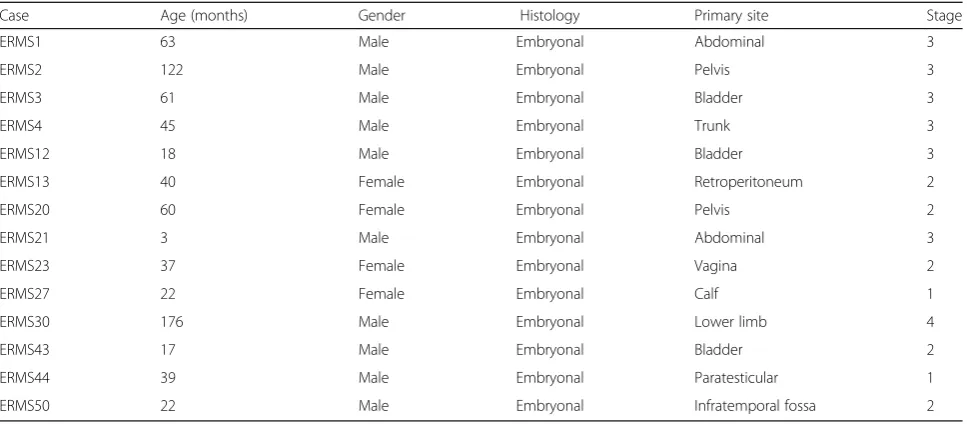
Fourteen ERMS primary tumour samples were obtained at diagnosis before any treatment from children admitted to the Department of Oncology at the Alder Hey Children’s NHS Foundation Trust, Liverpool. Histopathological diag-nosis was confirmed using immunohistochemistry. Details of the patients are reported in Table 1.
Institutional written informed consent was obtained from the patient’s parents or legal guardians. Control RNA was extracted from normal skeletal muscle (NSM) obtained from eight children undergoing surgery for non-oncological conditions. The study underwent ethical review and approval according to the local institutional guidelines (Alder Hey Children’s NHS Foundation Trust Ethics Committee, approval number 09/H1002/88).
RNA isolation and quantitative real-time PCR
Total RNA was isolated by tumour tissues ground under liquid nitrogen using 1 ml of TRIzol LS reagent (Invitro-gen, Carlsbad, CA) per 50–100 mg of sample according to the manufacturer’s protocol. RNA concentration and pur-ity were measured by NanoDrop 2000 (Thermo Fisher Scientific, Inc., Waltham, MA). Total RNA (2μg) was sub-jected to reverse transcription with High Capacity cDNA Reverse Transcription kit (Applied Biosystems, Foster City, CA) according to the manufacturer’s instructions. Quantitative real-time PCR (Q-PCR) for human EPH-A2 (Hs00171656) and Ephrin-A1 (Hs00358886) mRNAs was performed using the specific TaqMan Real Time Gene Ex-pression Assays (Applied Biosystems). Each sample was run in triplicate, in at least two independent experiments,
on a StepOne Real Time System (Applied Biosystems) machine. Transcript levels were normalized to the glyceraldehyde-3-phosphate dehydrogenase (GAPDH) mRNA, used as endogenous control. Relative quantifica-tion (RQ) of each mRNA in tumour samples in compari-son to NSM was calculated by the comparative Ct method (2-ΔΔCt), using the StepOne v2.3 software (Applied Biosys-tems). RQmax and RQmin, which are the maximum and
minimum limits of the RQ values based on the standard error of the meanΔCt values at 95% confidence interval, were used to plot error bars.
Cell lines and tumour sphere culture, pharmacological and radiation treatment
The human ERMS RD and TE671 cell lines were respectively obtained by American Type Culture Collec-tion (Manassas, VA) and Interlab Cell Line CollecCollec-tion (Genoa, Italy). Cell lines were maintained in high-glucose Dulbecco’s modified Eagle’s medium supple-mented with 10% foetal bovine serum (FBS), 1% v/v L -glutamine, 100 μg/ml streptomycin and 100 U/ml peni-cillin and grown at 37 °C in a humidified atmosphere of 5% CO2. DNA profiling using the GenePrint 10 System
(Promega Corporation, Madison, WI) was carried out to authenticate cell cultures, comparing the DNA profile of our cell cultures with those found in GenBank. Sphere-forming cells were obtained and radiation treatment performed as already described [22]. EPH receptor inhibitor GLPG1790 was provided by Galapagos NV [21], dissolved in DMSO and stored at −20 °C until use. Chemical structural data for GLPG1790 will be published shortly.
Table 1Clinicopathological features of the analysed tumour cases. Variables were categorized as follows: age at diagnosis (months), gender, histological subtype, primary site and clinical stage
Case Age (months) Gender Histology Primary site Stage
ERMS1 63 Male Embryonal Abdominal 3
ERMS2 122 Male Embryonal Pelvis 3
ERMS3 61 Male Embryonal Bladder 3
ERMS4 45 Male Embryonal Trunk 3
ERMS12 18 Male Embryonal Bladder 3
ERMS13 40 Female Embryonal Retroperitoneum 2
ERMS20 60 Female Embryonal Pelvis 2
ERMS21 3 Male Embryonal Abdominal 3
ERMS23 37 Female Embryonal Vagina 2
ERMS27 22 Female Embryonal Calf 1
ERMS30 176 Male Embryonal Lower limb 4
ERMS43 17 Male Embryonal Bladder 2
ERMS44 39 Male Embryonal Paratesticular 1
Cell viability, proliferation assay and FACS analysis Cells were seeded at a density of 105 cells/ml. After attachment, cells were treated with the indicated doses of GLPG1790 or DMSO for the indicated times. Cellular viability was measured using the trypan-blue exclusion assay (Life Technologies, Grand Island, NY) and the Countess II Automated Cell Counter (ThermoFisher Scientific, Waltham, MA). Data are expressed as average of triplicate ± standard deviation (SD). FACS analysis was performed as previously described [26]. Data were analysed using ModFit 3.1 software (BD Biosciences). Results were plotted as means ± SD of three separate experiments, having three determinations per assay for each experimental condition.
Transient transfection
RD and TE671 cells were seeded at 4 × 10⁵cells/well in 6-well plates; small interfering RNA (siRNA) against hu-man EPH-A2 (EPH-A2siRNA, sc-29304 by Santa Cruz Biotechnology, Dallas, TX) and/or EPH-A2 (EPH-B2siRNA, sc-39949 by Santa Cruz Biotechnology) or siRNA negative control (CTRsiRNA, sc-37007 by Santa Cruz Biotechnology) were combined with RNAiMAX (Invitrogen) and used at 60 nM final concentration; EPH-A2siRNAand EPH-B2siRNAare a pool of three target specific 19–25 nt siRNAs designed to specifically knock down the targeted genes. All the experiments were performed in proliferating growth medium, i.e. supple-mented with 10% FBS.
Morphological assessment
An Axio Vert.A1 microscope (Carl Zeiss Microscopy, Thornwood, NY), furnished with an AxioCam MRc5 camera (Carl Zeiss Microscopy), was used to observe the morphological changes of the cells treated with GLPG1790. Cells were photographed at 3 days post-treatment using a ×20 magnification. Images were analysed by using the ImageJ 1.47v software (http:// imagej.nih.gov/ij/).
Anchorage-dependent or anchorage-independent colony formation and wound healing assays
For anchorage-dependent colony formation assays, 4 × 10³cells were plated in 6-well plates, treated or not with GLPG1790 and cultured for 8 days. Colonies were fixed in 100% methanol, stained with 1% crystal violet, and photographed. To quantify colonies, crystal violet was dissolved in 30% acetic acid in water for 15 min at room temperature (rt) and absorbance was measured using the Biochrom Libra S22 UV/VIS spectropho-tometer (Biochrom, Berlin, Germany) at wavelength of 595 nm; 30% acetic acid in water was used as the blank. Colony formation capacity in GLPG1790 treated in comparison to untreated cells was
calculated as follow: (ODuntreated − ODGLPG1790 /
ODuntreated) × 100%, having three determinations per
experiment. For colony forming in soft agar assays, 2 × 10⁴ cells were resuspended in 4 ml of 0.33% spe-cial Noble agar (Difco, Detroit, MI) and plated (6-well plate) in growth medium-containing 0.5% soft agar. Colonies were photographed 14 days after plating.
For wound healing assays, RD and TE671 cells were plated in 6-well plates and incubated with or without GLPG1790 for 24 h. On the following day, a sterile pipette tip was used to scratch the cell monolayer (4– 5 parallel scratches/plate). Cells were washed with PBS, photographed to mark scratched tracks, and incubated for 24 additional hours to evaluate cell migration into the injured areas. Wound healing was quantified using ImageJ 1.47v software. Experiments were carried out in triplicate.
Western blot and immunofluorescence assays
experiments as previously described [28]. Experiments were replicated twice.
In vivo experiments, GLPG1790 and radiation treatment Female CD1-nu/nu mice, at 6 weeks of age, were pur-chased from Charles River (Milan, Italy) and maintained under the guidelines established by our institution (Uni-versity of L’Aquila, Medical School and Science and Technology School Board Regulations, complying with the Italian government regulation n.116 January 27, 1992 for the use of laboratory animals). Before any invasive manipulation, mice were anesthetized with a mixture of ketamine (25 mg/ml) and xylazine (5 mg/ml). For xenotransplants, exponentially growing TE671 cells were detached by trypsin-EDTA, washed twice in PBS and resuspended in saline solution at cell densities of 1 × 10⁶/200 μl. Xenotransplants were established by s.c. injecting in the leg of 45-day-old female nude CD1 mice 1 × 10⁷ tumour cells [23]. Treatments started when tumours reached a volume of 0.5–1 cm3.
GLPG1790 solution was prepared in 0.5% methylcellu-lose. The GLPG1790 dose used in the study had already been tested to be non-toxic to mice but effective in inducing inhibition of Ephrin signalling. GLPG170 was administered every day for 2 weeks and always before RT. Mice were irradiated using an Elekta 6-MV photon linear accelerator. Six fractions of 2 Gy were delivered at alternative days for a total dose of 12 Gy. A dose rate of 1.5 Gy/min was used with a source-to-surface distance (SSD) of 100 cm. Prior to irradiation, mice were anesthe-tized and were protected from off-target radiation by a 3-mm lead shield. Before tumour inoculation, mice were randomly assigned to four experimental groups. Each group was composed of 10 mice. One control group received 0.5% methylcellulose by oral gavage; one group received GLPG1790 solution at the dose of 30 mg/kg; one group received RT (six fractions of 2 Gy delivered three times/week to a total dose of 12 Gy); one group re-ceived GLPG1790 solution at the dose of 30 mg/kg and RT (six fractions of 2 Gy delivered three times/week to a total dose of 12 Gy) delivered 24 h after GLPG1790 treatment. Experiments were stopped 12 days after the last irradiation, and mice were sacrificed by carbon diox-ide inhalation. Tumours were directly frozen in liquid nitrogen for protein analysis and biochemical evaluation. The effects of different treatments on tumour growth were evaluated as follows: (1) by measuring tumour volume during and at the end of experiment (tumour volume was assessed every 4 days measurement with a Vernier calliper (length × width); the volume of the tumour was expressed in cubic millimetre according to the formulation 4/3πr3; (2) by measuring tumour weight at the end of the experiments; and (3) by determining the time to progression (TTP), tumour progression (TP)
defined as an increase of greater than 100% of tumour volume respect to the baseline. Tumour pieces were ho-mogenized by Precellys tissue homogenizer (Bertin In-struments, Montigny-le-Bretonneux, F) for grinding samples prior to protein analysis.
Statistical analysis
Data were expressed as mean ± standard deviation (SD) of each condition. Statistical significance between groups was assessed by Student’s t test, and probability (p) values of less than 0.05 were accepted as significant.
combination index (CI). If CI > 1, there are supra-additive effects and if CI < 1 infra-additive ones. Strictly additive ef-fects were observed if CI = 1. All statistical analyses were performed using the SPSS® statistical analysis software package, version 10.0.
Results
EPH-A2 and EPH-B signalling status in ERMS tumours and cell lines
EPH-A2 and EPH-B have been shown to be the EPH receptors most widely overexpressed in cancer [13].
a
b
c
Fig. 1Expression and activation status of EPH-A2, EPH-B and related Ephrin ligands in ERMS tumours and cell lines.aEPH-A2 andbEphrin-A1 transcript levels in 14 ERMS primary tumours and NSM, as measured by Q-PCR assays. GAPDH mRNA was used as an endogenous control. The relative mRNA expression levels are presented as the average fold changes (RQ) in tumour biopsies vs. NSM, set at 1. Error bars represent the RQmaxand RQminvalues of at least two independent assays, each performed in triplicate.cWestern blots showing the expression levels of
Upregulation of EPH-B receptors and Ephrin-B-related ligands has been found in RMS cells [18], whilst no data have yet been reported for EPH-A2- and Ephrin-A1-related ligand. The analysis of EPH-A2 and Ephrin-A1 transcript levels, performed in 14 ERMS primary tu-mours by using Real Time PCR, showed that both tran-scripts were significantly upregulated in all tumour samples in comparison to NSM (Fig. 1a, b). No statisti-cally significant correlations were found between EPH-A2 or Ephrin-A1 mRNA levels and gender or disease stage (EPH-A2 vs. gender: K-Tau = 0.0331, p = 0.9342, CI = −0.439 to 0.516; EPH-A2 vs. stage: r = −0.0164,
p = 0.9555, CI −0.542 to 0.519; Ephrin-A1 vs. gender: K-Tau = 0.0341, p = 0.9323, CI = −0.471 to 0.472; Ephrin-A1 vs. stage:r= 0.164,p= 0.5748, CI−0.401 to 0.639. Western blot experiments revealed that protein expression levels and/or phosphorylation status of EPH-A2, EPH-B, Ephrin-A1 (EPH-A2-related ligand) and Ephrin-B2 (EPH-B4-related ligand) were significantly increased (p< 0.01) in ERMS cell lines in comparison to NSM (Fig. 1c). Altogether, these results and the previ-ously reported data [18] indicate that EPH/Ephrin signalling is upregulated in ERMS.
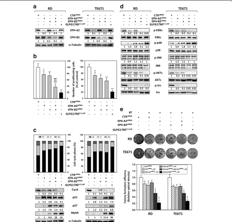
GLPG1790 affects viability of ERMS cell lines
The effects of GLPG1790, a pan-EPH receptor kinase in-hibitor [21], on RD and TE671 cell viability, were assessed by trypan blue dye exclusion test: cells were treated for 48 h with increasing doses (0–50μM) of GLPG1790. The drug significantly (p< 0.01) decreased ERMS cell viability in a dose-dependent manner, affecting on average the 50% of the cellular viability at a dose of 3.5μM on both cell lines (Fig. 2a). Immunoblotting experiments showed that in both ERMS cell lines, 3.5 μM GLPG1790 rapidly and persistently decreased the activation/phosphorylation sta-tus of EPH receptors, with a stronger action versus EPH-A2 than EPH-B (Fig. 2b). The effects of 3.5 μM GLPG1790 on ERMS proliferation rate and cell viability in adherent or non-adherent (by using Poly-Hema-coated plates) conditions were also investigated. In adherent con-ditions, GLPG1790 rapidly (1 day) and persistently (1 day to 6 days) inhibited tumour potential growth by 77.9 ± 4.9% on RD (p< 0.001) and 71.8 ± 5.1% on TE671 (p< 0.001) after 6 days of treatment (Fig. 2c, upper panel). A statistically significant increase of death by 21.8 ± 5.3 in RD (p< 0.001) and 17.3 ± 3.6 in TE671 (p< 0.001) was detected only 1 day after treatment (Fig. 2c, lower panel). In non-adherent conditions, GLPG1790 treatment drastic-ally reduced the ability of RD and TE671 to grow in sus-pension (Fig. 2d, upper panel) and resulted in almost total cell death (Fig. 2d, lower panel). Notably, GLPG1790-treated adherent cells exhibited a substantial change in their morphology, with more elongated cellular bodies already at 3 days post-exposure (Fig. 2e). Altogether, these
findings indicate that GLPG1790 has a dual action on ERMS population by inducing both growth arrest (cyto-static) and cytotoxic effects, which lead to a muscle-like differentiated phenotype.
GLPG1790 induces G1 cell cycle growth arrest and reverts ERMS transformed phenotype
Cell cycle distribution analysis, performed by flow cytometry on ERMS cells treated for 24 h with 3.5 μM GLPG1790, showed that this drug significantly reduced DNA replication (26.2% ± 2.1 RD and 22.5% ± 3.5 TE671 of S phase) by primarily arresting cells in the G1 phase (79.9% ± 4.7 RD and 86.6% ± 5.1 TE671) of the cell cycle, as reported in Fig. 3a. Consistent with the G1 arrest, GLPG1790 induced an early and persistent decrease of Cyclin A (CycA) and Cyclin B1 (CycB1) expression levels paralleled by the upregulation of p21 and p27 cell cycle in-hibitors and by downshift of the retinoblastoma tumour suppressor (Rb) (Fig. 3b), a master regulator of the G1-S transition. Unexpectedly, GLPG1790 rapidly and persist-ently upregulated the Cyclin D1 (CycD1) protein expres-sion levels whilst no modulation of Cyclin E (CycE) and c-Myc protein expression was observed (Fig. 3b). Immuno-fluorescence experiments also confirmed that GLPG1790 increases the expression of CycD1 and p21, whose distri-bution preferentially became nuclear (Fig. 3c). Then, the ef-fects of 3.5μM GLPG1970 on ERMS oncophenotype were investigated. Eight days after treatment, GLPG1790 reduced the ERMS ability to form colonies in comparison to untreated (DMSO) cells both in anchorage-dependent (78% ± 3.2 for RD and 82.4% ± 0.76 for TE671) and anchorage-independent (83.7% ± 6.1 for RD and 65.2% ± 2.1 for TE671) conditions (Fig. 4a, b, respectively). GLPG1790 also reduced ERMS cell migration as assessed by wound healing assays in which the same fields of con-fluent cells were pictured immediately after the scratch (time 0 h) and again after 24 h of GLPG1790 preincubation (Fig. 4c, left panel). Drug treatment decreased the level of wound closure to 37% for RD and 31% for TE671 of the control sample (Fig. 4c, right panel). Then, we investigated by Western blot analysis the GLPG1790 effects on the ex-pression levels of integrins, which are involved in cell adhe-sion and migration. Incubation with 3.5 μM GLPG1790 resulted in a decreased expression of integrin β1, β3 and β5 but not of integrinαV (Fig. 4d). Collectively, these re-sults suggest that the in vitro inhibition of EPH signalling is able to affect ERMS transformed phenotype by reducing cell substrate-dependent or cell substrate-independent pro-liferation as well as cell migration.
phenotype, the expression of specific skeletal muscle markers was evaluated by immunoblotting in ERMS cells treated for 72 and 144 h with GLPG1790 in com-parison to untreated control samples. As shown in
Fig. 5a, GLPG1790 induced a sustained increase of MYOD1, Myogenin and MyHC protein levels in both RD and TE671 cell lines. Notably, GLPG1790 reduced DNMT3B protein (Fig. 5a), whose expression we have
a
c
e
b
d
recently shown to restrain ERMS myogenic differenti-ation [28]. In immunofluorescence experiments, GLPG1790-treated cells displayed a myotube-like morphology with a strong fluorescence signal of MYOD1 and MyHC, this indicating that a proper myo-genic differentiation was triggered by the pan-EPH in-hibitor (Fig. 5b). The role of the EPH signalling in maintaining the ERMS stem-like population phenotype was also investigated. RD and TE671 cells were
cultured in stem cell medium (SC-medium) with or without GLPG1790. Drug treatment drastically reduced the rhabdosphere formation (Fig. 5c) as well as the per-centage of CD133 positive cells by 95.6% ± 4.2 in RD and 97.1% ± 6.1 in TE671, compared to the respective SC-DMSO samples (Fig. 5d). A significant decrement in the percentage of CXCR4 positive cells by 91.9% ± 7.7 in RD and 90.6% ± 8.2 in TE671 (Fig. 5e) as well as a downregulation of Nanog protein levels
a
c
b
(Fig. 5f ) were also evident in the GLPG1790 vs. SC-DMSO comparisons. Taken together, these results sug-gest that the inhibition of EPH signalling activates a sustained myogenic program in ERMS cells by inducing the sequential expression of myogenic genes and by concomitantly counteracting the ERMS stem-like cell phenotype.
GLPG1790 radiosensitizes ERMS cells by impairing the DNA double-strand break repair
We assessed whether GLPG1790 may sensitize ERMS cells to ionizing radiations by altering DNA damage and/ or impairing the molecular mechanisms of DSB (double-strand break) repair. For this purpose, ERMS cells were pretreated or not with GLPG1790 for 24 h and then
a
b
c
d
irradiated with a single dose of 4 Gy; after radiation treat-ment, GLPG1790 was washed out and colony formation assays were performed. As shown in Fig. 6a, GLPG1790 pretreatment significantly reduced ERMS ability to form colonies with a 85.9 ± 4.4% inhibition in RD and 84.6 ± 1.1% in TE671. Concentration of γ-H2AX, a bio-marker of DNA DSBs, and activation status and/or expres-sion levels of ATM and DNA-PKcs, which govern the DSB repair machinery, were determined in cell lysates obtained
after 2 h of a single dose of 4 Gy irradiation ± GLPG1790 (Fig. 6b). In the presence of GLPG1790 pretreatment for 24 h, γ-H2AX expression and phosphorylation levels of ATM at Serine 1981 did not increase with ionizing radia-tions in both cell lines (Fig. 6b). DNA-PKcs activation sta-tus (phosphorylation of Threonine 2609) was counteracted only in RD cells (Fig. 6b). These findings indicate that the inhibition of the EPH signalling in ERMS cells reduces H2AX accumulation and downstream DSB repair network.
a
b
c
d
e
f
Fig. 5GLPG1790 triggers myogenic differentiation and counteracts ERMS stem-like phenotype.aCell lysates from RD and TE671 cells untreated (DMSO) (−) or treated (+) with GLPG1790 for indicated times were analysed by immunoblotting with specific antibodies for indicated proteins;α-Tubulin expression shows the loading of samples. Representative of three independent experiments.bImmunofluorescence experiments showing the expression of MYOD1 and MyHC, at 72 h after GLPG1790 treatment. Images captured under ApoTome microscope at 40× magnification.cRepresentative microphotographs of RD and TE671 cells in adherent conditions (Adherent) and in stem cell (SC) medium after 15 days of incubation in the absence (SC-DMSO) or in the presence of 3.5μM GLPG1790 (SC-GLPG1790).dHistograms of percentage of CD133 positive cells determined by FACS analysis. Results represent the mean values ± SD of four independent experiments. Statistical significance: **p< 0.01, ***p< 0.001 vs. Adherent,$$$p< 0.001 vs. SC-DMSO.eHistograms of percentage of CXCR4 positive cells determined by FACS analysis. Results represent the mean value of four independent experiments ± SD. Statistical signifi-cance: **p< 0.01, ***p< 0.001 vs. Adherent,$$p< 0.01 vs. SC-DMSO.fWestern blot analysis of Nanog in protein lysates from RD and TE671 cells in adher-ent, SC-DMSO or SC-GLPG1790 cultured conditions for 15 days;α-Tubulin expression shows equal loading of samples. Representative of three
GLPG1790 effects on signal transduction in ERMS cells In order to correlate the effects of GLPG1790 on the ERMS phenotype with specific biochemical mechanisms, we investigated the activity of this compound on key sig-nal transduction pathways linked to RMS development,
muscle differentiation and EPH signalling. To this pur-pose, ERMS were treated with GLPG1790 at different times and Western blot analysis was performed. As shown in Fig. 6c, GLPG1790 treatment (i) rapidly and persistently inhibited the phosphorylation/activation of
a
c
b
ERKs, AKTs, mTOR and Src proteins, (ii) induced the rapid and persistent phosphorylation/activation of p38 in both RD and TE671 cell lines and (iii) was able to activate JNKs transiently in RD (from 3 to 12 h) and persistently in TE671 (from 3 to 72 h) cells.
Silencing EPH-A2 and/or EPH-B2 reproduces GLPG1790-induced effects in ERMS cells
To investigate if the GLPG1790-mediated effects in RD and TE671 cells were due to the suppression of EPH-A2 and/or EPH-B2 activity, we used specific small interfering RNAs (siRNAs) directed against the EPH-A2 or EPH-B2 subtypes, which share the highest biochemical selectivity profile versus GLPG1790 [21]. A sequence against theC.
elegans(CTRsiRNA) was used as a negative control. West-ern blotting analysis at 72 h after transfection revealed that EPH-A2 protein levels were specifically reduced in EPH-A2siRNA-transfected cells (Fig. 7a), whilst EPH-B2 knockdown was obtained only in EPH-B2siRNA-transfected samples (Fig. 7a). A significant reduction of both proteins was observed in EPH-A2siRNA/EPH-B2siRNA cells com-pared to those transfected with the negative control siRNA (CTRsiRNA) (Fig. 7a). GLPG1790 did not perturbate total levels of both EPH-A2 and EPH-B2 proteins (Fig. 7a). At 72 h subsequent to transfection, direct counting for liv-ing cells usliv-ing trypan blue dye exclusion test confirmed that EPH-A2, EPH-B2 and EPH-A2 + EPH-B2 depletion could significantly inhibit the proliferation potential of ERMS cells compared to CTRsiRNAcells (Fig. 7b). EPH-A2 silencing inhibited proliferation by 22% in RD and 24% in TE617 cells, EPH-B2 silencing by 24% in RD and 36% in TE671 whilst knocking down of both A2 and EPH-B2 was able to reduce cell number by 63% in RD and 44% in TE617 cells (Fig. 7b). To further determine whether the reduced ERMS cell growth was due to alterations in cell cycle progression, flow cytometry analysis was performed. Based on PI staining of cellular DNA content, EPH-A2 or EPH-B2 downregulation resulted in a significant GLPG1790-like increase of cell percentage in G1 phase with a concomitant decrease of cell percentage in S and G2 phases (Silencing EPH-A2-RD; G1 69.32 ± 1.9%, S 23.47 ± 2.4%, G2 7.2 ± 0.32%, Silencing EPH-B2-RD; G1 73.13 ± 3.6%, S 18.66 ± 1.5%, G2 8.2 ± 0.29%, Silencing EPH-A2-TE671; G1 66.54 ± 2.8%, S25.25 ± 1.5%, G2 8.2 ± 0.27%, Silencing EPH-B2-TE671; G1 67.32 ± 2.3%, S 26.45 ± 2.3%, G2 6.23 ± 0.17%), whilst CTRsiRNA -trans-fected cells rapidly divided and progressed through the cell cycle at high rates (RD G1 52.81 ± 2.5%, S 32.75 ± 2.3%, G2 13.44 ± 1.1%, TE671 G1 49.21.81 ± 1.4%, S 36.57 ± 2.4%, G2 14.22 ± 1.5%,) (Fig. 7c, upper panel). The concomitant silencing of EPH-A2 and EPH-B2 pro-duced the most prominent effects (Silencing EPH-A2 + EPH-B2-RD G1 76.0 ± 3.2%, S 16.21 ± 2.2%, G2 7.47 ± 0.31%, Silencing EPH-A2 + EPH-B2-TE671 G1
64.21 ± 7.8%, S 28.58 ± 4.5%, G2 7.12 ± 0.42%) although the percentage of cells in the different phases did not completely matched with the number obtained by 3.5μM GLPG1790 treatment (Fig. 7c, upper panel). Consistent with G1 arrest, the expression of different cell cycle regu-lators was modulated in a GLPG1790-related manner. As GLPG1790, the expression of Cyclin A was significantly downregulated by EPH-A2siRNA, EPH-B2 siRNA or EPH-A2siRNA/EPH-B2siRNAin both the cell lines (Fig. 7c, lower panel). As GLPG1790, the expression of the cell cycle in-hibitor p21 was increased by EPH-A2 and EPH-A2/EPH-B2 silencing in both cell lines, whilst EPH-EPH-A2/EPH-B2 had effect only in RD but not in TE671 cells (Fig. 7c, lower panel). The expression of the cell cycle inhibitor p27 was signifi-cantly increased by EPH-B2 and EPH-A2/EPH-B2 knock-ing down mainly in TE671 cells (Fig. 7c, lower panel). Heavy chain of sarcomeric myosin (MyHC) was increased in a GLPG1790-like manner only in the presence of EPH-A2 and EPH-B2 siRNA double transfection in both cell lines (Fig. 7c, lower panel). The effects of EPH-A2siRNA, EPH-B2 siRNA or EPH-A2siRNA/EPH-B2siRNA transfection in ERMS oncogenic signalling were also investigated (Fig. 7d). In a GLPG1790-like manner, (i) silencing of EPH-A2 and EPH-A2/EPH-B2 affected ERK phosphoryl-ation/activation in both RD and TE671 cell lines, whilst EPH-B2siRNAreduced ERK activity only in TE671(Fig. 7d); (ii) silencing of EPH-B2 and EPH-A2/EPH-B2 upregulated p38 phosphorylation/activation in both ERMS cell lines, whilst no effects on p38 phosphorylation/activation were observed by EPH-A2 silencing (Fig. 7d); (iii) phosphoryl-ation/activation status of JNKs was not affected by EPH-B2 and/or EPH-EPH-B2 knocking down; (iv) transient deple-tion of EPH-A2, EPH-B2 and EPH-A2/EPH-B2 expression was able to downregulate AKT and Src phosphorylation in both RD and TE671 cells (Fig. 7d). Concerning the pos-sible role of EPH-A2 and/or EPH-B2 in radiosensitizing ERMS cells, no significant effect was observed in EPH-A2siRNAor EPH-B2siRNAERMS cells treated with 4 Gy of RT (Fig. 7e), whilst silencing of both EPH-A2 and EPH-B2 radiosensitized ERMs cells but at a lesser extent than GLPG1790 exposure (Fig. 7e). Taken together, the cellular and molecular effects achieved by using EPH-A2 and EPH-B2 siRNA combined knockdown are comparable to GLPG1790 effects.
GLPG1790 inhibits tumour growth and radiosensitizes ERMS in xenograft mouse models
a
b
c
d
e
alternate days, for 2 weeks and for a total of six applica-tions. Tumour volumes were measured every 4 days for a period of 24 days in untreated (vehicle), GLPG1790-treated (GLPG1790), irradiated (RT) and GLPG1790/ir-radiated (GLPG1790 + RT) tumours (Fig. 8a). The rate of tumour growth was found to be markedly reduced by GLPG1790 treatment, with a 71% reduction in tumour growth being observed at the end of treatment (P< 0.001; Fig. 8a). Furthermore, GLPG1790 + RT com-bined treatment decreased growth by 83% versus RT
alone at end point (Fig. 8a). Tumour weights in mice treated with GLPG1790 decreased significantly ranging from 60 to 80% in GLPG1790(+)/RT(−) and from 80 to 90% in GLPG1790(+)/RT(+) in comparison to controls (Fig. 8b). The number of mice with tumour progression significantly differed across the groups, and this was confirmed by the median values of TTP (Fig. 8c). In the vehicle group, tumour progression occurred within 8 days after the beginning of treatment whilst in the RT group, tumour progression occurred within 12 days after
a
b
c
d
Fig. 8Effects of GLPG1790 combined or not with irradiation on in vivo tumour growth.aGrowth curve of tumour volumes from xenografted TE671 cell lines, untreated (vehicle), GLPG1790-treated (GLPG1790), irradiated (RT), GLPG1790-pretreated and irradiated (RT + GLPG1790). Tumour volumes were evaluated as describes in methods and represent the mean ± SEM of 10 mice. The upper panel shows the sequential treatments of xenografted mice started when tumours reached a volume of approximate 0.5 cm3. GLPG1790 (30 mg/kg) was administered 5 days a week for
the beginning of irradiation treatment. The treatment with GLPG1790 significantly improved the TTP com-pared to vehicle (p< 0.0001) or RT (p< 0.0001). In the GLPG1790 group, tumour progression occurred from the 16th day after the beginning of treatment and com-pleted within the day 24. The most evident improvement was documented when GLPG1790 was coupled with RT: in this group, no tumour progression occurred after the beginning of treatments. Immunoblotting on excised tumours showed that GLPG1790 treatment, combined or not with RT, downregulated EPH-A2, EPH-B, ERK and AKT phosphorylation and reduced Ki-67 expression compared with tumours from vehicle-treated mice (Fig. 8d). Thus, xenografted human TE671-derived tumours are still sensitive to GLPG1790 after 13 days of treatment (Fig. 8d).
Discussion
GLPG1790 is a selective and potent pan-inhibitor of the EPH receptors [21], which are overexpressed in many malignancies [12–16] and which are often associated with a poor clinical outcome [13] as well as with a resist-ance to chemo- [31, 32] and radio-therapy [33–35]. The present study has investigated, for the first time, the pos-sible in vitro antitumour activity of GLPG1790 in ERMS by dissecting the drug-mediated biological effects and molecular mechanisms in RD and TE671 human ERMS cell lines. GLPG1790 was able to decrease the phosphor-ylation/activation levels of EPH-A2 and EPH-B recep-tors, which are highly expressed and abnormally activated in ERMS cells lines and tumour biopsies (Fig. 1 for EPH-A2 and [18] for EPH-B). The in vitro inhibition of EPH signalling seems to be a crucial step in reverting ERMS cancer phenotype towards skeletal muscle differ-entiation, by restricting the expression of proliferative markers and by upregulating the expression of myogenic differentiation markers. Indeed, GLPG1790 treatment at low concentrations (≤3.5μM) induced a significant de-crease in cell proliferation and viability, primarily associ-ated with cell cycle arrest, this supporting a cytostatic activity of the drug. Accumulation at G1 phase occurred through several molecular mechanisms, including a sig-nificant reduction of both Cyclin A and Cyclin B1 levels, and a marked overexpression of p21 and p27 cell cycle regulators. Furthermore, GLPG1790 exposure led to the downregulation of Rb tumour suppressor, this impairing the transcriptional expression of proliferative genes. The incubation of RD and TE671 cells with GLPG1790 also promoted dramatic morphological changes, with the ap-pearance of more elongated and fused cellular bodies that was consistent with the induction of the myogenic program. In line with this observation, GLPG1790 in-duced a significant up-regulation of MYOD1 and myo-genin, followed by increased levels of MyHC protein, a
ERMS tumours. Differentiation therapy has been shown to have a significant clinical antitumour activity in acute promyelocytic leukemia, and promising preclinical activ-ity in liposarcoma and osteosarcoma [45, 46], this outlin-ing the importance to test the antitumour effects of this EPH inhibitor in xenograft models [21]. We also found that GLPG1790 exposure was able to significantly re-duce the migratory as well as the clonogenic capacity of RD and TE671 cells by altering the expression of specific proteins, including members of the integrin superfamily, this supporting the role of EPH signalling in regulating the migratory behaviour and metastatic potential of can-cer cells [6–8]. Combined siRNA knockdown of both
EPH-A2 and EPH-B2 genes, herein, shown and known [18] to be overexpressed in ERMS tumours, replicated many of the phenotypic effects observed in ERMS cells after drug exposure, confirming that GLPG1790 activity is mediated by the efficient impairment of EPH activity [21]. Finally, our data underline the role of GLPG1790 exposure in potentiating the effects of radiotherapy, which usually works by inducing DSBs as well as by inhibiting the non-homologous end joining (NHEJ) and the homologous recombination (HR) DNA repair path-ways in exposed tumour cells [47]. Indeed, GLPG1790 enhanced radiosensitivity of ERMS cell lines, as demon-strated by the clonogenic survival reduction of more
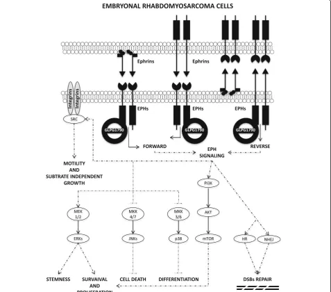
than 90%, altering the accumulation of DNA DSBs, as confirmed by the impaired expression of the phosphory-lated form of the H2AX histone [48]. Even if the rela-tionship between EPH/Ephrin signalling, DSB repair machinery and response to RT is still largely unknown; a correlation between EPH overexpression and the acqui-sition of a radioresistant phenotype has been reported in other solid tumours [34, 35, 49, 50]. Here, we showed that GLPG1790 abrogates the RT-induced ATM and DNA-PKcs phosphorylation, whose activation is linked to the HR and NHEJ pathways, respectively [47]. Aug-mentation of radiation response by GLPG1790 treatment was also confirmed by our preliminary in vivo experi-ments, in which the combination therapy with GLPG1790 and fractionated radiation was significantly more effective than the drug or the RT alone in reducing tumour masses. The current treatment for patients with RMS is a combin-ation of surgery, chemotherapy and/or radiotherapy. How-ever, the development of resistance to chemotherapy and radiotherapy is often a significant limiting factor, leading to therapeutic failures and poor survival [5]. Since EPH signalling seems to sustain the oncogenic and radioresis-tant phenotype of ERMS by regulating several molecular mechanisms [50] as schematized in Fig. 9, the combined use of GLPG1790 and RT may represent an attractive strategy to make clinical treatment of ERMS tumours more effective. Further studies in ERMS animal models will be necessary to assess if GLPG1790 has a significant activity in preventing the in vivo radioresistance. Further-more, since chemical inhibition of the DNA repair ma-chinery has been proposed as a novel strategy for cancer treatment, radiosensitizing effects conferred by GLPG1790 in ERMS cells may also open a new field of promising ap-proaches in the treatment of other cancer types that over-express EPH family members.
Conclusions
The results of this study demonstrate the preclinical in vitro and in vivo antitumour activity of GLPG1790, a new potent pan-inhibitor of EPH receptors, in human ERMS cells. In particular, GLPG1790 induces G1-growth arrest and com-mits ERMS cells towards skeletal muscle differentiation. Drug treatment prevents rhabdosphere formation and downregulates stem cell markers. GLPG1790 also radiosen-sitizes ERMS cells by impairing the DNA double-strand break repair pathway. Finally, RMS xenografts exhibited greater sensitivity to the combined GLPG1790 and radio-therapy treatment.
Abbreviations
ATM:Ataxia-telangiectasia mutated; DNA-PKcs: DNA-dependent protein kinase; EPH: Erythropoietin-producing hepatocellular; MyHC: Myosin heavy chain; MYOD1: Myoblast determination protein 1
Acknowledgements
We thank Olga Mannarino, Department of Paediatrics and Infantile Neuropsychiatry, Sapienza University of Rome, who provided useful insights and expertise.
Funding
This research did not receive any specific grant from funding agencies in the public, commercial, or not-for-profit sector. GLPG1790 was provided by Gal-apagos NV.
Authors’contributions
FM, GLG, CF and FMa conceptualized and designed the study. SC, SCe, VD, FP and FDF performed the experiments and acquired result data. GLG, CF, RS, RM, and AP analysed and interpreted the data. FM, FMa, FB, PP, LS, and EVdA drafted the manuscript. CM, CD, VT and HPM critically revised the manuscript. All authors read and approved the final version of the manuscript.
Ethics approval and consent to participant
The study on RMS patients underwent ethical review and approval according to the local institutional guidelines (Alder Hey Children’s NHS Foundation Trust Ethics Committee, approval number 09/H1002/88). Studies on animal models were performed according to the guidelines established by the University of L’Aquila, Medical School and Science and Technology School Board Regulations, in compliance with the Italian government regulation n.116 January 27, 1992 for the use of laboratory animals.
Competing interests
Filip Berinckx, Philippe Pujuguet, Laurent Saniere and Ellen Van der Aar are employers of Galapagos NV. The other authors declare that they have no competing interests.
Publisher’s Note
Springer Nature remains neutral with regard to jurisdictional claims in published maps and institutional affiliations.
Author details 1
Department of Paediatrics and Infantile Neuropsychiatry,“Sapienza” University of Rome, Rome, Italy.2Department of Biotechnological and
Applied Clinical Sciences, Division of Radiation Oncology, University of L’Aquila, L’Aquila, Italy.3Department of Experimental Medicine,“Sapienza”
University of Rome, Rome, Italy.4Multi-Factorial Disease and Complex Phenotype Research Area, Bambino Gesu Children’s Hospital, IRCCS, Rome, Italy.5Department of Experimental Medicine, Section of Biotechnology and Medical Histology and Embriology, Second University of Naples, Naples, Italy.
6
Division of Medical Oncology, Department of Clinical and Experimental Medicine and Surgery“F. Magrassi A. Lanzara”, Second University of Naples, Naples, Italy.7Department of Oncology, Alder Hey Children’s NHS Foundation Trust, Liverpool, UK.8Department of Perinatal and Paediatric
Pathology, Alder Hey Children’s NHS Foundation Trust, Liverpool, UK.
9Galapagos NV, Industriepark Mechelen Noord, General De Wittelaan L11 A3,
2880 Mechelen, Belgium.10Galapagos France, 102 avenue Gaston Roussel, 93230 Romainville, France.11Department of Biotechnological and Applied
Clinical Sciences, Division of Pharmacology, University of L’Aquila, L’Aquila, Italy.12Department of Radiological, Oncological and Pathological Sciences, “Sapienza”University of Rome, Rome, Italy.
Received: 21 June 2017 Accepted: 26 September 2017
References
1. Dagher R, Helman L. Rhabdomyosarcoma: an overview. Oncologist. 1999;4: 34–44.
2. Barr FG. Gene fusions involving PAX and FOX family members in alveolar rhabdomyosarcoma. Oncogene. 2001;20:5736–46.
3. Parham DM, Barr FG. Classification of rhabdomyosarcoma and its molecular basis. Adv Anat Pathol. 2013;20:387–97.
5. Hosoi H. Current status of treatment for pediatric rhabdomyosarcoma in the USA and Japan. Pediatr Int. 2016;58(2):81–7.
6. Pasquale EB. Eph receptor signalling casts a wide net on cell behaviour. Nat. Rev. Mol. Cell Biol. 2005;6:462–75.
7. Pasquale EB. Eph-Ephrin bidirectional signaling in physiology and disease. Cell. 2008;133:38–52.
8. Lisabeth EM, Falivelli G, Pasquale EB. Eph receptor signaling and ephrins. Cold Spring Harb Perspect Biol. 2013;5:a009159.
9. Binns KL, Taylor PP, Sicheri F, Pawson T, Holland SJ. Phosphorylation of tyrosine residues in the kinase domain and juxtamembrane region regulates the biological and catalytic activities of Eph receptors. Mol Cell Biol. 2000;20: 4791–805.
10. Palmer A, Klein R. Multiple roles of ephrins in morphogenesis, neuronal networking, and brain function. Genes Dev. 2003;17:1429–50.
11. Minami M, Koyama T, Wakayama Y, Fukuhara S, Mochizuki N. EphrinA/EphA signal facilitates insulin-like growth factor-I-induced myogenic
differentiation through suppression of the Ras/extracellular signal-regulated kinase 1/2 cascade in myoblast cell lines. Mol Biol Cell. 2011;22:3508–19. 12. Kania A, Klein R. Mechanisms of ephrin-Eph signalling in development,
physiology and disease. Nat Rev Mol Cell Biol. 2016;17:240–56. 13. Pasquale EB. Eph receptors and ephrins in cancer: bidirectional signalling
and beyond. Nat Rev Cancer. 2010;10:165–80.
14. Brantley-Sieders D, Schmidt S, Parker M, Chen J. Eph receptor tyrosine kinases in tumor and tumor microenvironment. Curr Pharm Des. 2004;10: 3431–42.
15. Arvanitis DN, Davy A. Regulation and misregulation of Eph/ephrin expression. Cell Adhes Migr. 2012;6:131–7.
16. Chen J. Regulation of tumor initiation and metastatic progression by Eph receptor tyrosine kinases. Adv Cancer Res. 2012;114:1–20.
17. Chen J, Song W, Amato K. Eph receptor tyrosine kinases in cancer stem cells. Cytokine Growth Factor Rev. 2015;26:1–6.
18. Berardi AC, Marsilio S, Rofani C, Salvucci O, Altavista P, Perla FM, et al. Up-regulation of EphB and ephrin-B expression in rhabdomyosarcoma. Anticancer Res. 2008;28:763–9.
19. Boyd AW, Bartlett PF, Lackmann M. Therapeutic targeting of EPH receptors and their ligands. Nat Rev Drug Discov. 2014;13:39–62.
20. Barquilla A, Pasquale EB. Eph receptors and ephrins: therapeutic opportunities. Annu Rev Pharmacol Toxicol. 2015;55:465–87.
21. Pujuguet P, Beirinckx F, Delachaume C, Huck J, Van der Aar E, Brys R, et al. Abstract 1753: GLPG1790: the first Ephrin (EPH) receptor tyrosine kinase inhibitor for the treatment of triple negative breast cancer. Cancer Res Am Association Cancer Res. 2014;74:1753.
22. Ciccarelli C, Vulcano F, Milazzo L, Gravina GL, Marampon F, Macioce G, et al. Key role of MEK/ERK pathway in sustaining tumorigenicity and in vitro radioresistance of embryonal rhabdomyosarcoma stem-like cell population. Mol Cancer. 2016;15:16.
23. Marampon F, Bossi G, Ciccarelli C, Di Rocco A, Sacchi A, Pestell RG, et al. MEK/ERK inhibitor U0126 affects in vitro and in vivo growth of embryonal rhabdomyosarcoma. Mol Cancer Ther. 2009;8:543–51.
24. Seki M, Nishimura R, Yoshida K, Shimamura T, Shiraishi Y, Sato Y, et al. Integrated genetic and epigenetic analysis defines novel molecular subgroups in rhabdomyosarcoma. Nat Commun. 2015;6:7557. 25. Jahangiri A, Weiss WA. It takes two to tango: dual inhibition of PI3K and
MAPK in rhabdomyosarcoma. Clin Cancer Res. 2013;19:5811–3. 26. Megiorni F, Cialfi S, McDowell HP, Felsani A, Camero S, Guffanti A, et al.
Deep sequencing the microRNA profile in rhabdomyosarcoma reveals down-regulation of miR-378 family members. BMC Cancer. 2014;14:880. 27. Marampon F, Gravina GL, Popov VM, Scarsella L, Festuccia C, La Verghetta
ME, et al. Close correlation between MEK/ERK and Aurora-B signaling pathways in sustaining tumorigenic potential and radioresistance of gynecological cancer cell lines. Int J Oncol. 2014;44:285–94.
28. Megiorni F, Camero S, Ceccarelli S, McDowell HP, Mannarino O, Marampon F, et al. DNMT3B in vitro knocking-down is able to reverse embryonal rhabdomyosarcoma cell phenotype through inhibition of proliferation and induction of myogenic differentiation. Oncotarget. 2016;7:79342–56. 29. Prewett MC, Hooper AT, Bassi R, Ellis LM, Waksal HW, Hicklin DJ. Enhanced
antitumor activity of anti-epidermal growth factor receptor monoclonal antibody IMC-C225 in combination with irinotecan (CPT-11) against human colorectal tumor xenografts. Clin Cancer Res. 2002;8:994–1003.
30. Marampon F, Gravina GL, Di Rocco A, Bonfili P, Di Staso M, Fardella C, et al. MEK/ERK inhibitor U0126 increases the radiosensitivity of
rhabdomyosarcoma cells in vitro and in vivo by downregulating growth and DNA repair signals. Mol Cancer Ther. 2011;10:159–68.
31. Alam S, Yadav V, Bajaj S, Datta A, Dutta S, Bhattacharyya M, et al. DNA damage-induced ephrin-B2 reverse signaling promotes chemoresistance and drives EMT in colorectal carcinoma harboring mutant p53. Nat Publ Gr. 2015;23:1–16.
32. Wang Y, Liu Y, Li G, Su Z, Ren S, Tan P, et al. Ephrin type-A receptor 2 regulates sensitivity to paclitaxel in nasopharyngeal carcinoma via the phosphoinositide 3-kinase/Akt signalling pathway. Mol Med Rep. 2015;11:924–30.
33. Wang L-F, Fokas E, Juricko J, You A, Rose F, Pagenstecher A, et al. Increased expression of EphA7 correlates with adverse outcome in primary and recurrent glioblastoma multiforme patients. BMC Cancer. 2008;8:79. 34. Bhatia S, Baig NA, Timofeeva O, Pasquale EB, Hirsch K, MacDonald TJ, et al.
Knockdown of EphB1 receptor decreases medulloblastoma cell growth and migration and increases cellular radiosensitization. Oncotarget. 2015;6:8929–46. 35. Charmsaz S, Beckett K, Smith FM, Bruedigam C, Moore AS, Al-Ejeh F, et al.
EphA2 is a therapy target in EphA2-positive leukemias but is not essential for normal hematopoiesis or leukemia. PLoS One. 2015;10:e0130692. 36. Stark DA, Karvas RM, Siegel AL, Cornelison DDW. Eph/ephrin interactions
modulate muscle satellite cell motility and patterning. Development. 2011; 138:5279–89.
37. Alonso-Martin S, Rochat A, Mademtzoglou D, Morais J, de Reyniès A, Auradé F, et al. Gene expression profiling of muscle stem cells identifies novel regulators of postnatal myogenesis. Front cell Dev Biol. 2016;4:58. 38. Palmqvist R, Stenling R, Oberg A, Landberg G. Expression of cyclin D1 and
retinoblastoma protein in colorectal cancer. Eur J Cancer. 1998;34:1575–81. 39. Comstock CES, Revelo MP, Buncher CR, Knudsen KE. Impact of differential
cyclin D1 expression and localisation in prostate cancer. Br J Cancer. 2007; 96:970–9.
40. Engert JC, Berglund EB, Rosenthal N. Proliferation precedes differentiation in IGF-I-stimulated myogenesis. J Cell Biol. 1996;135:431–40.
41. Petricoin EF, Espina V, Araujo RP, Midura B, Yeung C, Wan X, et al. Phosphoprotein pathway mapping: Akt/mammalian target of rapamycin activation is negatively associated with childhood rhabdomyosarcoma survival. Cancer Res. 2007;67:3431–40.
42. Marampon F, Ciccarelli C, Zani BM. Down-regulation of c-Myc following MEK/ERK inhibition halts the expression of malignant phenotype in rhabdomyosarcoma and in non muscle-derived human tumors. Mol Cancer. 2006;5:31.
43. Yeung CL, Ngo VN, Grohar PJ, Arnaldez FI, Asante A, Wan X, et al. Loss-of-function screen in rhabdomyosarcoma identifies CRKL-YES as a critical signal for tumor growth. Oncogene. 2013;32:5429–38.
44. Raimondi L, Ciarapica R, De Salvo M, Verginelli F, Gueguen M, Martini C, et al. Inhibition of Notch3 signalling induces rhabdomyosarcoma cell differentiation promoting p38 phosphorylation and p21(Cip1) expression and hampers tumour cell growth in vitro and in vivo. Cell Death Differ. 2012;19:871–81.
45. Haydon RC, Zhou L, Feng T, Breyer B, Cheng H, Jiang W, et al. Nuclear receptor agonists as potential differentiation therapy agents for human osteosarcoma. Clin Cancer Res. 2002;8:1288–94.
46. Charytonowicz E, Terry M, Coakley K, Telis L, Remotti F, Cordon-Cardo C, et al. PPARγagonists enhance ET-743–induced adipogenic differentiation in a transgenic mouse model of myxoid round cell liposarcoma. J Clin Invest. 2012;122:886–98.
47. Hein AL, Ouellete MM, Yan Y. Radiation-induced signaling pathways that promote cancer cell survival (review). Int J Oncol. 2014;45:1813–9. 48. Kuo LJ, Yang LX. Gamma-H2AX - a novel biomarker for DNA double-strand
breaks. In Vivo (Brooklyn). 2008;22:305–9.
49. Oweida A, Bhatia S, Hirsch K, Calame D, Griego A, Keysar S, et al. Ephrin-B2 overexpression predicts for poor prognosis and response to therapy in solid tumors. Mol Carcinog. 2017;56:1189–96.