Iran J Public Health, Vol. 47, No.11, Nov 2018, pp.1725-1733
Original Article
Sulfur Mustard-induced Changes in Blood Urea Nitrogen, Uric
Acid and Creatinine Levels of Civilian Victims, and Their
Correlation with Spirometric Values
Ensieh Sadat MIRSHARIF
1,
*Fatemeh HEIDARY
1, Mohammad Reza VAEZ MAHDAVI
1,
Reza GHAREBAGHI
1,2, Shahriar POURFARZAM
1, *Tooba GHAZANFARI
11. Immunoregulation Research Center, Shahed University, Tehran, Iran 2. International Virtual Ophthalmic Research Center, Tehran, Iran
*Corresponding Authors: Email: irrc@shahed.ac.ir
(Received 09 Aug 2017; accepted 17 Dec 2017)
Introduction
Exposure to Sulfur mustard (SM) as a chemical blistering weapon could induce a variety of mani-festations in mankind including dermal, ocular, pulmonary, gastrointestinal, hematological, and reproductive toxicity (1, 2). The molecular patho-genesis is the impairment of the major cellular
components such as DNA, RNA, lipids, carbohy-drates, and proteins, leading to biological conse-quences in the majority of tissues in the body (3, 4). Graphical studies of the whole body have de-tected SM in the liver and kidneys, following in-travenous and percutaneous injections (5).
Abstract
Background: The aim of this study was assessment of the chronic effects of sulfur mustard (SM) among vic-tims.
Methods: In this cohort study, 355 SM-exposed subjects from Sardasht, and 123 controls from Rabat, both
from West Azerbaijan Province, Iran were included. The spirometric evaluation and the global initiative for chronic obstructive lung disease (GOLD) classification were applied for all. Serum levels of urea, creatinine (Cr), and uric acid (UA) and glomerular filtration rate (GFR) were assessed. Data analysis was conducted using IBM SPSS.
Results: All were male, with a mean age of 43.7±10.7 and 41.6±9.9 years in case and control groups,
respec-tively. The case group had significantly higher values of Cr (P<0.001) and UA (P=0.018) than the control group. This was also the case in the Cr level (P<0.001) in subjects without pulmonary dysfunction, between both groups. There was significant difference in the GFR (P=0.047) between both groups and between sub-groups with pulmonary dysfunction in the case and control sub-groups (P=0.045), as well as between SM-exposed subjects with and without pulmonary dysfunction (P=0.009). Serum Cr, UA, sUA/Cr ratio, and BUN as well as the GFR did not have any significant correlation with forced expiratory volume in one second (FEV1), forced vital capacity (FVC), and FEV1/FVC ratio.
Conclusion: Despite significantly high levels of Cr and UA in the case group, no significant correlation was
found between serum Cr, UA, sUA/Cr ratio, BUN, and GFR with spirometric values. Further studies are re-quired to reveal the underlying molecular and clinical significance of these findings.
The kidney could be affected by reactive oxygen species (ROS) due to the profound amount of long-chain polyunsaturated fatty acids (LCPs) in renal lipids. Therefore, alkylating of subcellular components following SM exposure leads to met-abolic disruption and induces oxidative stress in the kidney. A molecular chain of this induced oxi-dative stress causes the formation of ROS, lipid peroxidation, and protein oxidation, and sets up a rise in antioxidant enzymes, such as glutathione-S-transferase, superoxide dismutase, and catalase (6). Serum concentrations of creatinine (Cr), uric acid (UA), and Blood urea nitrogen (BUN) reveal renal function. Among them, Cr is the one most com-monly used for evaluation of renal dysfunction (7). Moreover, UA is used not only for the assessment of renal function but also considered to be a marker reflecting hypoxic status (8). Alongside with application of these markers for renal func-tion, currently growing numbers of studies evalu-ating serum UA to Cr ratio (sUA/Cr ratio) as a marker in pulmonary disorders such as Chronic Obstructive Pulmonary Disease (COPD). A high-er level of this ratio was associated with abnormal-ly low spirometric values and a high level of dysp-nea. In addition, this ratio was higher in COPD patients than in healthy subjects (9, 10).
Despite extensive research for evaluating the dermal, pulmonary, and ocular complications of SM toxicity, there are few studies about its renal side effects. To the best of our knowledge, no study has been conducted yet to assess the corre-lation between renal function biomarkers with spirometric values in SM-exposed patients. Therefore, the aim of current study was to assess the chronic effects of SM in victims of this war-fare agent, in the 20 years after the first exposure.
Materials and Methods
Study Design, Participants, and Clinical Evaluation
We have explained study design in detail in our publication on the Sardasht-Iran Cohort Study (SICS) (11). We included 355 SM-exposed sub-jects from the city of Sardasht (Sardasht County,
West Azerbaijan Province, Iran) and 123 control subjects from the city of Rabat (Central District of Sardasht County, West Azerbaijan Province, Iran) in 2006, after a full explanation to all sub-jects and signing of written consent by all. An approval of this study was received from the Ministry of Health and Medical Education (Iran) and the Research Board for Ethics in Research Center for Janbazan Medical and Engineering. All subjects underwent spirometric evaluation based on the American Thoracic Society criteria (12), using spirometry (HI-801; Central Sports, Tokyo, Japan) by a senior trained nurse. The measurements were repeated three times for each case and the best one was chosen for the final analysis. Global initiative for chronic obstructive lung disease (GOLD classification) was applied to categorize the severity of pulmonary function in the subjects (13, 14).
Serum Collection and Measurement of Bi-omarkers
With use of a 21-gauge needle blood sample was taken and placed into Vacutainer blood collecting tubes (BD Biosciences). Following 20 min of centrifugation of the blood samples at 2000×g (4 °C), serum was retrieved, aliquoted, and frozen at -80 °C for future laboratory measurements of biomarkers. Serum levels of urea, Cr, and UA were assessed by suitable assay kits (Pars Azmoon-Co, Tehran, Iran). Values of urea were converted to BUN for better clinical evaluation.
Statistical Analysis
sig-nificance of this analysis were reported. Sensitivity and specificity were reported for biochemical pa-rameters. Significance level defined as P-values less than 0.05. Data analysis was performed using IBM SPSS 21 (Chicago, IL, USA).
Results
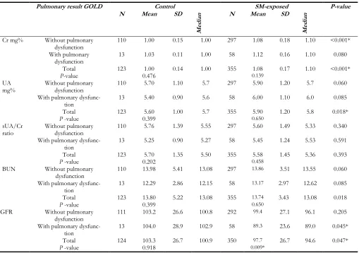
Comparison of serum levels of BUN, Cr, UA, and GFR among two groups with and with-out pulmonary dysfunction
Out of 478 study subjects, 355 were from the case group (the SM-exposed) and 123 were from the control group. All subjects were male, with a mean
age of 43.7±10.7 years in the case group and 41.6±9.9 years in the control group. The case group had significantly higher values of Cr (P<0.001), UA (P=0.018), and BUN (P=0.018) as compared to the control group. This significant difference was noted in the Cr level (P<0.001) in subjects without pulmonary dysfunction, between two groups. Subjects in the case group had an insignificant lower serum sUA/Cr ratio (P=0.393) (Table 1).
Using an independent t-test, a significant differ-ence was revealed in the GFR (P=0.047) between the study groups (Table 1).
Table 1: Comparison of the serum levels of BUN, Cr, UA and GFR in study groups with and without pulmonary
dysfunction; Using GOLD stage for disease severity
Pulmonary result GOLD Control SM-exposed P-value
N Mean SD
Med
ian N Mean SD
Med
ian
Cr mg% Without pulmonary
dysfunction 110 1.00 0.15 1.00 297 1.08 0.18 1.10 <0.001*
With pulmonary
dysfunction 13 1.03 0.11 1.00 58 1.12 0.16 1.10 0.080
Total 123 1.00 0.14 1.00 355 1.08 0.17 1.10 <0.001*
P-value 0.476 0.139
UA
mg% Without pulmonary dysfunction 110 5.70 1.10 5.7 297 5.90 1.20 5.7 0.060
With pulmonary
dysfunc-tion 13 5.40 0.90 5.6 58 6.00 1.10 6.0 0.085
Total 123 5.60 1.00 5.7 355 5.90 1.20 5.8 0.018*
P -value 0.399 0.650
sUA/Cr
ratio Without pulmonary dysfunction 110 5.76 1.39 5.55 297 5.60 1.49 5.33 0.340
With pulmonary
dysfunc-tion 13 5.25 0.90 5.27 58 5.45 1.24 5.53 0.591
Total 123 5.70 1.35 5.50 355 5.58 1.45 5.36 0.393
P -value 0.202 0.458
BUN Without pulmonary
dysfunction 110 13.98 5.41 13.08 297
13.86 3.51 13.55 0.060
With pulmonary dysfunc-tion
13 12.29 2.86 12.15 58 13.17 2.97 12.62 0.085
Total 123 13.80 5.22 13.08 355 13.74 3.43 13.08 0.018
P -value 0.399 0.650
GFR Without pulmonary
dysfunction 111 103.2 26.6 100.8 292
99.4 27.1 96.1 0.205
With pulmonary
dysfunc-tion 13 104.0 28.9 102.9 58
89.3 23.6 89.0 0.045*
Total 124 103.3 26.7 100.9 350 97.7 26.7 94.6 0.047*
P -value 0.918 0.009*
GOLD= Global Initiative for Chronic Obstructive Lung Disease; Cr= Creatinine; UA= Uric acid; sUA/Cr ratio= Serum Uric acid to creatinine ratio; BUN= blood urea nitrogen; GFR: glomerular filtration rate; N= number; SD= standard deviation; mg:
This was also the case between subgroups with pulmonary dysfunction in the control and case groups (P=0.045), and between case group with and without pulmonary dysfunction (P=0.009).
Correlation of serum levels of BUN, Cr, UA, and GFR with spirometry values
Serum Cr, UA, sUA/Cr ratio, BUN, and GFR did not have any statistically significant correla-tion with all measured spirometric values (Table 2). These values included FEV1 (Forced Expira-tory Volume in One Second), FVC (Forced Vital Capacity), and FEV1/FVC in both groups.
Performance of biochemical factors; sensitiv-ity and specificsensitiv-ity in detection of pulmonary dysfunction
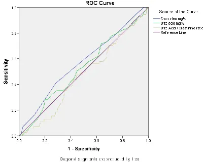
A ROC analysis was done in evaluating the per-formance of biochemical factors in the detection of pulmonary dysfunction (Fig. 1). The AUC and significance of this analysis (P-value) were 0.567 (P=0.07), 0.524 (P=0.527), and 0.469 (P=0.409) for Cr, UA and the sUA/Cr ratio respectively. The largest AUC belonged to Cr and none of them were significantly related to the presence of pulmonary dysfunction.
Table 2: Correlation between serum Cr, UA, sUA / Cr ratio, BUN and GFR with spirometry values
Variable Control SM-exposed
FVC
% FEV1 % FEV1/FVC % FVC % FEV1 % FEV1/FVC %
Cr mg% r
-0.004 0.023 0.014 0.021 0.000 -0.076
P -value 0.964 0.804 0.913 0.697 0.993 0.261
UA mg% r 0.098 0.066 0.189 -0.054 -0.091 -0.049
P -value 0.281 0.469 0.125 0.306 0.088 0.464
sUA/Cr ratio r 0.080 0.042 0.133 -0.051 -0.058 0.020
P -value 0.379 0.648 0.283 0.335 0.277 0.765
BUN r 0.098 0.066 0.189 -0.054 -0.091 -0.049
P -value 0.281 0.469 0.125 0.306 0.088 0.464
GFR r 0.015 -0.082 -0.067 0.045 0.036 0.023
P -value 0.870 0.368 0.591 0.399 0.498 0.735
Abbreviations: Cr= Creatinine; UA= Uric acid; sUA/Cr ratio= Serum Uric acid to creatinine ratio; BUN= blood urea nitrogen; GFR: glomerular filtration rate; FVC= Forced vital capacity; FEV1= forced expiratory volume in one second
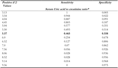
For pulmonary dysfunction based on GOLD classification, UA alone was more specific than the sUA/Cr ratio, with similar sensitivity at high-er cutoff values and more sensitivity with similar specificity at lower cutoff values (Table 3, 4). This was applicable to all study groups.
Discussion
In this study, the difference in the sUA/Cr ratio between two groups was insignificant. However, the levels of Cr, UA, and BUN were significantly higher in the case group. This significant differ-ence seen in the Cr levels of subjects without pulmonary dysfunction, between two groups.
Fig. 1: Receptor Operation Curve (ROC) curve for
Table 3: Cutoff values of Uric Acid ratio for pulmonary dysfunction (GOLD)
Positive if ≥
Values Sensitivity Specificity
Uric Acid*
3.00 1 0.002
3.55 0.972 0.022
4.05 0.93 0.052
4.55 0.873 0.118
5.05 0.803 0.236
5.35 0.718 0.364
5.55 0.592 0.435
6.05 0.423 0.6
6.55 0.282 0.779
7.05 0.127 0.86
7.55 0.042 0.939
8.05 0.028 0.961
8.60 0 0.978
9.05 0 0.988
Note: *The Area under the curve (AUC): 0.524, 95% Confidence interval (CI): 0.451-0.596
Table 4: Cutoff values of Serum Uric acid to creatinine ratio for pulmonary dysfunction (GOLD)
Positive if ≥
Values Sensitivity Specificity
Serum Uric acid to creatinine ratio*
3.13 1 0.005
3.54 0.944 0.022
4.04 0.887 0.091
4.45 0.803 0.187
5.04 0.577 0.351
5.50 0.493 0.514
5.57 0.465 0.558
6.0 0.254 0.678
6.52 0.127 0.806
7.0 0.07 0.862
7.56 0.056 0.926
8.06 0.028 0.936
8.52 0.028 0.956
9.14 0.014 0.968
9.56 0 0.975
Note: * The Area under the curve (AUC): 0.469, 95% Confidence interval (CI): 0.399-0.5398
In addition, there were significant differences in the GFR between study groups among subgroups with pulmonary dysfunction in control and SM-exposed groups, and also between the case group with and without pulmonary dysfunction.
Cr, UA, sUA/Cr ratio, and BUN did not have any significant correlation with FVC, FEV1, and FEV1/FVC in both groups.
Studies have shown both short- and long-term side effects in a variety of body organs after SM exposure (16). Similarly, our results showed significantly higher levels in renal function bi-omarkers, including Cr, UA, and BUN, as well as reduced GFR in the case group, as compared to the control group. Abundance of LCPs in the renal tissue makes this vital organ susceptible to ROS. Therefore, SM-induced oxidative stress led to an increase in the Cr level and a reduction of GFR, which by itself could impair renal function (6). GFR reduction in chronic kidney disease (CKD) frequently causes Hyperuricemia (HUA). Since two-thirds of daily UA excretion is through the renal tissue, more than 90% of HUA is caused by renal impairment. Studies revealed that induced oxidative stress following the rise in UA level may lead to glomerular and systemic hyper-tension (17).
Despite a comprehensive number of reported studies about the short-term effects of SM on various organs (18-23), few studies have been conducted on the long-term effects of exposure to this gas on organs, mainly the kidneys. The delayed biochemical complications of SM were investigated in 42 severely toxic veterans whose disability was more than 40%, 23 years after ex-posure. BUN, Cr, and UA levels did not show any significant differences between the case and control groups. However, significant reductions in serum albumin and total protein levels were found between the two groups. This reduction could be due to renal losses (16). In contrast, in current study, there were significantly higher levels of Cr, UA, and BUN in the case group than the control group. This contradiction may lie in the recruitment of their study sub-jects—who were severely injured—in contrast to our subjects. Furthermore, we found significant differences in the GFR between the study groups, parallel to higher values of serum bi-omarkers, which could justify the significantly higher levels of Cr, UA, and BUN in our study. However, the former study did not present the
GFR of their study subjects, which makes it im-practical to have further comparisons and expla-nation of differences between the findings of the two studies.
The renin-angiotensin-aldosterone system (RAAS), having various regulatory roles in the body, is closely related to renal and pulmonary functions (24). Serum uric aicd and sUA/Cr ratio were significantly higher in patients with COPD with systemic disease like hypertension and malignancy, compared to healthy controls (10). Moreover, their ROC curve analysis re-vealed the importance of the sUA/Cr ratio in prediction of COPD exacerbation, especially in higher values, as compared to the sUA level per se. In contrast, despite significantly higher levels of UA and Cr in the case group than the control group, we did not find a significant link between the sUA/Cr ratio with pulmonary dysfunction and spirometric values in both groups. Although the ROC curve analysis showed high specificity in the higher cutoff value and higher sensitivity in the lower cutoff value for the UA level as com-pared to the sUA/Cr ratio, the UA level had no diagnostic value because of the insignificance of these results.
The chronic obstructive pulmonary disease pa-tients, with a sUA/Cr ratio higher than median value, had lower FVC and FEV1 spirometric val-ues and a higher rate of dyspnea (9). The sUA/Cr ratio correlated with FVC, FEV1, and dyspnea, and also, that this ratio could be considered a valuable predictive parameter in COPD patients. In our study, despite a significantly higher level of sUA in the case group than the control group, no significant relationship has been noted with the sUA/Cr ratio in both groups. Moreover, we did not find any significant relation between spiro-metric values and serum levels of UA, Cr, and the sUA/Cr ratio. This difference could be because of the distinct underlying pathophysiology mech-anisms of SM-induced pulmonary dysfunction and COPD.
signifi-cant rise in BUN and BUN to Cr ratio was found. Tissue catabolism or starvation may lead to Cr reduction and that pre-renal azotemia caused a significant rise in BUN and BUN to Cr ratio (25). In current study, we found significantly higher levels of Cr, UA, BUN, and GFR, in con-junction with an insignificant lower sUA/Cr ra-tio, in the case group. In fact, different patho-physiological manifestations of acute NM-exposure (in the former study) and chronic SM-exposure (in the latter study) could explain the differences in blood biomarkers in these two studies. Moreover, these contrary levels were seen in inflammatory biomarkers following acute (26,27) and chronic (28-30) SM exposure.
Renal structure could be injured by inflammatory mediators or immune-mediated factors linked with primary lung pathology. Otherwise, quite the opposite, it could be kidney disease that de-termines successive pulmonary damage. Hight rate of chronic renal failure (CRF) and microal-buminuria among COPD patients has been re-ported, lately. Which may suggest a correlation between these two entities. Moreover, levels of Cr, cysteine, and brain natriuretic peptide were significantly high in COPD subjects (31).
Studies revealed a higher risk of CRF in COPD patients and acute kidney injury (AKI) following lung transplantation and a strong positive correla-tion was noted between the serum Cr level and pulmonary function (32-35). In contrast, in current study, in spite of significantly higher lev-els of UA, Cr, and BUN in the case group, no significant relation of serum Cr, UA, sUA /Cr ratio, BUN, and GFR with pulmonary dysfunc-tion and all measured spirometric values was found in both groups.
By evaluating the gender difference in correlation between pulmonary function abnormalities and eGFR using spirometric parameters, a parallel reduction was found in eGFR with a reduction in FEV1/FVC in both sexes and a reduction in FVC in males (36). Although, we found a signifi-cant difference in the GFR between the two groups and between subgroups with pulmonary dysfunction in both groups, the correlation
be-tween the GFR and spirometric parameters was insignificant.
Other studies have shown a reduction in the GFR following an increase in UA and Cr levels, which is parallel with the findings of our study (17, 37). An increase in UA level by inducing oxi-dative stress and endothelial dysfunction led to systemic and glomerular hypertension, which subsequently causes reduced renal blood perfu-sion and GFR which, decreases renal function (17). This postulated pathophysiological mecha-nism may account for changes in renal function biomarkers and reduction in the GFR of our SM-exposed subjects.
Conclusion
Despite significantly higher levels of Cr and UA, and a significant reduction in the GFR of the case group than the control group, no significant correlation was found between serum Cr, UA, sUA/Cr ratio, BUN, and GFR with spirometric values. Further studies are required to reveal the underlying molecular and clinical significance of these findings.
Ethical considerations
Ethical issues (Including plagiarism, informed consent, misconduct, data fabrication and/or fal-sification, double publication and/or submission, redundancy, etc.) have been completely observed by the authors.
Acknowledgements
Immunoregulation Research Center of Shahed University carried out this study and acknowledg-es the spiritual and financial support of Shahed University, Janbazan Medical and Engineering Research Center (JMERC), Iranian Foundation of Martyr and Veterans Affairs, and the Ministry of Health and Medical Education, Tehran, Iran.
Conflict of interest
References
1. Batal M, Boudry I, Mouret S et al (2014). DNA damage in internal organs after cutaneous ex-posure to sulphur mustard. Toxicol Appl
Phar-macol, 278(1):39-44.
2. Nadoushan MRJ, Ghazanfari T, Yaraee R et al (2009). Total serum bilirubinemia and intensi-ty of sulfur mustard exposure in Iranian chemical victims 20 years after exposure.
Tox-in Reviews, 28)1): 44-47.
3. Liu F, Jiang N, Xiao ZY et al (2016). Effects of poly (ADP-ribose) polymerase-1 (PARP-1) inhibition on sulfur mustard-induced cutane-ous injuries in vitro and in vivo. PeerJ, 4:e1890. 4. Fan LJ, Bernstein IA (1991). Effect of
bis(beta-chloroethyl)sulfide (BCES) on base mismatch repair of DNA in monkey kidney cells. Toxicol
Appl Pharmacol, 111(2):233-41.
5. Clemedson CJ, Kristoffersson H, Soerbo B, Ullberg S (1963). Whole body autoradio-graphic studies of the distribution of sulphur 35-labelled mustard gas in mice. Acta Radiol
Ther Phys Biol, 1:314-20.
6. Ozbek E (2012). Induction of oxidative stress in kidney. Int J Nephrol, 2012:465897.
7. Jayasundera S, Macnab R (2012). Laboratory tests of renal function. Anaesthesia & Intensive
Care Medicine, 13(7):328-331.
8. Kadowaki T, Hamada H, Yokoyama A et al (2007). Significance of serum uric acid in pa-tients with chronic respiratory failure treated with non-invasive positive pressure ventila-tion. Intern Med, 46(11):691-7.
9. Garcia-Pachon E, Padilla-Navas I, Shum C (2007). Serum uric acid to creatinine ratio in patients with chronic obstructive pulmonary disease. Lung, 185(1):21-4.
10. Kocak ND, Sasak G, Akturk UA et al (2016). Se-rum Uric Acid Levels and Uric Ac-id/Creatinine Ratios in Stable Chronic Ob-structive Pulmonary Disease (COPD) Pa-tients: Are These Parameters Efficient Predic-tors of Patients at Risk for Exacerbation and/or Severity of Disease? Med Sci Monit, 22:4169-4176.
11. Ghazanfari T, Faghihzadeh S, Aragizadeh H et al (2009). Sardasht-Iran cohort study of
chemi-cal warfare victims: design and methods. Arch
Iran Med, 12(1):5-14.
12. No authors listed (1995). Standardization of Spi-rometry, 1994 Update. American Thoracic Society. Am J Respir Crit Care Med, 152(3):1107-36.
13. Pauwels RA, Buist AS, Calverley PM et al (2001). Global strategy for the diagnosis, manage-ment, and prevention of chronic obstructive pulmonary disease. NHLBI/WHO Global Initiative for Chronic Obstructive Lung Dis-ease (GOLD) Workshop summary. Am J
Respir Crit Care Med, 163(5):1256-76.
14. Pourfarzam S, Ghazanfari T, Merasizadeh J et al (2009). Long-term pulmonary complications in sulfur mustard victims of Sardasht, Iran.
Toxin Rev, 28(1):8-13.
15. Cockcroft DW, Gault MH (1976). Prediction of creatinine clearance from serum creatinine.
Nephron, 16(1):31-41.
16. Keramati MR, Balali-Mood M, Mousavi SR et al (2013). Biochemical and hematological find-ings of Khorasan veterans 23 years after sul-fur mustard exposure. J Res Med Sci, 18(10):855-9.
17. Sah OSP, Qing YX (2015). Associations Be-tween Hyperuricemia and Chronic Kidney Disease: A Review. Nephrourol Mon, 7(3):e27233.
18. Pant SC, Vijayaraghavan R (1999). Histomor-phological and histochemical alterations fol-lowing short-term inhalation exposure to sul-fur mustard on visceral organs of mice. Biomed
Environ Sci, 12(3):201-13.
19. Hassan ZM, Ebtekar M, Ghanei M, Taghikhani M et al (2006). Immunobiological conse-quences of sulfur mustard contamination. Iran
J Allergy Asthma Immunol, 5(3):101-8.
20. Azizi F, Amini M, Arbab P (1993). Time course of changes in free thyroid indices, rT3, TSH, cortisol and ACTH following exposure to sulfur mustard. Exp Clin Endocrinol, 101(5):303-6.
21. Joseph LB, Composto GM, Heck DE (2016). Tissue injury and repair following cutaneous exposure of mice to sulfur mustard. Ann N Y
Acad Sci, 1378(1):118-123.
23. Mei YZ, Zhang XR, Jiang N et al (2014). The in-jury progression of T lymphocytes in a mouse model with subcutaneous injection of a high dose of sulfur mustard. Mil Med Res, 1:28. 24. Visconti L, Santoro D, Cernaro V et al (2016).
Kidney-lung connections in acute and chronic diseases: current perspectives. J Nephrol, 29(3):341-348.
25. Goswami DG, Kumar D, Tewari-Singh N et al (2015). Topical nitrogen mustard exposure causes systemic toxic effects in mice. Exp
Toxicol Pathol, 67(2):161-170.
26. Anand T, Vijayaraghavan R, Bansal I, Bhattacharya BK (2009). Role of inflammato-ry cytokines and DNA damage repair pro-teins in sulfur mustard exposed mice liver.
Toxicol Mech Methods, 19(5):356-62.
27. Wormser U, Brodsky B, Proscura E, Foley JF, Jones T, Nyska A (2005). Involvement of tumor necrosis factor-alpha in sulfur mustard-induced skin lesion; effect of topical iodine.
Arch Toxicol, 79(11):660-70.
28. Ghassemi-Broumand M, Aslani J, Emadi SN (2008). Delayed ocular, pulmonary, and cuta-neous complications of mustards in patients in the city of Sardasht, Iran. Cutan Ocul Toxicol, 27(4):295-305.
29. Ghasemi H, Ghazanfari T, Yaraee R et al (2009). Evaluation of relationship between the serum levels of inflammatory mediators and ocular injuries induced by sulfur mustard: Sardasht-Iran Cohort Study. Int Immunopharmacol, 9(13-14):1494-8.
30. Yaraee R, Ghazanfari T, Ebtekar M et al (2009). Alterations in serum levels of inflammatory cytokines (TNF, 1alpha, 1beta and IL-1Ra) 20 years after sulfur mustard exposure:
Sardasht-Iran cohort study. Int
Immunopharma-col, 9(13-14):1466-70.
31. Yoshizawa T, Okada K, Furuichi S et al (2015). Prevalence of chronic kidney diseases in pa-tients with chronic obstructive pulmonary disease: assessment based on glomerular fil-tration rate estimated from creatinine and cys-tatin C levels. Int J Chron Obstruct Pulmon Dis, 10:1283-9.
32. Hopkins D, Daniels T, Marinio L, Parsons E (2015). The association between nutritional status, serum creatinine and lung function in adults with cystic fibrosis. J Cyst Fibros, 14(Suppl 1):S117.
33. Incalzi RA, Corsonello A, Pedone C et al (2010). Chronic renal failure: a neglected comorbidity of COPD. Chest, 137(4):831-7.
34. Chen CY, Liao KM (2016). Chronic Obstructive Pulmonary Disease is associated with risk of Chronic Kidney Disease: A Nationwide Case-Cohort Study. Sci Rep, 6:25855.
35. Wehbe E, Brock R, Budev M et al (2012). Short-term and long-Short-term outcomes of acute kidney injury after lung transplantation. J Heart Lung
Transplant. 31(3):244-51.
36. Kim YS, Kim HY, Ahn HS et al (2016). Glo-merular filtration rate affects interpretation of pulmonary function test in a Korean general population: results from the Korea National Health and Nutrition Examination Survey 2010 to 2012. Korean J Intern Med, 31(6):1101-1109.