EUKARYOTICCELL, Dec. 2007, p. 2332–2342 Vol. 6, No. 12 1535-9778/07/$08.00⫹0 doi:10.1128/EC.00143-07
Copyright © 2007, American Society for Microbiology. All Rights Reserved.
Formation of Atroviridin by
Hypocrea atroviridis
Is Conidiation
Associated and Positively Regulated by Blue Light and the
G Protein GNA3
䌤
Monika Komon-Zelazowska,
1Torsten Neuhof,
1,3Ralf Dieckmann,
4Hans von Do
¨hren,
2Alfredo Herrera-Estrella,
3Christian P. Kubicek,
1* and Irina S. Druzhinina
1Technische Universitat Wien, Institut fu¨r Verfahrenstechnik, Umwelttechnik, und Technische Biowissenschaften, FB Gentechnik und Angewandte Biochemie, Getreidemarkt 9-166, 1060 Wien, Austria1; Technische Universitat Berlin, Institut fu¨r Chemie,
FG Biochemie, und Molekulare Biologie, Franklinstr. 29, 10587 Berlin, Germany2; National Laboratory of
Genomics for Biodiversity, Cinvestav Campus Guanajuato, Apartado Postal 629, Irapuato 36500, Mexico3; and Anagnostec GmbH, Am Mu¨hlenberg 11, 14476, Potsdam, OT Golm, Germany4
Received 25 April 2007/Accepted 24 September 2007
Species of the mycoparasitic fungal genusHypocrea/Trichodermaare prominent producers of peptaibols, a class of small linear peptides of fungal origin. Some of these peptaibols have been shown to act synergistically with cell-wall-degrading enzymes in the inhibition of the growth of other fungi in vitro and in vivo. Here we present the structure of theHypocrea atroviridispeptaibol synthetase gene (pbs1), deduced from the genome sequence ofH. atroviridis. It consists of 19 typical peptide synthetase modules with the required additional modifying domains at the N and C termini. Phylogenetic and similarity analyses of the individual amino acid-activating modules is consistent with its ability to synthesize atroviridins. Matrix-assisted laser desorp-tion ionizadesorp-tion–time of flight mass spectrometry of surface-grown cultures of H. atroviridisshowed that no peptaibols were formed during vegetative growth, but a microheterogenous mixture of atroviridins accumu-lated when the colonies started to sporulate. This correlation between sporulation and atroviridin formation was shown to be independent of the pathway inducing sporulation (i.e., light, mechanical injury and carbon starvation, respectively). Atroviridin formation was dependent on the function of the two blue light regulators, BLR1 and BLR2, under some but not all conditions of sporulation and was repressed in apkr1(regulatory subunit of protein kinase A) antisense strain with constitutively active protein kinase A. Conversely, however, loss of function of the G␣-protein GNA3, which is a negative regulator of sporulation and leads to a hypersporulating phenotype, fully impairs atroviridin formation. Our data show that formation of atroviridin byH. atroviridisoccurs in a sporulation-associated manner but is uncoupled from it at the stage of GNA3.
Peptaibols, a class of linear peptides of fungal origin with 7 to 20 residues, have three structural characteristics: (i) a high proportion of dialkylated amino acids with an abundance of
␣-aminoisobutyric acid (Aib); (ii) an N-acyl (usually acetyl) terminus; and (iii) a C-terminal amino alcohol, such as pheny-lalaninol or leucinol. Peptaibols naturally occur as mixtures of isoforms, and more than 300 sequences are now known (http: //www.crvstbbk.ac.uk/peptaibol/home.shtml). They are divided into three subclasses, which are (i) the long-sequence peptai-bols such as alamethicins or trichorzianins, which contain 18 to 20 amino acid residues; (ii) the short-sequence peptaibols such as harzianins or zervamicins containing 11 to 16 residues; and (iii) the lipopeptaibols with 7 or 11 residues and whose N terminus is acylated by a short fatty acid chain (e.g., trichogin A [11, 43]. Peptaibols generally exhibit antimicrobial activity against gram-positive bacteria and fungi but have also been implicated in the interaction with mammalian cells (39). Their biological activities are believed to be due to their
membrane-modifying properties and ability to form transmembrane volt-age-dependent channels (23).
Fungi of the anamorphic fungal genusTrichoderma( Hypo-creale, Ascomycota), which contains cosmopolitan soilborne fungi with economic importance as biocontrol agents (13, 27), are well known as producers of peptaibols. It has been shown in vitro that peptaibols act synergistically with the cell-wall-degrading enzymes secreted by Trichoderma to inhibit the growth of fungal pathogens (22, 32). This antagonism is thought to be due to the action of the peptaibols on the mem-brane of the target fungus, thereby inhibiting memmem-brane-asso- membrane-asso-ciated enzymes involved in cell wall synthesis.
Despite their attractive potential as antimycotica and their potential importance in the physiology of the fungus, little information is available on the physiological and molecular regulation of peptaibol formation. Such a knowledge would in turn be a prerequisite to understanding the role of these com-pounds for the fungus. Like other fungal peptides that are products of secondary metabolism, peptaibols are synthesized by nonribosomal peptide synthetases, and genes encoding the respective peptaibol synthetases are known fromTrichoderma virens(44, 45) andT. harzianum(42). Due to the large molec-ular mass of the transcript, which makes its intact isolation difficult, almost no studies have been carried out on the regu-lation of transcript formation. Recently, Vizcaino et al. (42)
* Corresponding author. Mailing address: Technische Universitat Wien, FB Gentechnik und Angewandte Biochemie, Institute of Chem-ical Engineering, Getreidemarkt 9-166, A-1060 Wien, Austria. Phone: (11) 43-1-58801-17250. Fax: (11) 43-1-581-6226. E-mail: ckubicek @mail.zserv.tuwien.ac.at.
䌤Published ahead of print on 12 October 2007.
2332
on September 8, 2020 by guest
http://ec.asm.org/
reported that the peptaibol synthetase transcript ofT. harzia-numcan only be detected under nitrogen starvation.
In the present study, we used a recently developed method for studying peptaibol formation from low amounts of fungal biomass using matrix-assisted laser desorption ionization–time of flight (MALDI-TOF) mass spectrometry (MS) (25) to in-vestigate the formation ofH. atroviridispeptaibols. This fungus has been chosen because it is a strong biocontrol agent, and its peptaibols may play a role in its antagonistic and mycoparasitic abilities. In order to correctly align the observed masses with the peptaibols, we have cloned in silico theH. atroviridis pbs1
gene and deduced its protein structure predicting that it pro-duces atroviridin. Here we show that the formation of atroviri-din byH. atroviridisoccurs by a sporulation-coupled pathway, whose physiological implications are discussed. In addition, we show that atroviridins belong to the group of secondary me-tabolites which require positive regulation by G proteins.
MATERIALS AND METHODS
Nomenclature.The nomenclature for the genes and proteins described here has been partially revised in order to follow the recommendations of the
con-sortium annotating theTrichodermagenomes to follow theNeurospora
/pyreno-mycete nomenclature, and—if already known—to use the name of the respective
Neurosporaorthologue. Where this led to a change in the name of an already-cited gene or protein is indicated at the first use.
Reagents.2,5-Dihydroxibenzoic acid from Sigma Chemicals (St. Louis, MO) was used as matrix for MALDI-TOF experiments. Trifluoroacetic acid, ethanol, acetonitrile, and methanol from Merck were used as solvents.
Strains and culture conditions.The wild-type strains ofH. atroviridisused
throughout the present study were IMI 206040 and ATCC 74058 (⫽P1). The
following mutants, prepared from these strains, were used: the blue-light
regu-lator (BLR1 and -2)⌬blr-1, and⌬blr-2delta strains (5), the G-␣protein
GNA-delta⌬gna3strain (⫽tga3) (48) and strain AS-pkr1, in which the protein kinase
A (PKA) regulatory subunit (PKR1) gene is inactivated by expression of an
antisense copy (6). With the exception of thegna3strain, which was derived from
H. atroviridisP1, all other mutants were from strain IMI 206040. Cultures were routinely grown at 25°C on malt agar plates. To test the influence of exogenous cyclic AMP (cAMP; Sigma), cAMP was added to potato dextrose agar cooled to 48°C after autoclaving.
Colonies were induced to conidiate as previously described (5) by exposure to
white light (25mol photons m⫺2s⫺1; 1,800 lx). In case of dark-grown cultures,
harvesting was done under red safety light, which has been shown to have no effect on the fungus for the short period applied (1 to 2 min).
To induce conidiation by carbon starvation, fungal colonies were grown in
darkness for 48 h in MM (1.66 mM MgSO4, 5.16 mM K2HPO4, 2.68 mM KCl,
12.5 mM NH4NO3, 7.19M FeSO4, 6.95M ZnSO4, 10.1M MnCl2), 111 mM
glucose (6), and a piece of agar with mycelium and then transferred to glucose-free MM medium and allowed to grow for a further 24 h in either complete darkness or under illumination. For injury-induced conidiation (6), fungal colo-nies were grown in total darkness at 25°C for 72 h, cut in strips with a scalpel, and incubated for an additional 24 h in the dark at 25°C.
Extraction and preparation of mycelia for MALDI-TOF analysis.A few
mi-crograms of fungal mycelia, corresponding to 5 mm2of the edge of the colonies,
were suspended in acetonitrile-methanol-water (1:1:1), and 1l of the
suspen-sion was directly spotted onto target wells of a 100-position sample plate and
immediately mixed with 1l of matrix solution (10 mg of 2,5-dihydroxybenzoic
acid/ml in acetonitrile-methanol-water [1:1:1] and 0.3% trifluoroacetic acid). The sample matrix mixture was allowed to air dry prior to analysis. Alternatively, freeze-dried mycelium obtained from shaken cultures or fungi grown on plates
was homogenized in 60% ethanol and centrifuged. Then, 1l of the protein
solution was spotted onto a MALDI target plate and mixed with matrix.
MS analysis of low-molecular-mass peptides.Measurements were performed in the delayed extraction mode, allowing the determination of monoisotopic mass values. A low mass gate of 800 Da improved the measurement by filtering out the most intensive matrix ions. The mass spectrometer was used in the positive ion detection and reflector mode.
Semiquantitative analysis of peptaibol formation.Because the biomass weight of the part of the fungal colony that was prepared for analysis could not be measured, the values obtained cannot be related to the biomass amount, and the method therefore does not allow a quantitative analysis. Consequently, only very strong differences (present versus not present or present versus almost not present) were considered relevant and used for the interpretation of results.
Bioinformatic methods.A full-length copy ofH. atroviridis pbs1was identified
in its genome sequence by TBLASTN search with theT. virens pbs1gene as a
probe. The amino acids activated by individual ATP-binding (amino acid acti-vating)-domains were identified by neighbor joining (NJ) analysis (MEGA 3.1)
(20) of individual A domains ofH. atroviridis pbs1and those fromT. virens(44),
T. harzianum(42), andH. jecorina(T. reesei[unpublished data]) (Table 1). For this purpose, the amino acid sequences were aligned with CLUSTAL X; the alignment was manually improved in GENEDOC and then exported to MEGA 3.1. The NJ tree was constructed with 1,000 bootstrap replicas.
RESULTS
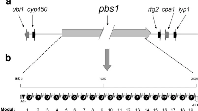
In silico characterization of the H. atroviridis 19-residue peptaibol synthetase and its putative product. Using the T. virens tex1sequence as a query in TBLASTN, we identified a continuous open reading frame in the genome sequence ofH. atroviridisIMI 206040, which, when translated, would give rise to a 21,879-amino-acid (aa) protein. This deduced polypeptide consists of 19 complete peptide synthetase modules and the respective additional acetyltransferase and alcohol dehydroge-nase domains at the N and C termini of the predicted protein (Fig. 1a), thus clearly identifying it as a peptaibol synthetase. It is the only large peptaibol synthase gene in the genome ofH. atroviridis. The genes flankingpbs1inH. atroviridisare shown in Fig. 1b: the immediate downstream region contains se-quences putatively encoding proteins similar to S. cerevisiae
Rtg2p and anN. crassacalcium/proton exchanger in the same order and orientation as immediately downstream ofT. virens tex1andT. harzianum salps2(42, 46) (Fig. 1a). The upstream region contains genes encoding proteins of the cytochrome P450 subfamilies and a prenyltransferase (Fig. 1). Both genes are also found in the same orientation upstream of the harzi-anin synthetase gene ofH. jecorina(unpublished data) but are absent from the 5⬘flanking area ofT. virens tex1. In fact, these two genes occur at two different scaffolds of the T. virens
genome database (scaffolds 6 and 17), which are both different
TABLE 1. Comparison ofH. atroviridisPBS1 to other peptaibol synthases fromTrichoderma/Hypocrea
Organism Gene/protein Product name Product length Source or reference
H. atroviridis pbs1/PBS1 Atroviridin 19-mer, 20-mer This study
H. virens tex1/TEX1 Trichorzin 18-mer 46
H. jecorina par1/PAR1 Paracelsin 20-mer Unpublished
har1/HAR1 Harzianin 11-14-mer Unpublished
H. lixii salps2/SALPS2 Trichorzin (?) 18-mer (?) 42a
a
Only three terminal modules were sequenced; however, because of the presence of a C-terminal alcohol dehydrogenase domain and the amino acid activated by the last three A domains, its identity as a large peptaibol synthase was concluded (42).
on September 8, 2020 by guest
http://ec.asm.org/
from that containing tex1 (scaffold 12 [unpublished data]). These comparisons show that the locus encoding the large peptaibol synthetases in Trichoderma apparently underwent major reorganization during evolution.
Peptide synthetases consist of a conserved iteration of mod-ules, each consisting in 533 order of an ATP-binding domain, an amino acid thioesterification domain, and a condensation domain (37). Therein, the ATP-binding domains specify the substrate specificity and thus the sequence of the formed pep-tide. In order to predict the latter, we first performed a phy-logenetic analysis of all 19-aa activating domains and corre-sponding domains identified inT. virensTEX1 andH. jecorina
paracelsin and harzianin synthetases (PAR1 and HAR1, re-spectively; H. von Do¨hren et al., unpublished data) for which the amino acid sequences of the respective peptaibol products are known (Table 1). The rationale for this was the hypothesis that domains activating the same amino acid may be more similar to each other within differentTrichodermaspp. than to other domains of the same protein. The resulting tree (Fig. 2) verifies this hypothesis in part: there are consistent and well-supported clades for the P-and Q-activating domains, and— albeit poorly resolved at the central nodes—several terminal clades for Aib and Ala. However, a number of clades were mixed. We also noted that domains which activate the same amino acids but are close to either the N or the C terminus of the resulting peptaibol consistently formed different clusters. This analysis allowed some identification but left several do-mains unidentified.
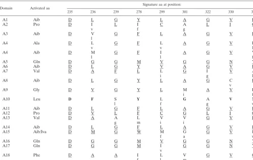
Therefore, the domains were further analyzed with respect to the presence of the signature sequences proposed by Stach-elhaus et al. (38) and Challis et al. (7). For this purpose, we
compared them to those present inH. jecorinaPAR1 because the respective paracelsin is structurally most similar to atro-viridin (Table 2) (25). Modules putatively acting on the same aa indeed showed conserved signature sequences, differences often being only conserved changes (e.g., L3V). Based on these combined analyses in Fig. 2 and Table 2, the peptaibols “encoded” byH. atroviridiswould be compatible with that of atroviridins from a bona fide strain ofH. atroviridis(26), Ac-UPUAQUVUGLUPVU(U/J)QQF-OH, with the exception that the U between A4 and Q5 is missing.
Identification of atroviridins formed by H. atroviridis. In order to compare the structure of the peptaibols putatively formed by PBS1 to those actually formed byH. atroviridisIMI 206040 and P1, we used MALDI-TOF analysis of surface-grown cultures ofH. atroviridis.The rationale for this was that a review of the literature about peptaibol formation by Tri-chodermarevealed that most researchers have used either sur-face cultures or submerged cultures, to which solid compo-nents had been added (19). The results from such an experiment, which yielded consistent results for both strains, are shown in Fig. 3: as long as the culture ofT. atroviridewas in the state of vegetative hyphal growth, no peptaibol forma-tion was detected (data not shown). However, as soon as the fungus started to sporulate, peptaibol formation was evident by the observation of a characteristic island of several mass peaks in the range of 1,935 to 2,010 Da. Table 3 compares the masses of these peaks with those of atroviridins and neoatroviridins of
H. atroviridis: the main peak atm/z1,963 corresponds to pro-tonated atroviridin A. Masses at 1,920 and 1,934 correspond to atroviridins A and B without the Aib in position 6 and for which the respective module is missing in PBS1. Masses at
FIG. 1.pbs1locus ofH. atroviridisand modular organization of the predicted PBS1 protein. (a) Chromosomal arrangement of thepbsllocus:
ubi1, UbiA-like prenyltransferase;cyp450, cytochrome P450 subfamily protein;pbs1l, 19-mer peptaibol synthetase;rgt2, Rtg2-like protein; cpa1, Ca2⫹/H⫹antiporter;lyp1, lysine permease. With the exception ofpbs1, gene lengths are drawn to scale. (b) Modules within the PBS1 protein,
numbered consecutively from 1 to 19. The domains within the modules are indicated as follows: gray cylinder, ketoacyl synthetase; black triangle, acyl transferase; white square, condensation domain; black circles: adenylation domains; gray triangles, thiolation domain; white arrow, alcohol dehydrogenase. The amino acids activated by the individual adenylation domains are indicated in the one-letter code (U,␣-aminoisobutyric acid; J, isovaline).
2334 KOMON-ZELAZOWSKA ET AL. EUKARYOT. CELL
on September 8, 2020 by guest
http://ec.asm.org/
1,949, 2,003, and 2,017 may represent new atroviridin variants because they can be explained by the loss or gain of one or two methylene groups from atroviridin A, respectively. While the identification of all of the components of the peptaibol mixture was beyond the scope of the present study, these findings confirm that this “mass island” corresponds to the atroviridins and that both the 20-residue and the 19-residue peptaibols (which would correlate with the module structure) are detect-able. We have thus taken the abundance of this peak island, which did not change in its shape (i.e., ratio of individual peaks to each other) throughout this investigation as a semiqualita-tive measure (see above) for the formation of peptaibols.
Peptaibol formation depends on the BLRl and BLR2 pro-teins and is stimulated by light.Since atroviridin formation was only detected under conditions of conidiation, we investi-gated this in more detail. The process of conidiation by fungi is subject to several regulatory influences (47), and the correla-tion observed above may therefore be simply coincidence, i.e., independent regulation by the same physiological event, or due to a common signal upstream of the conidiation event. We therefore investigated whether different methods for promot-ing conidiation would always correlate with peptaibol forma-tion or whether the latter specifically occurs only under one of these conditions. As a first condition, we chose white light, which is known to trigger photoconidiation inH. atroviridisvia the function of the blue light regulator proteins BLR1 and BLR2 (5). Figure 4a shows that the triggering of conidiation by light was indeed paralleled by a strong accumulation of pep-taibols, and only traces were seen in dark cultivated cultures. The latter may have been due to some sporulation, since they were completely absent in a parallel set of experiments (data not shown). This accumulation was completely blocked in the
blr1andblr2delta mutants. These data indicate that peptaibol formation under conditions of light-induced sporulation de-pends on the BLR1/2 proteins.
Starvation-induced sporulation triggers peptaibol forma-tion independently of light and the BLR proteins. Another universal inducer of conidiation in fungi is nutrient starvation, and we therefore tested whether the accumulation of peptai-bols would also be stimulated under these conditions. Figure 4b shows that carbon starvation indeed led to the accumulation of peptaibols and that under these conditions the accumulation was independent of theblrlandblr2genes and, consistent with this, was also independent of light. This would in theory be in contrast to previous reports that carbon deprivation-induced sporulation depends on the BLRl/BLR2 proteins (6). How-ever, more recent data have shown that the BLR1/2-dependent response is only observed when cultures are subjected to a starvation for not more than 12 h and absent during incubation under starvation conditions for a longer time (A.
Herrera-FIG. 2. Phylogenetic relationships of the different adenylation do-mains of PBS1 to the adenylation dodo-mains found in theH. jecorina
paracelsin synthetase (PAR [unpublished data]), theH. jecorina har-zianin synthetase (HAR [unpublished data]), andT. virensTEX1 (45). Modules in PBS1 (named “ATRO” in this figure) are followed by the respective module number and are shaded in gray. TEX1 is named “TVA” in this figure. In PAR, HAR, and TVA the numbers indicate the positions of the respective modules in the protein, but in addition the amino acid (indicated in the three-letter code) specifies the
acti-vated amino acid. When more than a single amino acid is given (linked by an asterisk [*]), this indicates that the module accepts more than a single amino acid. The tree was constructed by NJ analysis, using 1,000 bootstrap replications, whose statistics (percentage of occurrence of the branch in 1,000 trees) are given at the respective nodes. The bracket summarizes all branches leading to modules involved in ad-enylation of␣-aminoisobutyric acid.
on September 8, 2020 by guest
http://ec.asm.org/
Estrella, unpublished data). The present data therefore show that the response to light observed above (i.e., peptaibol accu-mulation and dependence on the BLR proteins) is due to its sporulation-associated nature and not because of a direct reg-ulatory role of the light triggering signal cascade on peptaibol synthetase.
Induction of conidiation-associated peptaibol formation by mycelial injury is light dependent but BLR independent.As a third, independent approach to study the correlation of pep-taibol formation with conidiation, we used mechanical stress (injury with a scalpel (see reference 5) of the mycelia. This conidiation pathway has also been shown to be independent of the BLR proteins (5), and consequently there is also sporula-tion in the darkness. Interestingly, however, peptaibol forma-tion under these condiforma-tions inH. atroviridis was completely dependent on the presence of light, and virtually no peptaibols were detectable in its absence in spite of sporulation; essen-tially the same findings were also observed in theblrlandblr2
mutants (data not shown). While this is consistent with the above findings that injury-promoted conidiation is BLR inde-pendent, it documents that there is also a BLR-independent pathway of stimulation of peptaibol formation by light under stress.
The hypersporulating H. atroviridis delta-gna3 mutant is defective in peptaibol formation. Conidiation in Neurospora
crassais negatively regulated by the G␣-protein Gna3 (17), and consistent data have also been reported also forH. atroviridis
(48). Consequently, agna3-delta mutant leads to hypersporu-lation. We wondered whether this mutant would consequently overproduce peptaibols. However, in contrast to these expec-tations, the results shown in Fig. 5 document that this mutant is almost completely impaired in peptaibol formation and that this impairment is apparent under all conditions tested above, i.e., light, carbon starvation, and mechanical injury, while the hypersporulating phenotype indeed formed under all of these (with the exception of starvation) conditions. As has been reported previously (48), the addition of exogenous cAMP (1 and 5 mM) did not alter the slowly growing, hypersporulating phenotype of the mutants. We therefore tested whether the addition of cAMP would rescue peptaibol formation in these mutants, but we found that this also was not the case (data not shown). Consequently, these data imply that peptaibol forma-tion is positively controlled by GNA3 via a pathway that must be different from that repressing conidiation and that this control overrides the correlation with sporulation.
Constitutive activation of PKA leads to impairment of pep-taibol formation.Despite of the lack of influence of addition of exogenous cAMP, the disparity of the effect of loss of function of GNA3 on conidiation and peptaibol formation prompted us to investigate whether the effect on peptaibol formation would
TABLE 2. Signature sequences of the A domains ofH. atroviridisPBS1 compared toH. jecorinaPAR1a
Domain Activated aa
Signature aa at position:
235 236 239 278 299 301 322 330 331
A1 Aib D L G Y L A G V F
A2 Pro D I L I C A L I C
v f g
A3 Aib D V G F L A G V F
l
A4 Ala D L G F L A G V F
v v l
A4 Aib D M G F I A G V V
l l f
A5 Gln D G G M V G G N Y
A6 Aib D L G Y V A G V F
A7 Val D A F L L G I V A
i g l
A8 Aib D L G Y L A G C F
v
A9 Gly D V G Y L M A V L
i f
A10 Leu D F S Y L G A V M
l f g v
A11 Aib D L G F L A G V F
A12 Pro D V L F C G L I C
A13 Val D A A L V V G V F
g m i i
A14 Aib D L G F L A G V F
A15 Aib/Iva D M G W M G G V I
f a v
A16 Gln D G G M V G G N Y
A17 Gln D G G M I G G N Y
v
A18 Phe D A A I L V G V G
f l m
aChallis et al. (7). Identical signature amino acids are underlined. If the corresponding signature amino acid is different in PAR1, the respective amino acid is
indicated by a lowercase letter below that of PBS1. Since no activation domain for leucine (A10) is present in PAR1, its signature was taken from TEX1, and identical amino acids are indicated in boldface to illustrate this difference. aa, amino acids.
2336 KOMON-ZELAZOWSKA ET AL. EUKARYOT. CELL
on September 8, 2020 by guest
http://ec.asm.org/
involve PKA (PKA1). G-protein-mediated inhibition of sporu-lation is known to involve the downstream action of PKA (35). To this end, we used a strain overexpressing the antisense version of the regulatory subunit of PKA1 (6) and whose PKA1 activity is thus constitutively activated. Consistent with what is known in other fungi, sporulation in this strain was essentially absent (see also reference 6). Also consistent with the findings that the addition of cAMP had no effect, peptaibol formation was not observed in this strain, irrespective of the conditions used (light/darkness, mechanical injury, starvation, etc. [data not shown]). Therefore, the positive action of GNA3 on pep-taibol formation does not involve PKA1.
DISCUSSION
Peptaibols are among the largest peptide-like components formed by fungi and also have a number of interesting appli-cations. However, apart of their isolation and chemical char-acterization, little information has thus far become available about the reasons for their biodiversity and regulation of for-mation. Here we used in silico analysis to predict the structure of the peptaibol synthetase ofH. atroviridis, and we report that
the formation of the respective peptaibols—atroviridins—par-tially correlates with sporulation and is GNA3 dependent. To facilitate the subsequent discussion, these findings and the interaction of components are summarized in Fig. 6.
Although we have not produced a delta mutant ofH. atro-viridis pbs1, in silico analysis predicts that the respective gene encodes all of the domains necessary for synthesizing at least 19-residue peptaibols. Amino acid residues 232 to 430 ofH. atroviridisPBS1 showed 52% identity over 80% of the length of the ketoacyl synthetase domain ofH. jecorinaPAR1, and res-idues 431 to 815 were 74% identical over 100% of the length of the PAR1 acyltransferase domains and were also highly similar toT. virensTEX1. These domains therefore encode the pro-teins acetylating of the peptaibol N terminus. Amino acid res-idues 22694 to 23110, on the other hand, were 73% identical over 100% of the length of the alcohol dehydrogenase domain (pfam00106) of PAR1, which is necessary for the reductive cleavage of the final amino acid to generate the C-terminal alcohol. The same domain has been found at the same place in
T. virensTEX1 andT. harzianumSALPS2 (42, 46). Therefore, together with the 19-aa activating, transferring, and condensa-tion domains, the deduced PBS1 protein theoretically contains all of the enzymatic activities necessary and typical to produce peptaibols.
Peptaibols are notoriously microheterogeneous. Wiest et al. (46) emphasized that this multiplicity of products is likely a result of the ability of the activating modules to bind multiple substrates. In addition, this ability may be reflected in different
Kmvalues for different amino acids, because it is known that
the microheterogeneity can be influenced by supplementing cultures with a specific amino acid, thereby likely increasing its intracellular concentration (see reference 19). The possibility that multiple peptaibol synthetases would be responsible for generation of this microheterogeneous mixture has been ruled out by Wiest et al. (46), who showed that atex1knockout inT. virenseliminated the formation of all 18-residue peptaibols. In
FIG. 3. Detailed view of a MALDI-TOF mass spectrum from a peptaibol producing sporulating culture of the wild-type strainH. atroviridisIMI 206040, grown in the presence of light. Them/zvalues of the individual peaks are given.
TABLE 3. Interpretation of theH. atroviridisatroviridin by MALDI-TOF analysis
m/zvalue(s) Compound
identity Explanation
1,920 Atroviridin A —Ala⫹H⫽1919; position 6 deletion
1,934 Atroviridin B –Ala⫹H⫽1933, position 6 deletion
1,948, 1,949 New atroviridin –CH2(1961 minus 14)
1,963 Atroviridin A (1961)⫹H⫽1962 1,987, 1,988 ?
2,001 ?
2,003, 2,004 New atroviridin ⫹CH2, 1989⫹14
2,017, 2,018 New atroviridin ⫹2CH2, 1989⫹28
on September 8, 2020 by guest
http://ec.asm.org/
2338
on September 8, 2020 by guest
http://ec.asm.org/
addition, Wei et al. (45) proved that the small 11-14-mer pep-taibols are products of a second, smaller peptaibol synthase. Our genome sequence data support such a claim: in fact,pbs1
is the only gene in theH. atroviridis genome that encodes a peptaibol synthetase consisting of the required number of
modules for synthesis of peptaibols with more than 16 residues (C. P. Kubicek, unpublished data). Similar findings have also been made for H. jecorina (H. von Doehren, unpublished data). However, our data contribute a new aspect on the mul-tiplicity of peptaibol production: the atroviridins formed con-tain the sequence Aib5-Aib6-Gln7. while the structure of PBS1 does not contain a second Aib domain preceding the Gln domain. The only explanation that can be offered for this finding is that the U5 module acts twice, forming a hexapeptide intermediate. Such cases of the iterative use of peptide syn-thetase modules are known in fungal nonribosomal peptide synthetases (43) but have thus far not been reported for pep-taibol synthetases. Our data suggest that this could be a further mechanism contributing to peptaibol heterogeneity.
The phylogenetic analysis, which was performed to identify
FIG. 4. BLR1- and BLR2-dependent formation of atroviridin inH. atroviridisby different sporulation-inducing pathways. Graphs show the MALDI-TOF spectra of cultures of the wild-type strain (IMI), the delta-blr1mutant strain (blr1), and the deltablr2mutant strain (blr2) under illumination (L) and in darkness (D) on ME agar (a), under conditions of starvation (S) (b), and under conditions of mechanical injury (i) (c). See Materials and Methods for details. The ranges of the intensity of the MALDI-TOF spectra were 0 to 28,000 (a), 0 to 23,000 (b), and 0 to 25,000 (c). The insets show photographs of the respective cultures immediately before peptaibol extraction. Data consistent with the claims drawn were obtained in at least one additional, separate experiment.
FIG. 5. GNA3 is required for peptaibol production byH. atroviri-dis. H. atroviridisP1, and its⌬gna3mutant were used. Other symbols are used as explained in Fig. 4. Only experiments performed with illuminated cultures are shown since none of the nonilluminated cul-tures of the delta-gna3mutant showed any peptaibol production. The range of intensity is 0 to 18,000 to make the traces of peptaibols which are still formed more visible. The experiment shown is typical for three independent experiments, whose results were consistent with the data shown here.
FIG. 6. Schematic summary of the regulation of peptaibol biosyn-thesis, based on results from the present study. (a) Summary of pep-taibol formation under the various conditions in the mutants tested. L, light; D, dark. Strains are abbreviated as indicated in Materials and Methods. “Y” (yes) indicates peptaibol formation; “N” (no) indicates no peptaibol formation. Conditions in which peptaibol formation cor-relates with sporulation are in white on a black background. (b) Model for the putative interaction of the factors, as studied here, on sporu-lation and peptaibol formation. Red arrows indicate signaling mecha-nisms in which peptaibol formation is uncoupled from sporulation.⫹, activation;⫺, inhibition. The dotted arrow indicates the possibility of BLR1 and BLR2 action via GNA3 (unpublished data), for which no evidence is presented here.
on September 8, 2020 by guest
http://ec.asm.org/
the individual activating domains, revealed a very high diversity and, with the exception of the domains specific for Pro, Gln, Phe, and in some cases Aib, phylogeny was unable to predict the amino acids bound by these modules. Similar difficulties were observed with the signature sequences proposed by Chal-lis et al. (7) based on a limited set of domains. This high amino acid diversity contrasts with that of the otherwise rather highly conserved transfer and condensation domains (⬎80% iden-tity). This and the fact that some of the modules activating the same amino acid (e.g., Aib or Ala) occur in different clades of the NJ tree suggests a history of gene duplication and a high mutation rate that ultimately leads to an alteration of the substrate specificity. In addition, the fact that the pbs1locus has only partially maintained synteny inTrichodermasuggests the possibility of recombination as an additional origin of the diversity of peptaibols in this fungal genus.
Thepbs1mRNA, in a stretched form, must be about 10m long, and it was already emphasized (46) that the mechanism for the transcription of such large mRNAs in eukaryotes is unknown, and the correlation between transcript abundance and expression level is questionable. Therefore, rather than quantifying the pbs1 transcript, we used MALDI-TOF MS identification of the peptaibols as a means to learn about their regulation of formation. Also, the MALDI-TOF procedure has the advantage that it allows the analysis of a very small part of the growing colony, thereby ensuring the analysis of a homog-enous fungal tissue. Unfortunately, it also has the drawback that absolute quantification is impossible since the weight of the fungal material that was extracted could not be deter-mined. Consequently, only extreme changes (present or not present) are used for interpretation. However, using this semi-quantitative approach, we could show that the formation of “long” peptaibols by the wild-type ofH. atroviridisis associated with conidiation. Such an association is not without precedent: as an example, sporulation-deficient mutants ofAspergillusspp. (2), Claviceps purpurea (28), or Fusarium verticillioides (34) have also been shown to be defective in the production of their respective secondary metabolites. Calvo et al. (4) grouped sec-ondary metabolites associated with sporulation into three broad categories: (i) metabolites that are required for the sporulation process (for example, the sporulation-activating linoleic acid-derived compounds produced byA. nidulans) (8– 10), (ii) metabolites that are required for sporulation struc-tures (for example, pigments such as melanins, which are re-quired for the integrity of sexual and asexual spores) (1, 15), and (iii) metabolites that are toxic and whose formation coin-cides with the approximate time of sporulation (for example, the biosynthesis of mycotoxins) (14, 41). Based on their fungi-cidal action, peptaibols would fit into the last group. However, an important difference is that they are not secreted but remain bound to the spores ofTrichoderma. So could they be impor-tant for sporulation in the sense of groups i and ii listed above? Peptaibol synthetase-delta mutants ofT. virens(46) and ofT. harzianum (D. Keszenman-Pereyra et al., unpublished data) still sporulate, so the answer would be no. However, both the
T. harzianum and the T. virens delta mutants still form the 11-14 residue peptaibols (46; Keszenman-Pereyra et al., un-published), and they could compensate for the loss of forma-tion of the 18- to 20-residue peptaibols. In addiforma-tion, the pep-taibols might have subtle effects on sporulation that escape
inspection of the morphology only. For example, they are known to function as voltage-gated membrane channels, pri-marily transporting K⫹(11, 12, 18, 33), which could be impor-tant for ion transport and/or homeostasis during sporulation. In fact, gramicidin, another ion channel-forming peptide anti-biotic, induces sporulation in its producerBacillus subtilis(24, 29). The necessity for the presence of specific membrane chan-nels in fungal spores has recently been stressed by Sidoux-Walter et al. (36), who showed that sporulation inS. cerevisiae
is accompanied by the expression of a specific aquaporin, which is responsible for water outflow from spores. Thus, in analogy, one could speculate that the peptaibols are important for K⫹and other monovalent cation homeostasis during the conidiation ofTrichoderma. We have been unable to find a report describing the effect of peptaibols on the producer or-ganisms, which in the light of the present findings clearly war-rants investigation.
Sporulation ofH. atroviridisis dependent on light (4, 6), and it was therefore not unexpected that the formation of a conidiation-associated secondary metabolite would conse-quently respond to light as well. However, the present data show that the relationship between light and peptaibol forma-tion is more complex: the fact that mechanical injury can over-ride control by BLR1 and -2 but is still affected by light (i.e., no peptaibol formation was observed in the dark, despite sporu-lation) suggests that there is a second, light-dependent mech-anism that is essential for peptaibol formation. The occurrence of light stimulation of gene expression by an BLR1 and two independent pathways inH. atroviridishas been recently shown (31), and it is possible that this pathway may contribute to peptaibol formation under conditions of mechanical injury-induced sporulation.
The G-protein signaling pathway has been shown to be in-volved in the control of secondary metabolism and conidiation inAspergillusspecies, particularly inA. nidulans(4), but there is also evidence indicating the regulation of trichothecene pro-duction inFusarium(40) and␥-pentylpyrone formation inH. atroviridis(30) by a similar pathway. Interestingly, not all ef-fects of G proteins (as would be expected from a sporulation-associated process) are negative: while inA. nidulansthe dom-inant-activating fadAG42R allele represses conidiation and
sterigmatocystin biosynthesis, it stimulates penicillin biosyn-thesis at the same time (4). Introduction of thefadAG42Rallele
intoF. sporotrichioidesresults in reduced conidiation but in-creased trichothecene production (40). It is also worth noting that all of the reports on a role of G proteins in fungal sec-ondary metabolism were done with theAspergillusG-protein FadA or itsTrichodermaorthologue TGA1 (GNA1). In con-trast, the present study was undertaken with another G pro-tein, GNA3. Although both GNA1 and GNA3 (previously termed TGA3 [48]) are negative regulators of conidiation inH. atroviridis(30, 48),N. crassaGNA1 is regulated by the trans-membrane receptor protein GPR-4, which is responsible for carbon source signaling (21). The receptor to which GNA3 binds is not known yet. Thus, our data show that not only FadA/GNA1 mediate secondary metabolism, but at least GNA3 also does so. It is tempting to speculate that all G proteins might be able to regulate secondary metabolism, be-cause null mutants in all of them affect sporulation (16). This is understandable in view of the multiple signals (pH, sugar,
2340 KOMON-ZELAZOWSKA ET AL. EUKARYOT. CELL
on September 8, 2020 by guest
http://ec.asm.org/
nitrogen content, light, and many others) that determine whether a fungal cell can maintain a vegetative mode of growth or whether it is advisable to conidiate.
The subsequent steps required for triggering of peptaibol formation by GNA3 are unclear. If this occurred via activation of adenylate cyclase and thus the formation of cAMP, one would assume that theH. atroviridisstrain harboring the anti-sense gene for the regulatory subunit of PKA and thus bearing a constitutively active PKA would overproduce peptaibols. This was shown not to be the case, and indeed, at the level of PKA, peptaibol formation again strictly correlated with sporu-lation and was therefore absent in the PKA-overproducing mutant. The finding of loss of sporulation is consistent with the hypersporulating phenotype of the delta-gna3strain and sug-gests that the pathway triggering sporulation inH. atroviridisis subject to a negative control by a G-protein/PKA pathway similar to that inA. nidulans(35). Hence, the positive effect of GNA3 on peptaibol biosynthesis must involve another signal-ing pathway for which, thus far, only the requirement for GNA3 is known.
ACKNOWLEDGMENTS
This study was supported by the Fifth (EC) Framework program (Quality of Life and Management of Living Resources; project EUROFUNG 2 [QLK3-1999-00729]) and by a grant from the Austrian Science Foundation (P 17325-B17) to C.P.K. The genomic sequence forH. atroviridiswas provided by the DOE Joint Genome Institute, Walnut Creek, CA. This study was performed under the auspices of the U.S. Department of Energy’s Office of Science, Biological, and Environmental Research Program and the by the University of Cali-fornia, Lawrence Livermore National Laboratory, under contract W-7405-Eng-48; the Lawrence Berkeley National Laboratory under contract DE-AC03-76SF00098; and the Los Alamos National Labora-tory under contract W-7405-ENG-36.
We thank S. Zeilinger for thetga3-delta strain ofH. atroviridisand S. Baker for help with theH. atroviridisgenome sequence.
REFERENCES
1.Alspaugh, J. A., J. R. Perfect, and J. Heitman.1997.Cryptococcus
neofor-mansmating and virulence are regulated by the G-protein alpha subunit
GPA1 and cAMP. Genes Dev.11:3206–3217.
2.Bennett, J. W., and K. E. Papa.1988. The aflatoxigenicAspergillus, p. 264–
280.InD. S. Ingram and P. A. Williams (ed.), Genetics of plant pathology,
vol. 6. Academic Press, London, United Kingdom. 3. Reference deleted.
4.Calvo, A. M., R. A. Wilson, J. W. Bok, and N. P. Keller.2002. Relationship between secondary metabolism and fungal development. Microbiol. Mol.
Biol. Rev.66:447–459.
5.Casas-Flores, S., M. Rios-Momberg, M. Bibbins, P. Ponce-Noyola, and A. Herrera-Estrella.2004. BLR-1 and BLR-2, key regulatory elements of
pho-toconidiation and mycelial growth inTrichoderma atroviride. Microbiology
150:3561–3569.
6.Casas-Flores, S., M. Rios-Momberg, T. Rosales-Saavedra, P. Martinez-Her-nandez, V. Olmedo-Monfil, and A. Herrera-Estrella.2006. Cross talk be-tween a fungal blue-light perception system and the cyclic AMP signaling
pathway. Eukaryot. Cell5:499–506.
7.Challis, G. L., J. Ravel, and C. A. Townsend.2000. Predictive, structure-based model of amino acid recognition by nonribosomal peptide synthetase
adenylation domains. Chem. Biol.7:211–224.
8.Champe, S. P., and A. A. E. El-Zayat.1989. Isolation of a sexual sporulation
hormone fromAspergillus nidulans. J. Bacteriol.171:3982–3988.
9.Champe, S. P., P. Rao, and A. Chang.1987. An endogenous inducer of
sexual development inAspergillus nidulans. J. Gen. Microbiol.133:1383–
1388.
10.Chang, P.-K., J. W. Cary, D. Bhatnagar, T. E. Cleveland, J. W. Bennett, J. E. Linz, C. P. Woloshuk, and G. A. Payne.1993. Cloning of theAspergillus parasiticus apa-2gene associated with the regulation of aflatoxin
biosynthe-sis. Appl. Environ. Microbiol.59:3273–3279.
11.Chugh, J. K., and B. A. Wallace.2001. Peptaibols: models for ion channels.
Biochem. Soc. Trans.29:565–570.
12.Duclohier, H., G. M. Alder, C. L. Bashford, H. Bruckner, J. K. Chugh, and
B. A. Wallace.2004. Conductance studies on trichotoxin_A50E and
impli-cations for channel structure. Biophys. J.87:1705–1710.
13.Harman, G. E., and C. P. Kubicek.1998.TrichodermaandGliocladium, vol. 2. Enzymes, biocontrol, and commercial applications. Taylor & Francis, London, United Kingdom.
14.Hicks, J., J.-H. Yu, N. Keller, and T. H. Adams.1997.Aspergillussporulation and mycotoxin production both require inactivation of the FadA G-alpha
protein-dependent signaling pathway. EMBO J.16:4916–4923.
15.Kawamura, C., T. Tsujimoto, and T. Tsuge.1999. Targeted disruption of a melanin biosynthesis gene affects conidial development and UV tolerance in
the Japanese pear pathotype of Alternaria alternata. Mol. Plant-Microbe
Interact.12:59–63.
16.Kays, A. M., and K. A. Borkovich.2004. Severe impairment of growth and
differentiation in aNeurospora crassamutant lacking all heterotrimeric G
alpha proteins. Genetics166:1229–1240.
17.Kays, A. M., P. S. Rowley, R. A. Baasiri, and K. A. Borkovich.2000.
Regu-lation of conidiation and adenylyl cyclase levels by the G␣protein GNA-3 in
Neurospora crassa. Mol. Cell. Biol.20:7693–7705.
18.Kropacheva, T. N., and J. Raap.2002. Ion transport across a phospholipid membrane mediated by the peptide trichogin GA IV. Biochim. Biophys. Acta1567:193–203.
19.Kubicek, C. P., M. Zelazowska-Komon, E. Sandor, and I. S. Druzhinina.
2007. Facts and challenges in the understanding of the biosynthesis of
pep-taibols byTrichoderma. Chem. Biodiv.4:1068–1082.
20.Kumar, S., K. Tamura, and M. Nei.2004. MEGA3: integrated software for molecular evolutionary genetics analysis and sequence alignment. Brief.
Bioinform.5:150–163.
21.Li, L., and K. A. Borkovich.2006. GPR-4 is a predicted G-protein-coupled receptor required for carbon source-dependent asexual growth and
devel-opment inNeurospora crassa. Eukaryot. Cell5:1287–1300.
22.Lorito, M., V. Farkas, S. Rebuffat, B. Bodo, and C. P. Kubicek.1996. Cell
wall synthesis is a major target of mycoparasitic antagonism byTrichoderma
harzianum. J. Bacteriol.178:6382–6385.
23.Lucaciu, M., S. Rebuffat, C. Goulard, H. Duclohier, G. Molle, and B. Bodo.
1997. Interaction of the 14-residue peptaibols, harzianins HC, with lipid bilayers: permeability modifications and conductance properties. Biochim.
Biophys. Acta1323:85–96.
24.Marahiel, M. A., M. M. Nakano, and P. Zuber.1993. Regulation of peptide
antibiotic production inBacillus. Mol. Microbiol.7:631–636.
25.Neuhof, T., R. Dieckmann, I. S. Druzhinina, C. P. Kubicek, and H. von Do¨hren.2007. Intact-cell MALDI-TOF mass spectrometry analysis of
pep-taibol formation by the genusTrichoderma:can molecular phylogenic
knowl-edge predict peptaibol structures? Microbiology153:3417–3437.
26.Oh, S. U., B. S. Yun, S. J. Lee, J. H. Kim, and I. D. Yoo.2002. Atroviridins
A-C and neoatroviridins A-D, novel peptaibol antibiotics produced by
Tri-choderma atrovirideF80317. I. Taxonomy, fermentation, isolation, and
bio-logical activities. J. Antibiot.55:557–564.
27.Papavizas, G. C.1985.TrichodermaandGliocladium: biology, ecology, and
potential for biocontrol. Annu. Rev. Phytopathol.23:23–54.
28.Pazoutova, S., V. Pokorny, and Z. Rehacek.1977. The relationship between
conidiation and alkaloid production in saprophytic strains ofClaviceps
pur-purea. Can. J. Microbiol.23:1182–1187.
29.Pschorn, W., H. Paulus, J. Hansen, and H. Ristow. 1982. Induction of
sporulation inBacillus brevis. 2. Dependence on the presence of the peptide
antibiotics tyrocidine and linear gramicidin. Eur. J. Biochem.129:403–407.
30.Reithner, B., K. Brunner, R. Schuhmacher, I. Peissl, V. Seidl, R. Krska, and S. Zeilinger.2005. The G protein alpha subunit Tga1 ofTrichoderma atro-virideis involved in chitinase formation and differential production of
anti-fungal metabolites. Fungal Genet. Biol.42:749–760.
31.Rosales-Saavedra, T., E. U. Esquivel-Naranjo, S. Casas-Flores, P. Martinez-Hernandez, E. Ibarra-Laclette, C. Cortes-Penagos, and A. Herrera-Estrella.
2006. Novel light-regulated genes inTrichoderma atroviride: a dissection by
cDNA microarrays. Microbiology152:3305–3317.
32.Schirmbo¨ck, M., M. Lorito, Y.-L. Wang, C. K. Hayes, I. Arisan-Atac, F. Scala, G. E. Harman, and C. P. Kubicek. 1994. Molecular mechanisms
involved in biocontrol byTrichoderma harzianum: co-induction and
syner-gism of hydrolytic enzymes and pentaibol antibiotics. Appl. Environ.
Micro-biol.60:4364–4370.
33.Shenkarev, Z. O., T. A. Balashova, Z. A. Yakimenko, T. V. Ovchinnikova, and A. S. Arseniev.2004. Peptaibol zervamicin IIb structure and dynamics
refinement from transhydrogen bond J couplings. Biophys. J.86:3687–3699.
34.Shim, W.-B., and C. P. Woloshuk.2001. Regulation of fumonisin B1
biosyn-thesis and conidiation inFusarium verticillioidesby a cyclin-like (C-type)
gene,FCC1. Appl. Environ. Microbiol.67:1607–1612.
35.Shimizu, K., and N. P. Keller. 2001. Genetic involvement of a cAMP-dependent protein kinase in a G-protein signaling pathway regulating
mor-phological and chemical transitions inAspergillus nidulans. Genetics157:
591–600.
36.Sidoux-Walter, F., N. Pettersson, and S. Hohmann.2004. The Saccharomy-ces cerevisiaeaquaporin Aqy1 is involved in sporulation. Proc. Natl. Acad.
Sci. USA101:17422–17427.
37.Stachelhaus, T., and M. A. Marahiel.1995. Modular structure of genes
on September 8, 2020 by guest
http://ec.asm.org/
encoding multifunctional peptide synthetases required for non-ribosomal
peptide synthesis. FEMS Microbiol. Lett.125:3–14.
38.Stachelhaus, T., H. D. Mootz, and M. A. Marahiel.1999. The specificity-conferring code of adenylation domains in nonribosomal peptide
syntheta-ses. Chem. Biol.6:493–505.
39.Szekeres, A., B. Leitgeb, L. Kredics, Z. Antal, L. Hatvani, L. Manczinger, and C. Vagvolgyi.2005. Peptaibols and related peptaibiotics ofTrichoderma:
a review. Acta Microbiol. Immunol. Hung.52:137–168.
40.Tag, A., J. Hicks, G. Garifullina, C. Ake, T. D. Phillips, M. Beremand, and N. P. Keller.2000. G-protein signaling mediates differential production of
toxic secondary metabolites. Mol. Microbiol.38:658–665.
41.Trail, F., N. Mahanti, and J. Linz.1995. Molecular biology of aflatoxin
biosynthesis. Microbiology141:755–765.
42.Vizcaino, J. A., R. E. Cardoza, L. Dubost, B. Bodo, S. Gutierrez, and E. Monte.
2006. Detection of peptaibols and partial cloning of a putative peptaibol synthetase
gene fromTrichoderma harzianumCECT 2413. Folia Microbiol.51:114–120.
43.von Do¨hren, H.2004. Biochemistry and general genetics of non-ribosomal
peptide synthetases in fungi. Adv. Biochem. Eng. Biotechnol.88:217–264.
44.Wallace, B. A.2000. Common structural features in gramicidin and other ion
channels. Bioessays22:227–234.
45.Wei, X., F. Yang, and D. C. Straney.2005. Multiple non-ribosomal peptide
synthetase genes determine peptaibol synthesis inTrichoderma virens. Can. J.
Microbiol.51:423–429.
46.Wiest, A., D. Grzegorski, B. W. Xu, C. Goulard, S. Rebuffat, D. J. Ebbole, B. Bodo, and C. Kenerley.2002. Identification of peptaibols from Tri-choderma virensand cloning of a peptaibol synthetase. J. Biol. Chem.
277:20862–20868.
47.Yoder, O. C., and B. G. Turgeon.2001. Fungal genomics and pathogenicity.
Curr. Opin. Plant Biol.4:315–321.
48.Zeilinger, S., B. Reithner, V. Scala, I. Peissl, M. Lorito, and R. L. Mach.
2005. Signal transduction by Tga3, a novel G protein alpha subunit of
Trichoderma atroviride. Appl. Environ. Microbiol.71:1591–1597.
2342 KOMON-ZELAZOWSKA ET AL. EUKARYOT. CELL