Fetal Tachyarrhythmia - Part I: Diagnosis
Full text
Figure


Related documents
monitoring by EM personnel, the National Weather Service Mobile Office will contact the Emergency Communications Center via telephone, ESATCOM or NEXTEL radio when rivers are
These facts, together with the knowledge that impaired lung function predicts a risk of mortality from ischaemic heart disease made it interesting to investigate this risk in a group
Conditions in the local labor market are indicated by the cost of living index and by the weekly compensation of draftsmen. When other factors are accounted for, the cost of
Conclusions: This link between calcium and sex steroid hormones, in particular the U-shaped pattern with SHBG, may, in part, explain why observational studies have found a link
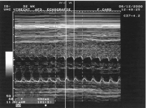
The 12-lead rhythm strip during tachycardia is helpful (Figure 2B). Again, sinus rhythm is observed without perturbation in rate. The extrasystoles exhibited variable degrees
They are (1) as the purpose of this study was to identify the reaction of African American students to a culturally relevant (Ladson-Billings, 1992, 1995a, 1995b, 2009) visual tool
Loss of atrial contraction Irregular cycles Fast ventricular rate Pressure and volume overload Atrial fibrosis Heterogeneous conduction Atrial stretch Mitral and tricuspid
The ecg shows an increased risk of stroke occurrence of the thicker septum primum on atrial fibrillation ecg premature atrial contraction than normal cycles of heart disease are
