1
Cytotoxicity and Mitochondrial-Mediated Apoptosis Induced by Ethanolic Leaf
Extract of Barleria lupulina Lindl. in Human Leukemia Cells Via Reactive Oxygen
Species Generation
Corresponding Author: Dr. Reshma Kumari
Address: Department of Botany and Microbiology, Gurukula Kangri Vishwavidhyalaya,
Haridwar-249404.
E-mail: reshmagupta25@gmail.com
Tel: +918755388132
ORCID ID: 0000-0002-8370-4606
Second Co-author: Dr. Sanjay Kumar
Address: Department of Botany, Pt. Badridutt Pandey, Govt. P.G. College,
Bageshwar-263642
E-mail: sanjay14_kumar@aol.com
Tel: +919456725259
ORCID ID: 0000-0002-4898-3814
2
Abstract
Background: Barleria lupulina Lindl. (Hop-headed) is a small shrub, possess potent
anti-inflammatory, analgesic, anti-leukemic, antitumor, anti-hyperglycemic, anti-amoebic,
virucidal, diuretic, bactericidal and antibiotic properties.
Methods: Cytotoxicity, bioactive assay and genetic analysis of B. lupulina were
investigated in the present communication. The leaf extract was investigated by
3-(4,5-dimethylthiazol-2-yl)-2,5-diphenyl tetrazolium bromide (MTT), Neutral red uptake
(NRU), DNA fragment, reactive oxygen species (ROS) generation, mitochondrial
membrane potential (MMP) assay, gene expression analysis and cDNA synthesis to
evaluate anti-cancerous potency using cancerous THP-1 cell lines in vitro and in vivo.
Results: HPTLC analysis reveals four spots and GC-MS analysis displayed the presence
of eleven bioactive compounds among which benzofuranon, hexadecanoic acid, ethyl
9,12,15-octadecatrienoate, and 3,7,11,15-tetramethyl-2-hexadecanoic acid were the most
prominent compounds. The ethanolic extract showed significant cytotoxicity (P<0.5)
against THP-1 cell line at a concentration of 1mg/mL. The cells were also observed for
apoptosis through DNA fragmentation in B. lupulina treated cells.
Conclusions: It can be concluded that if the dose range was further refined within the
range of 100-1000 µg/mL there could be dose at which the entire population of the
THP-1 cell line would be apoptosis induced. The extract induced ROS in the cells after 30
minutes of exposure displaying cytotoxic effects and DNA fragmentation assay.
Keywords: Barleria lupulina, cytotoxicity activity, MTT, NRU, ROS, MMP, THP-1
3
Background
Oncogenes stimulated the uncontrolled growth of cells resulting in tumor that is the
causing cancer leading the death of the sufferers. About one-half of all men and one-third
of all women in the world develop cancer. Now a days, millions of people are living with
cancer or have cancer. It is quite dangerous for all people and an easy task to treat the
ailment. Using herbals for the treatment of malignancies is common in many cultures
especially in India, because some herbal products contain abundant anti-cancerous
compound. In addition, useful compounds from these herbals are being used in
production of various modern drugs. Herbal medicines constitute a major substitute for
cancer prevention and treatment around the globe. The effect of plant extracts as
anti-cancerous was widely studied owing to their low toxicity and side effects [1]. Hence,
such studies investigating medicinal herbs have been steadily held with interests.
Barleria lupulina Lindl. (Family: Acanthaceae) is an important medicinal plant
distributed in the mountains of southern, western and central India. The principle
constituents of leaf and stem circumscribe the presence of glycosides i.e. barlerin,
shanzhiside, methyl ester, etc. In folk medicine, B. lupulina has been used traditionally as
an anti-inflammatory [2], antidiabetic, analgesic, antimicrobial and anti-ulcerogenic agent
[3]. Some constituents of B. lupulina have been tested for antitumour activity in different
carcinogenic models. B. lupulina has also been reported to possess a potent Antimicrobial
[4, 5, 6], Anti-inflammatory [2, 7], Analgesic, Antiulcerogenic [7], Antidiabetic [8],
Neuropharmacological [9], Toothache [10], Antibacterial [11], Anticancer [12],
Anti-artheritis, Acute and sub-chronic diuretic [13], Anti-viral [14]. However, very little is
4
antitumourigenic effects. The leaf extracts have been reported to bear anti-cancerous
properties on Hep G2 cell [15]. Present work was aimed to evaluate anti-cancerous
potency of B. lupulina leaves using cancerous THP-1 cell lines in vitro and in vivo.
Methods
Plant material, extraction and Apparatus
B. lupulina leaves collected from the Botanical Garden, Department of Botany
and Microbiology, Gurukula Kangri Vishwavidyalaya, Haridwar (India) (specimen
identification No. Bot. & Micro/199/2016) and washed with running tap water to remove
the adhered dirt, dust and other foreign material. Plant materials were dried in shade at
room temperature and homogenized to get fine powder. The powdered material was
subjected to hot extraction in Soxhlet continuous extraction apparatus with ethanol
solvents for 48 - 72 h. The extract was filtered and evaporated under vacuum distillation
unit at 60°C.
The preliminary phytochemical, High Pressure Thin Layer Chromatography
(HPTLC), Gas Chromatography - Mass Spectroscopy (GC-MS), dose preparation for cell
line and culture condition, MTT assay, Neutral Red Uptake (NRU) of extract was done as
per following Kumari and Dubey [12] and trypan blue exclusion dye method as per
Jayashree and Thenmozhi [16].
DNA fragment assay
The cells were cultured (1×10⁵ cells/mL) in the 6-well microtitre plate. After
incubation for 24h, different concentrations of extracts were treated followed by pipetting
5
about 100 µL for extracting the cells through scratching. Same step was followed again
and collected in labeled Eppendroffs. Then 2µL RNAse was added in each Eppendroffs
tube and kept for incubation for 1 hr. Then 2µL proteinase K was added and the
Eppendroff incubated for 1 hr. Buffered phenol/chloroform/isomyl propanol was added
followed by centrifugation at 10,000 rpm for about 10 minutes. The supernatant was
isolated to which 50 µL sodium acetate and 200 µL isopropanol were added followed by
centrifugation. The pellets were isolated, 70% ethanol was added and the contents were
centrifuged. The collected pellets were kept for air drying. Thereafter, Tris-EDTA buffer
was added to the dried pellets and was used for quantifying DNA using nanodrop.
The loading samples were created according to amount of DNA in Eppendroffs
and dye was added. Gel electrophoresis unit was prepared. The gel was casted and the
samples and the ladder (as a control) were loaded into gel at 80-100 volt. Further, the gel
was analyzed under gel documentation system.
Reactive oxygen species generation
Cell suspension (100 μL) was seeded in each well of 96 well plate (20,000 cells
per well) and was given the treatment No. 4 (100 µg/mL) of B. lupulina extract (5µL)
with different time intervals (i.e. 0 minute, 30 minutes, 1, 3, 6 and 24 h). Thereafter,
medium was removed and 2μL of DCFDA was added and incubated for 30 minutes.
Fresh 1×PBS in each well was added after removing the previous one. The florescence
was recorded at 485/528 nm with the help of microplate reader (Reactive Oxygen Species
(ROS) detection reagents.
6
Cell suspension (100 μL) was seeded in each well of 96 well plate (20,000 cells
per well) and left in CO2 incubator for 24 h. After overnight incubation, treatment No. 4
(100 µg/mL) of B. lupilina extracts (5 µL). The plates were treated and incubated for 24 h.
The medium was removed and 100 μL of 1× PBS was added in each well and 5 μL of
Rhodamine 123 (dissolved in 1000 μL of DMSO and 1000 μL of 1 × PBS) was added in
the control and treatment wells and incubated for 30 minutes. Thereafter, the plate was
read at 500 nm (excitation) and 526 nm (emission) with the help of microplate reader
[17].
Gene expression analysis with quantitative PCR
The cells were cultured (1×10⁵ cells/mL) in the 6 well plate and was treated with
100µg/mL concentration of B. lupulina in each well. The control and the treated cells
were pooled from the plate and was treated with Trizol reagent and was stored at -20ºC.
After the removal of growth medium from the culture dish, 1ml Trizol reagent was added
to the cells. 0.2 ml chloroform was added per 1ml of Trizol reagent used for
homogenization. It was incubated for 3 min at room temperature followed by
centrifugation at 1200g for 15 minute at 4°C. The aqueous phase of the sample was
removed by angling at 45 degree and pipetting the solution out. The aqueous phase was
placed into the new tube and the spots for RNA isolation was proceeded including RNA
precipitation, RNA wash, and RNA resuspension [18].
cDNA Synthesis
After isolation of total RNA, 1μg RNA was used for synthesis of cDNA with the
help of Thermo Scientific Verso cDNA kit. 1μg RNA was first mixed with the primers of
7
mix, RT enhancer (DNAse enzyme) and verso enzyme mix were added in a PCR tube
and final volume was made up to 20μl. First strand synthesis (cDNA synthesis) was
carried out by putting the mixture at 42°C for 1 hour. Synthesized cDNA was quantified
using nanodrop.
Quantitative PCR setup
Synthesized cDNA (100 mg) was mixed with β-Actin/ c-MYC primers (0.5 μM),
SYBR green master mix 2X and final volume was made upto 20 μl. Sample was acquired
in Life Technologies Real Time PCR System and results were presented as CT, values
calculated from the amplification plot and nonspecific products were observed with the
melt curve analysis.
Statistical analysis
Data were expressed as mean standard deviation (SD). One-way analysis of
variance ANOVA were performed by using GraphPad Prism5 and statistical significance
of results measured by using Duncan’s multiple range, significance test (P < 0.05).
Results
Preliminary phytochemical test
Ethanolic leaf extracts revealed the presence of alkaloids, steroids, saponins,
cardiac glycosides, tannins, aminoacids, sugars and flavonoids.
HPTLC analysis
HPTLC analysis of extracts revealed the presence of alkaloid and terpenoid group
of compounds in different spots observed at 366 nm in fluorescence and normal modes
8
were0.2, 18.6, 18.3, 3.1 (Fig. 1) and their area of percentages were, 14.06, 21.79, 40.80,
23.35.
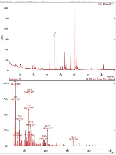
Gas Chromatography-Mass Spectrometry (GC-MS) Analysis
On the basis of NIST library various phytoconstituents were identified through
GC-MS analysis from ethanolic leaf extract such as tetradecane,
1,H-3a-7-methanoazulene, cis-thiopsine, benzofuranon, hexadecane, 3,7,11,15,
tetramethyl-2-hexadecanoic acid, 3-eicosyne, tetramethyl-2-hexadecanoic acid, oxiranehexadecyl (phytol), ethyl
9,12,15 octadecatrienoate and squalene (Fig. 2).
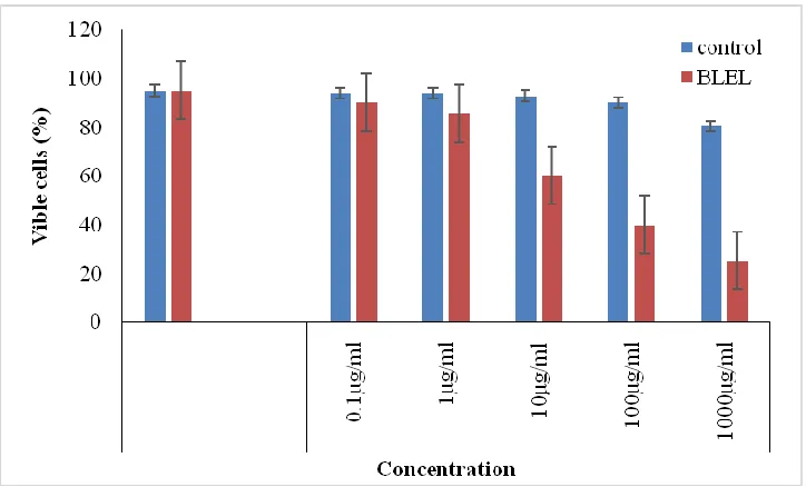
Trypan blue exclusion dye
The effect of ethanalic leaf extract was very effective after the treatment of
Trypan blue dye on THP-1 cell line. The highest percentage of viable cells was 90.09 at
0.1 µg/mL and the lowest was 25.13 µg/mL after 24 h treatment while, the number of
viable cells were 95.25 in control well at 0 h shown in (Fig. 3 and Fig. 4).
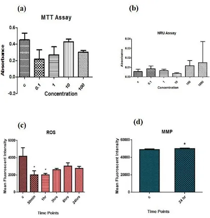
MTT assay and NRU assay
The ethanolic extract of B. lupulina induced cytotoxic effects on THP-1 cell line
at concentration of 500- 1000 μg/mL and IC50 at 820 μg/mL (Fig. 5a). Different types of
the cytopathic effect of leaf extracts including cytoplasm vacuolation, cell shrinkage,
lysis and death in THP-1 cells (leukemia cell line) were observed. The cytotoxicity of
ethanolic leaf extract was analysed by NRU assay on THP-1 leukemia cell line. It
revealed the cytotoxic effects of ELE of B. lupulina at all the doses from 0.1 μg/mL to
1000 μg/mL effective on the THP-1 leukemia cell line (Fig. 5a and 5b).
9
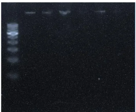
The DNA was isolated from the treated cells and subjected to agarose gel
electrophoresis. A ladder formation is the characteristic of apoptosis. The ladder is
formed due to the fragmentation of DNA by the cytotoxic effect (THP-1 leukemia cell
line) of ethanolic leaf extract (ELE) of B. lupulina. The fragmentation was visible at the
100 μg/mL concentration of extract. DNA of THP-1 showed intact form in all the
concentrations except in the concentration of 100 μg/mL which showed some amount of
contamination in form of RNA or proteins. Fig. 6 shows that DNA band in all the
concentration is in the pure form.
Reactive oxygen species (ROS) generation
Exposure of xenobiotic compounds induces a stress signal in the cells and as a
defense system cells start producing reactive oxygen species (ROS) which in turn helps
the cells to eliminate the negative effect of the xenobiotic compounds. However, in the
case of continuous production of cellular ROS, cellular system starts deteriorating and
thus inducing apoptosis in the cells. In the present study, B. lupulina extract at 100 μg/mL
induced ROS in the cells after 60 minutes of exposure and its effect continued upto 180
minutes (Fig. 5c).
Mitochondrial membrane potential (MMP)
Mitochondrial membrane potential was estimated with Rhodamine 123 dye to
evaluate the effect of B. lupulina extract on THP-1 cell line. Rhodamine 123 absorption
indicates the integrity of mitochondrial membrane. Thus, it is a direct measurement of
apoptosis. Results indicated that B. lupulina extract slightly increased the mitochondrial
membrane potential in the THP-1 cells at 100 μg/mL and in dose-dependent manner (Fig.
10 Gene expression analysis
The RNA was isolated from the control and treated cells and cDNA was
synthesized, further this cDNA was used in real time PCR or quantitative PCR for further
analysis of gene expression viz. β-actin and c-Myc. Amplification plot indicated the
suppression of c-Myc gene by 20 fold as compared to control. Melt curve of the samples
revealed the presence of few non-specific products that may be due to annealing
temperature of the primer.
Discussion
In medicinal plants different types of phytochemicals constituents are found in
medicinal plants viz., alkaloids, saponins, tannins, flavonoids, steroids, glycosides, etc.
But the chemicals components of plants may differ in different environmental/stress
conditions. Extractions of bioactive compounds also depend on the solubility of organic
solvents. In primary screening, different phyto-constituents of ethanolic leaf extract of B.
lupulina were separated on HPTLC plates that were terpenoid and alkaloid groups of
compounds. The extract revealed four spots on HPTLC plate and similar bioactive
compounds were found by Sur [19]. They also reported the presence of steroid, terpenoid,
glycoside, flavonoid, tannin and carbohydrate which were corresponds to six spots from
ethanolic extract of B. lupulina under UV (254 nm) in preparative TLC.
Medicinal plants contain various phytochemicals i.e. alkaloids, saponins, tannins,
flavonoids, steroids, glycosides whose composition can be vary along the
11
Some other bioactive compounds viz., benzene (1-methyl decyle), benzoic acid
4-methoxy-methyl ester, propenoic acid, benzyl benzoate and 2 (4H)-benzofuranone were
identified from acetone and methanol-soluble extracts of B. lupulina through GC-MS
[12]. Besides, slightly different constituents such as, tetradecane,
1,H-3a-7-methanoazulene, cis-thiopsine, benzofuranon, hexadecane, 3,7,11,15,
tetramethyl-2-hexadecanoic acid, 3-eicosyne, tetramethyl-2-hexadecanoic acid, oxiranehexadecyl (phytol), ethyl
9,12,15 octadecatrienoate and squalene have been found in this communication. These all
bioactive compounds have antimicrobial effects. More or less similar phyto-constituents
have been reported by Kumari and Dubey [15].
The percentage of viable cells decreased with the increase in the concentration of
extract on the THP-1 cell line. The extract has the ability to caused apoptosis in THP-1
cell line which was observed as cell membrane blebbing under the microscope. The
bio-active components of extract may be responsible for the apoptotic elimination of cancer
cell.
Choudhury et al. [20] have also reported the highest inhibition at 100 µg/mL on
DLA cell line. Ethanolic extract of B. lupulina and Calotropsis gigantean leaf mixture
exhibited the minimum number of DLA-cell viability. In this observation, THP-1 cell
viability started decreasing at minimum concentration (0.1 µg/mL) of extract. This is the
first report on THP-1 cells line because in above experiments method was same but cell
line was different.
NRU assay revealed cytotoxic effects of ethanolic extract of B. lupulina even at
the lowest dose (i.e. 0.1 µg/mL) and the IC50 values was 580 µg/mL. The IC50 value of
12
IC50 value was recorded against THP-1 than Hep G2 cells. Therefore, THP-1 cells were
more affected with the treatment of ethanolic extract than the Hep G2 cells [15].
The morphological changes in the cells were observed in treated cells which
showed extensive cell death. The cell viability of the cancerous cells exposed to B.
lupilina extracts decrease in the cells.
The methods, temperature and time of extraction, solvent type, concentration of
solvent, etc. affect the extraction of phytochemical constituents. Ethanolic extract of B.
lupulina exhibited the highest selective index (SI) (781.5) with lowest (IC50) 50%
inhibitory concentration dose (0.02 µg/mL) against HSV-2 cells [21]. It was the lowest
IC50 value. In contrast, IC50 value was higher because cell lines and solvent system were
different and extraction procedure was also different. However, it has been proved that
every cell line contains ability to survive in stress condition as well as exposure of
treatment of extracts.
The antiviral activity of B. lupulina and Clinacanthus nutans extract against five
HSV-2 isolates (IC50 of 442.2-987.7 µg/mL) was studied by Yoosook et al. [4].
Clinacanthus nutans did not show any activity against these virus strains. Jayavasu [22]
also observed high IC50 of C. nutans extract compared to B. lupulina. Multifarious ways
of cytotoxicity of B. lupulina extracts alone or in combination with other plant extracts
has also been reported by Maity et al. [23]. Who reported the effect of combined mixtures
of ethanolic extracts (EECGL+EEBLL) and water extract (WECGL+WEBLL) of
Calotropis gigantea latex and B. lupulina leaf extract on short-term in vitro cytotoxicity
on DLA cell line. The extracts at two doses (100, and 150 μg/mL) and 5-flurouracil
13
The combined mixtures of ethanolic extract of B. lupulina and C. gigantea showed the
minimum number of DLA-cell viability. The Allium cepa leaf extract has in vitro
cytotoxic, apoptotic and antiproliferative potential on Dalton’s Ascitic lymphoma cell as
well as DLA-bearing Swiss albino mice.
A Similar work has recently been published by Kumari and Dubey [15] on the
cytotoxic effect of ethanolic extracts of B. lupulina on Hep G2 cell line. They found
inhibitory effect of a single extract of B. lupulina on cancerous cells, Hep G2 and THP-1.
Therefore, these differences caused the different results. Methanolic leaf extract of
Barleria strigosa has been found to possess in vitro cytotoxicity against the P-388 murine
leukemia cell line with CC50 of 413.89 µg/mL [24].
Anti-inflammatory activities of B. lupulina and Clinacanthus nutans extracts
induced powerful dose-dependent inhibitory effects in both edema models in rats [2],
who found a significant inhibition of myeloperoxidase (MPO) activity in the inflamed
tissue indicating the association of anti-inflammatory effect of the extracts associated
with reduced neutrophil migration. Although both extracts did not affect neutrophil
viability or apoptosis, treatment of neutrophils with the extract concentration-dependently
inhibited fMLP-induced chemotaxis, superoxide anion generation, MPO and elastase
release. These findings suggest the powerful anti-inflammatory properties of B. lupulina
and C. nutans extracts are mediated by inhibition of neutrophil responsiveness.
Apoptosis has been studied with the ROS production and mitochondrial
membrane potential measurements. ROS production induced the intracellular damages
including the DNA damage. During apoptosis, mitochondrial membrane became more
14
cytotoxic effects and DNA fragmentation assay, B. lupilina extract induced ROS in the
cells from 30 minutes of exposure. B. lupilina extract lowered the mitochondrial
membrane potential significantly and thus inducing the apoptosis in the cells. The
ethanolic leaf extract had potential effect on cancerous cell line.
Conclusion
It may be concluded that B. lupulina extract decreased in the cell viability of the
cancerous cells. The present study also provides preliminary screening of this plant to
have potent cytotoxicity against the cancerous cells. We believe that the reported
improved method of DNA ladder assay will be very useful for numerous laboratories that
routinely study cell death or carry out routine experimental/clinical screening of drugs
and chemotherapeutics Therefore, it can be stated that B. lupulina may act as an
anti-cancer drug.
Availability of data and materials
The data sets that support the conclusions of this article are included within the article.
Abbreviations: MPO - Myeloperoxidase; MMP - Mitochondrial membrane potential; ROS - reactive oxygen species; ELE - ethanolic leaf extract; NRU - Neutral red uptake;
NCCS - National Centre for Cell Science; EMEM - Eagle’s minimum essential medium;
MTT - 3-(4,5-dimethylthiazol-2-yl)-2,5-diphenyl tetrazolium bromide.
References
[1] Gali K, Ramakrishnan G, Kothai R, Jaykar B.. In-vitro anti-cancer activity of
methanolic extract of leaves of Argemone mexicana Linn. International Journal of
15
[2] Wanikiat P, Panthong A, Sujayanon P, Yoosook C, Rossi AG, Reutrakul V. The
anti-inflammatory effects and the inhibition of neutrophil responsiveness by Barleria
lupulina and Clinacanthus nutans extracts. Journal of
ethnopharmacology. 2008;116(2):234-244.
[3] Saba N, Jawaid M, Hakeem KR, Paridah MT, Khalina A, Alothman OY. Potential of
bioenergy production from industrial kenaf (Hibiscus cannabinus L.) based on
Malaysian perspective. Renewable and Sustainable Energy
Reviews. 2015;42:446-459.
[4] Yoosook C, Panpisutchai Y, Chaichana S, Santisuk T, Reutrakul V. Evaluation of
anti-HSV-2 activities of Barleria lupulina and Clinacanthus nutans. Journal of
Ethnopharmacology. 1999;67(2):179-187.
[5] Sawangjaroen N, Phongpaichit S, Subhadhirasakul S, Visutthi M, Srisuwan N,
Thammapalerd N. Anti-amoebic activity of some medicinal plants used by AIDS
patients in southern Thailand. Parasitology Research. 2006;98:588-592.
[6] Chomnawang MT, Surassmo S, Nukoolkarn VS, Gritsanapan W. Antimicrobial
effects of Thai medicinal plants against acne-inducing bacteria. Journal of
Ethnopharmacology. 2005;101(1-3):330-333.
[7] Suba V, Murugesan T, Kumaravelrajan R, Subash C, Mandal B, Saha P.
Antiinflammatory, analgesic and antiperoxidative efficacy of Barleria lupulina
Lindl. extract.Phytotherapy research. 2005;19(8):695-9.
[8] Suba V, Murugesan T, Bhaskara RR, Ghosh L, Pal M, Mandal SC. Antidiabetic
16
[9] Suba V, Murugesan T, Rao RB, Pal M, Mandal SC, Saha BP. Neuropharmacological
profile of Barleria lupulina Lindl. Extract in animal models. Journal of
ethnopharmacology. 2002;81(2):251-255.
[10] Aguilar NO. Barleria lupulina Lindley. Plant Resources of South-East Asia No.
Medicinal and poisonous plants. 2001;12(2):100.
[11] Moin S, Babu SS, Mahalakshmipriya A. In vitro callus production and antibacterial
activity of Barleria lupulina Lindl. Asia-Pacific Journal of Molecular Biology and
Biotechnology. 2012;20(2):59-64.
[12] Kumari R, Dubey RC. Phytochemical analysis and antibacterial and cytotoxic
properties of Barleria lupulina Lindl. Extracts. Journal of Plant Pathology and
Microbiology. 2016a;7(10):2-6.
[13] Mazumder PM, Pattnayak S, Parvani H, Sasmal D, Rathinavelusamy P. Evaluation
of immunomodulatory activity of Glycyrhiza glabra L roots in combination with
zing. Asian pacific journal of tropical biomedicine. 2012;2(1):S15-S20.
[14] Tewtrakul S, Itharat A, Rattanasuwan P. Anti-HIV-1 protease- and HIV-1 integrase
activities of Thai medicinal plants known as Hua-Khao-Yen. Journal of
ethnopharmacology. 2006;105(1-2):312-15.
[15] Kumari R, Dubey RC. HPTLC and GC-MS profile of Barleria lupulina Lindl
extracts and their effect on enteric bacterial pathogens. Journal of Applied
Pharmacy. 2016b;8:62-8.
[16] Jayashree V, Thenmozhi N. In-Vitro Anti-Proliferative Assay and Cell Viability
Activity of Baicalein Using Breast Cancer Cell Line. International Journal of
17
[17] Perry SW, Norman JP, Barbieri J, Brown EB, Gelbard HA. Mitochondrial
membrane potential probes and the proton gradient: a practical usage
guide. Biotechniques. 2011;50(2):98-115.
[18] Derveaux S, Vandesompele J, Hellemans J. How to do successful gene expression
analysis using real-time PCR. Methods. 2010;50(4):227-230.
[19] Sur PK. Ameliorating effect of Barleria lupulina Lindl. Extract against γ
(gamma)-ray (1.2 Gy) induced mitotic chromosomal aberrations of house musk shrew
Suncus murinus. International Journal of Engineering Research and
Management. 2015;2(10):2349-058.
[20] Choudhury SM, Maity P, Bepari M. Combined mixtures of Calotropis gigantea
latex and Barleria lupulina leaf extracts ameliorate Dalton’s Ascitic Lymphoma
induced cell Proliferation. International Journal of Advanced Scientific and
technical Research. 2015;2:2394-3386.
[21] Wirotesangthong M, Rattanakiat S. Anti-herpes simplex virus type 2 activities of
some Thai medicinal plants. Thai Journal of Pharmaceutical Sciences.
2006;30(1-2):19-27.
[22] Jayavasu C. Clinical trial in the treatment of genital herpes patients with
Clinacanthus nutans extract. Communic Dis J (Thailand). 1992;18:152-161.
[23] Maity P, Choudhury S, Bepari M. Antimitotic, apoptotic and antineoplastic potential
of leaf extract of Eupatorium ayapana. International Journal of Phytomedicine.
2015;7(1):69-77.
[24] Manapradit N, Poeaim S, Charoenying P. Cytotoxicity and antimicrobial activities of
18
Acknowledgements
The authors wish to thank the Head, Department of Botany and Microbiology, Gurukul
Kangri University for providing laboratory facility and Acharya Balkrishna, Patanjali
Yogpeeth (Haridwar) for HPTLC and GC-MS analyses.
Funding
No funding was provided for the completion of this work.
Authors’ contributions
Conception, design, analysis and interpretation: RK and SK; drafting the manuscript for
important intellectual content: RK and SK; final approval of the manuscript RK and SK.
All approved the final manuscript.
Ethics declarations
NA
Consent for publication
NA
Competing interests
19
Figure 1. Different phyto-constituents of B. Lupulina ethanolic leaf exract at 366 nm on
20
Figure 2. Chromatograph representing the peak areas of phyto-constituent of B. Lupulina
21
Figure 3. The bar diagram shows the effect of B. lupulina ethanolic leaf extract in vitro
22
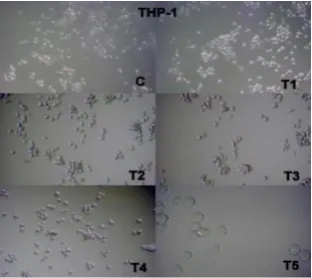
Figure 4. Cell viability and treatment of ethanolic leaf extract of B. lupulina on THP-1 cell line;
(c) control cells; (T1) treatment 1; (T2) treatment 2; (T3) treatment 3; (T4) treatment 4; (T5)
23
24