ischemic compression therapy for chronic carpal
tunnel syndrome
Guy Hains DC*
Martin Descarreaux DC, PhD†
Anne-Marie Lamy DC*
François Hains DC, FCCS(C), MSc*
* Private practice,
†Professor, Université du Québec à Trois Rivières, Québec Corresponding author:
Guy Hains, DC
2930 Côte Richelieu, Trois-Rivières, Québec, Canada, G8Z 3Y8
Phone: 819-375-5600,
E-Mail: [email protected]
Fax: 819-379-4397
© JCCA 2010
Méthodologie : essai clinique randomisé.
Objectif : le but de la présente étude est d’évaluer l’effet de la compressothérapie ischémique dans le traitement du syndrome du canal carpien chronique.
Méthode : Cinquante-cinq patients souffrant du syndrome du canal carpien ont été séparés aléatoirement en deux groupes. Trente-sept patients ont reçu 15
traitements expérimentaux, qui consistaient en des compressions ischémiques à des points gâchettes situés dans le creux axillaire de l’épaule, le long du biceps, à l’aponévrose bicipitale et au muscle rond pronateur situé dans le creux du coude. Dix-huit patients ont reçu le traitement contrôle, qui comprenait des compressions ischémiques sur des points gâchettes situés dans le muscle deltoïde, le muscle sus-épineux et le muscle sous-épineux. Des 18 patients du groupe contrôle, 13 ont accepté de recevoir les traitements expérimentaux à la suite des 15 traitements contrôles. Les mesures des résultats incluent un questionnaire validé de 18 questions servant à évaluer la gravité des symptômes et les capacités fonctionnelles relativement au syndrome du canal carpien, ainsi qu’une quantifi cation des améliorations perçues par les patients, au moyen d’une échelle allant de 0 % à 100 %. Les évaluations mesurant les résultats ont été effectuées au niveau de base, après 15 traitements, 30 jours après le dernier traitement et six mois plus tard.
Résultats : du côté du questionnaire sur l’invalidité,
Study Design: Randomized clinical trial.
Objective: The aim of this study was to evaluate the effect of ischemic compression therapy in the treatment of chronic carpal tunnel syndrome.
Method: Fifty-fi ve patients suffering from carpal tunnel syndrome were randomized to two groups. Thirty-seven patients received 15 experimental treatments which consisted of ischemic compressions at trigger points located in the axilla of the shoulder, the length of the biceps muscle, at the bicipital aponeurosis and at the pronator teres muscle in the hollow of the elbow. Eighteen patients received the control treatment involving ischemic compression on trigger points located in the deltoid muscle, supraspinatus muscle and infraspinatus muscle. Of the 18 patients forming the control group, 13 agreed to receive the experimental treatments after the 15 control treatments. Outcome measures included a validated 18-question questionnaire to assess the severity of symptoms and functional status in carpal tunnel syndrome, and a quantifi cation of the patients’ perceived improvement, using a scale from 0% to 100%. Outcome measures evaluations were completed at baseline, after 15 treatments, 30 days following the last treatment, and 6 months later.
Introduction
Carpal tunnel syndrome (CTS) is one of the most common and most clinically signifi cant of all nerve entrapment syndromes.1 Numbness and paresthesia along the
distri-bution of the median nerve in the hand, i.e. the thumb, index, major and half the ring fi nger are common symp-toms related to CTS. Sympsymp-toms and concurrent discom-fort often peak at night and may wake the patient several times. To ease pain and discomfort, the patient will shake the affected hand(s) and fl ex the fi ngers vigorously.2
Point prevalence of CTS is estimated at 2.7% and it is typically diagnosed in adults over the age of 30.3 The
symptoms generally originate from a nerve compression occurring when the median nerve runs through a fi brous
or fi broosseous tunnel or switches direction around a fi -brous or muscular band.2,4
Forty-seven percent of CTS cases can be related to the patient’s occupation. Over the last decades, there has been a major increase in work-related CTS cases.5,6
Compres-sion or entrapment may be present at a number of sites along the median nerve.7,8 To describe such phenomenon,
Leahy uses the expression “the whole nerve syndrome”.7
Conservative allopathic treatment usually includes wrist support, change in activities and anti-infl ammatory medication. If symptoms are not relieved by a conserva-tive approach within a six-month period, cortisone injec-tions may be used. Wrist surgery (carpal tunnel release) is considered where symptoms remain pronounced and
treatments it was 18.6 (SD, 7.0). The control group outcome at baseline was 36.3 (SD, 15.2); after 15 treatments it was 26.4 (SD, 9.9) and after the crossover (15 control treatments plus 15 experimental treatments) 20.2 (SD, 12.2). A signifi cant between group difference (P < 0.021) was noted in the patients’ perceived improvement after 15 treatments: 67 (SD, 26) percent and 50 (SD, 25) percent respectively for the experimental and control groups.
Conclusion: This practice-based clinical trial suggests that myofascial therapy using ischemic compression the length of the biceps, at the bicipital aponeurosis, at the pronator teres and at the subscapularis muscles could be a useful approach to reduce symptoms associated with the carpal tunnel syndrome. Patients’ perceived improvement in functional capacities persisted over a 6-month period.
(JCCA 2010; 54(3):155–163)
k e y w o r d s: chiropractic, myofascial trigger
points, ischemic compression, carpal tunnel syndrome, randomized clinical trial
une diminution signifi cative des résultats n’a été remarquée que chez le groupe expérimental. Pour le groupe expérimental, le résultat au niveau de base était de 33,5 (écart-type : 10,3); après 15 traitements, il était de 18,6 (écart-type : 7,0). Le résultat du groupe contrôle au niveau de base était de 36,3 (écart-type : 15,2); après 15 traitements, il était de 26,4 (écart-type : 9,9) et après le traitement croisé (15 traitements contrôles et 15 traitements expérimentaux) de 20,2 (écart-type : 12,2). Une différence signifi cative entre les groupes (p < 0,021) a été notée dans la perception qu’ont les patients de leur amélioration après 15 traitements : 67 % type : 26) et 50 % (écart-type : 25) respectivement pour le groupe expérimental et le groupe contrôle.
Conclusion : Cet essai clinique fondé sur la pratique suggère que la thérapie myofasciale au moyen de compressions ischémiques le long du biceps, à
l’aponévrose bicipitale, au muscle rond pronateur et aux muscles subscapulaires pourrait s’avérer un moyen utile de réduire les symptômes associés au syndrome du canal carpien. L’amélioration des capacités fonctionnelles, telle que perçue par les patients, a persisté pendant 6 mois.
(JCCA 2010; 54(3):155–163)
m o t s c l é s : chiropratique, point gâchette myofascial,
motor and sensitive functions decline.4,9 There are three
main reasons why patients agree to undergo surgery for CTS: (1) relief of night pain (36% of surgical patients), (2) relief of hand numbness (21%), (3) relief of daytime pain (13%).10
Furthermore, almost a third of the patients who have undergone CTS surgery experience persistent or recurrent symptoms after surgery and report that the initial improve-ment associated with carpal tunnel surgery is lost within less than two years.10 The most signifi cant discomfort
de-scribed by patients after carpal tunnel surgery is pain in the area of the scar, and a weakened hand.10 On average,
two years following surgery, 30% of patients characterize their results as being poor to medium.11
Natural history of CTS
When CTS is not treated surgically, the symptoms usu-ally disappear after nine months in the case of one half of those patients who do not move on to a surgical pro-cedure. However, 22% of such patients continue to have symptoms eight years later.12
Self-rating scales represent the most valid assess-ment method for CTS.13 When clinical symptoms are not
conclusive and common CTS diagnostic procedures are unable to confi rm the presence of median nerve compres-sion, it may be necessary to use electrodiagnostic proce-dures.13–15 These procedures are rarely appropriate for
initial CTS assessment, but are essential when it comes to pre-surgery examinations.13–15
Carpal tunnel syndrome is commonly treated in chiro-practic. In 1988, the number of cases of CTS declared by various specialists broke down as follows: chiropractors (23%), specialists in internal medicine (19%), neurolo-gists (14%), and family physicians (9%).5
A CTS survey study involving 254 physicians was car-ried out in 74 outpatient sentinel practices in 30 US states and three Canadian provinces. The authors of the study collected data from 552 CTS patients.16 Of this number,
23.5% were women, 70.4% were aged between 30 and 49, and 61.4% said that their work involved physical strain or repetitive movements. Clinicians determined that 43.1% of these cases were caused by the work itself. These prac-titioners rarely used electrodiagnostic procedures, prefer-ring conservative initial treatment such as wrist support and anti-infl ammatory medication, while cortisone injec-tions were rarely used.16 Another study showed that 40%
of 125 CTS sufferers who received conservative treat-ments over a period of 30 months said that they were will-ing to put up with their low-level residual symptoms for the rest of their life.17
Rationale for using ischemic compression therapy in the treatment of carpal tunnel syndrome
Since, in patients suffering from carpal tunnel syndrome, the median nerve is more than twice (2.1 times) its normal size when it enters the carpal tunnel,18 the authors of the
present trial hypothesized that part of the cause of the re-lated oedema could be noxious myofascial sites along the median nerve course. Along its course, part of this nerve enters the axilla of the shoulder, runs immediately adja-cent to the biceps, and descends within the hollow of the elbow under the pronator teres muscle and the bicipital aponeurosis. Other authors suggest that compression or entrapment may be present at a number of sites along the median nerve.7–8 In the present trial, the clinicians found
hypertonicity and trigger points (TrPs) along the biceps of every participant. Trigger points in the hollow of the elbow were also present in all cases. It was suspected that eliminating the trigger points located along the median nerve course would diminish the CTS symptoms with or without normalizing the size of the median nerve.
Figure 1 illustrates the trigger point locations along the biceps, at the bicipital aponeurosis and in the pronator teres muscle. In skeletal muscles the blood fl ow is extremely variable and it is tied to the activity level. At rest, only 25% of their capillaries are open.19 With exercise the blood
fl ow can increase up to 10 times, at which point almost all the capillaries open up to admit more blood.19 In the
present trial, the affected biceps (principally) was in par-tial and continual contraction because of TrPs. It is known that TrPs in a muscle cause a partial contraction.20,21 This
Our primary hypothesis of interest was that private clinic patients with CTS who are treated with ischemic compression on TrPs localized along the biceps, in the axilla and in the hollow of the elbow would exhibit more signifi cant reduction in the severity of symptoms and im-provement in functional status in comparison with patients treated with ischemic compression on TrPs localized in the deltoid, supraspinatus and infraspinatus muscles.
Methods
Participants
This prospective randomized clinical trial was conducted in a private clinic located in Trois-Rivières, Québec. The study was approved by the ethics committee of the Uni-versité du Québec à Trois-Rivières.
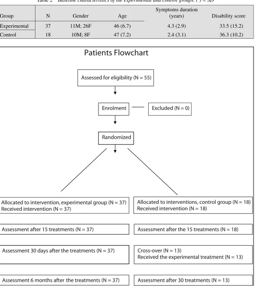
An advertisement was placed in a local newspaper on three different occasions offering CTS sufferers the op-portunity to take part in this research project. The fi rst 55 eligible patients were included in the study and un-derwent a course of 15 chiropractic treatments at a rate of three treatments per week (see Table 1). Thirty-seven patients received the experimental treatment; eighteen were given the control treatment (see Patient Flowchart). Patients accepted into the study were required to read and sign an informed consent form.
Randomization procedure
Each subject was randomly assigned to either the experi-mental group or the control group at a 2:1 ratio using a table of random numbers. Sixty numbers (2/3 even, 1/3 odd) were mixed in an envelope, and an independent re-search assistant drew a number for each participant, who was then allocated accordingly.
Treatment protocols
All the patients included in this study presented multiple trigger points (TrPs) and taut bands along the biceps and at the bicipital aponeurosis. TrPs at the pronator teres muscle were also common clinical fi ndings, but were not present in two patients. Twenty patients had TrPs in the axilla of the shoulder. All patients were examined for TrPs in these four areas while in a supine position, the arm supine and spread along the body or, in the case of the axilla of the shoulder, the hand of the patient under his head.
Patients were advised to stop any treatments other than that provided by the chiropractor treating their CTS. During the treatment, at each visit, pressure was applied for 5–15 seconds to each of the identifi ed trigger points. Thumb tip pressure (one thumb over the other) was then applied for 5 seconds every 2 cms, along the biceps. For the TrPs located in the hollow of the elbow (pronator teres, biceps aponeurosis) and in the axilla (subscapularis), the pressure was maintained for 15 seconds. Trigger points were treated using a light pressure, which was gradually increased until it reached the participant’s maximum pain tolerance level. The patients were blinded to treatment al-location and therefore did not know whether they were in the control or the experimental group.
Seventeen patients received 15 control treatments con-sisting of ischemic compressions of latent or active trig-ger points located in the posterior region of the clavicle (supraspinatus area), on the deltoid (anterior and lateral region), and on the center of the shoulder blade (infra-spinatus area). Since TrPs are often found in these loca-tions, this control treatment would appear plausible to the patient and the authors believed that it would not induce signifi cant clinical changes as concerned CTS symptoms. Following the control treatment phase of the study, the 18 patients who received fi fteen control treatments were offered the opportunity to receive fi fteen further treat-ments. They were still blinded to the kind of treatment they would receive. Thirteen agreed to continue with the treatment and, this time, only the experimental treatment was given.
Outcome measures
In order to quantify the severity of symptoms and the func-tional status of patients, a standard validated question-naire20 specifi c to patients suffering from CTS was used.
The scales used in this questionnaire are highly reprodu-cible (Pearson correlation coeffi cient, R = 0.91 and 0.93 for severity of symptoms and functional status, respect-ively) and internally consistent (Cronbach alpha, 0.89 and 0.91 for severity of symptoms and functional status, respectively).20 This type of questionnaire was chosen
be-cause carpal tunnel patients generally consult their clin-icians due to the severity of the symptoms experienced and the diffi culty they have in carrying out normal daily tasks. The fi rst part of the questionnaire defi nes the func-tional status of the patient while carrying out eight daily activities. The second part, using 10 simple questions,
de-fi nes the severity of the symptoms experienced by the pa-tient. All participants completed the questionnaire before and after the treatment protocol. A numerical scale where patients could rate their perceived improvement from 0% to 100% was also used. For the experimental group the questionnaire and numerical scale were also completed 30 days after the last treatment and 6 months later. For the control group, the questionnaire and the numerical scale were also completed after the crossover. The question-naires were fi lled in without the assessor being present.
Statistical analysis
To test the effects of experimental treatment over the Time, a repeated-measures one-way ANOVA was per-formed. The same analysis was completed to test the cross-over effect in the control group. When a main effect of Time was observed for the experimental group, post hoc comparisons were performed using Tukey tests. A t-test for independent samples was used to compare the perceived improvement percentage between the control and experimental groups after 15 treatments. For all an-alyses statistical signifi cance was set at p < 0.05.
Results
All 55 participants received the initial fi fteen treatments and completed the questionnaires as intended. Fifty-fi ve patients were randomly assigned to one of the two treat-ment groups. The t-test showed no statistically signifi cant difference between the two groups’ baseline characteris-tics (see Table 2).
The experimental group symptoms and functional sta-tus scale questionnaire mean scores (and standard devia-tions) were 33.5 (SD, 10.3) at baseline; 18.6 (SD, 7.0)
Inclusion Exclusion
• Be between 20 and 60 years old.
• Suffer from numbness in the hand affecting the thumb, the index fi nger, the middle fi nger and one half of the ring fi nger. • Have suffered on a daily basis for at least 3 months.
• Agree to a course of 15 chiropractic treatments at no cost. • All patients had to show at least 2 of the following physical
signs: a Tinnel positive sign, a Phallen positive sign, sleep problems caused by hand discomfort.
Table 2 Baseline characteristics of the experimental and control groups. ( ) = SD
Group N Gender Age
Symptoms duration
(years) Disability score
Experimental 37 11M; 26F 46 (6.7) 4.3 (2.9) 33.5 (15.2)
after 15 treatments; 17.5 (SD, 6.1) thirty days following the last treatment, and 20.7 (SD, 7.4) at 6 months. The experimental group maintained a signifi cant reduction in the symptoms and functional status scale questionnaire scores at both follow-up evaluations (One-way ANOVA: F (3, 108) = 39.2, p < 0.0001). Conversely, the
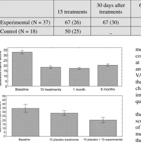
repeated-measure ANOVA yielded a signifi cant decrease for the control group only after the cross-over: 36.3 (SD, 15.2) at baseline; 26.4 (SD, 9.9) after 15 placebo treatments; and 20.2 (SD, 12.2) after the cross-over (One-way ANO-VA: F (SD, 2, 24) = 10.1, p < 0.001). Figure 2 illustrates the symptoms and functional status scale questionnaire changes in both groups throughout the trial. Mean (SD) improvement in the symptoms and functional status scale questionnaire are presented in Table 3.
A signifi cant difference (p < 0.021) was noted between the two groups regarding the perceived improvement scores after 15 treatments. The mean perceived percentage of improvement was 67 (26) and 50 (25) for the experi-mental and control groups respectively. Table 4 presents the mean scores from the perceived improvement numeri-cal snumeri-cale for both groups throughout the experiment.
Discussion
In this study, patients’ symptoms associated with CTS improved in the majority of patients who received is-chemic compression therapy in the axilla of the shoul-der, the length of the biceps, at the bicipital aponeurosis and at the pronator teres muscle. The data from the two questionnaires showed an improvement in both groups, but the improvement was signifi cantly greater in the ex-perimental group than in the control group. Moreover, a signifi cant reduction in pain and improved functional status were noted after the crossover (75% improvement) when the participants in the control group received the Table 3 Mean (SD) improvement in severity of symptoms and functional status (%).
15 treatments
30 days after treatments
6 months after treatments
15 control plus 15 experimental treatments
Experimental (N = 37) 42 (21) 45 (21) 36 (23)
Control (N = 18) 26 (18) _ _ 48 (15)
Table 4 Mean (SD) score from the perceived improvement numerical scale (%).
15 treatments
30 days after treatments
6 months after treatments
15 control plus 15 experimental treatments
Experimental (N = 37) 67 (26) 67 (30) 56 (35)
Control (N = 18) 50 (25) _ _ 75 (21)
Baseline 15 treatments 1 month 6 months
Baseline 15 placebo treatments 15 placebo + 15 experimental 35
30 25 20 15 10 5 0
Questionnaire score
Questionnaire score
50 45 40 35 30 25 20 15 10 5 0
experimental treatment. Even though the study protocol included 15 treatments, many patients (89%) in the ex-perimental group reported improvement within six treat-ments. They either said so spontaneously or when asked by the clinician during the 6th visit. In this study, only
ischemic compression therapy was used, but one may suppose that the results could be improved if such ther-apy was combined with ergonomic recommendations per se.23 No side effects were reported during the treatments,
except for a slight sensitivity reported by a small number of patients after the fi rst few treatments.
The most pathognomonic symptom of myofascial pain syndrome is the presence of pressure-sensitive palpa-ble nodules that reproduce the chief complaint: they are called trigger points.24 These TrPs may be located in
mus-cles, ligaments, tendons, fascias and articular capsules.25
Ischemic compressions are amongst the most popular methods of treatment used by chiropractors for patient care of the myofascial pain syndrome. The National Board of Chiropractic Examiners 2005 Job Analysis re-ported that over 91% of chiropractors use trigger point therapy for passive adjustive care.26
Overuse of the biceps can cause myofascial irritations and subsequent hypertonicity. Gerwin22 claims that a
my-ofascial trigger point refers to a zone of intense pain in a hardened muscle band that triggers pain when mechani-cally stimulated by plucking it manually. He added that there is a segmental hyper-contraction within the muscle fi ber. The present authors speculate that the hypertonic-ity of the biceps, pronator teres and subscapularis mus-cles can irritate the median nerve and may cause local oedema. Consequently, the nerve may be pinched when it runs through the narrow space of the carpal tunnel, and this would result in numbness and impairment of distal motor and sensory functions. The longer this process lasts (months-years), the more severe the neuropathy becomes, causing muscular weakness in the hand. We would argue that eliminating the TrPs along the median nerve relaxes the muscles and removes a source of irritation to the me-dian nerve.
The treatment of the whole median nerve was used effectively in a case study by Leahy.7 The median nerve
may be damaged along its whole length, from its root, between the cervical vertebrae, down to and including the wrist.8 According to Bonebrake et al.,27
conserva-tive treatment of CTS is intended to lessen muscular
and fi brous restriction. In their study, treatment was ap-plied along the whole median nerve and, amongst other techniques, they used ischemic compression. In a recent trial by George,28 fi ve patients suffering from CTS were
treated three times weekly for two weeks using the Active Release Technique (ART) with a protocol designed to af-fect the median nerve. Using the Boston Questionnaire, they concluded that ART offered a signifi cant reduction of the symptom severity and improvement of the functional status of the patients.
Davis29 published a randomized clinical trial that
showed a signifi cant improvement in CTS syndrome amongst the patients. Myofascial massage along the me-dian nerve was used with the chiropractic group but, since there were other modalities involved, the authors could not assess which was the active component of their inter-vention.
Limitations of the study
The total number of participants was small and there were only two treating chiropractors. The number of patients in the control group was small, compared with the number of those in the treatment group. The reason for this was that the treating clinicians found it diffi cult to construct a practice-based study that provided a group with what they considered would be a near placebo treatment. There was only a short-term follow-up comparison of the two group results and therefore it is unclear whether the re-sults reported in this study would persist beyond the point of treatment cessation. The compression sites treated by the clinicians were considered very important on the basis of clinical experience, though very few others have treat-ed these sites in the context of carpal tunnel syndrome. Finally, the control group was crossed over immediately after the 15 initial control treatments. In the absence of a wash-out period, a potential nonspecifi c effect of the pla-cebo intervention could have carried over into the active treatment period among those patients who did participate in the crossover portion of the study.
Conclusion
tunnel syndrome. Patients’ perceived improvement in functional capacities persisted over a six-month period. This last observation is based only on the before-and-after analysis of within group data. Future research on CTS should include a larger number of participants, a parallel placebo treatment group and long-term assessments.
References
1 Nordstrom DL, Vierkant RA, DeStefano F, Layde PM. Risk factors for carpal tunnel syndrome in a general population. Occup Environ Med. 1997; 54:734–40. 2 Phalen GS. The carpal-tunnel syndrome. Clinical
evaluation of 598 hands. Clin Orthop Relat Res. 1972; 83:29–40.
3 Atroshi I, Gummesson C, Johnson R, et al. Prevalence of carpal tunnel syndrome in the general population. JAMA. 1999; 282:153–158.
4 Practice parameter for carpal tunnel syndrome (summary statement). Report of the Quality Standards Subcommittee of the American Academy of Neurology. Neurology. 1993;
43:2406–9.
5 Occupational disease surveillance: carpal tunnel syndrome. MMWR Morb Mortal Wkly Rep. 1989; 38:485–9.
6 Einhorn N, Leddy JP. Pitfalls of endoscopic carpal tunnel release. Orthop Clin North Am. 1996; 27:373–80. 7 Leahy M, Mock L. Myofascial release technique and
mechanical compromise of peripheral nerves of the upper extremity. Chiropr Sports Med. 1992; 6:139–50.
8 Normand MC, Descarreaux M. Est-ce vraiment un syndrome du canal carpien? J Can Chiropr Assoc. 2000; 44:149–56.
9 Keller RB, Largay AM, Soule DN, Katz JN. Maine Carpal Tunnel Study: small area variations. J Hand Surg [Am]. 1998; 23:692–6.
10 Bessette L, Keller RB, Liang MH, Simmons BP, Fossel AH, Katz JN. Patients’ preferences and their relationship with satisfaction following carpal tunnel release. J Hand Surg [Am]. 1997; 22:613–20.
11 Cotton P. Symptoms may return after carpal tunnel surgery. JAMA. 1991; 265:1922, 1925.
12 DeStefano F, Nordstrom DL,Vierkant RA. Long-term symptom outcomes of carpal tunnel syndrome and its treatment. J Hand Surg [Am]. 1997; 22:200–10.
13 Katz JN, Gelberman RH, Wright EA, Lew RA, Liang MH. Responsiveness of self-reported and objective measures of disease severity in carpal tunnel syndrome. Med Care. 1994; 32:1127–33.
14 Phalen GS. The birth of a syndrome, or carpal tunnel revisited. J Hand Surg [Am]. 1981; 6:109–10.
15 Atcheson SG. Carpal tunnel syndrome: is it work-related? Hospital Pract (Off Ed). 1999; 34:49–56; quiz 147. 16 Miller RS, Iverson DC, Fried RA, Green LA, Nutting PA.
Carpal tunnel syndrome in primary care: a report from ASPN. Ambulatory Sentinel Practice Network. J Fam Pract. 1994; 38:337–44.
17 Katz JN, Larson MG, Sabra A, Krarup C, Stirrat CR, Sethi R, Eaton HM, Fossel AH, Liang MH. The carpal tunnel syndrome: diagnostic utility of the history and physical examination fi ndings. Ann Intern Med. 1990; 112:321–7. 18 Mesgarzadeh M, Schneck CD, Bonakdarpour A. Carpal
tunnel: MR imaging. Part I. Normal anatomy. Radiology. 1989; 171:743–8.
19 Marieb EN. Essentials of human anatomy and physiology. 2nd ed. 1988, Menlo Park, Calif.; Don Mills, Ont.: Benjamin/Cummings Pub. Co. xii, 417.
20 Levine DW, Simmons BP, Koris MJ, Daltroy LH, Hohl GG, Fossel AH, Katz JN. A self-administered questionnaire for the assessment of severity of symptoms and functional status in carpal tunnel syndrome. J Bone Joint Surg Am. 1993; 75:1585–92.
21 Simons DG. Review of enigmatic MTrPs as a common cause of enigmatic musculoskeletal pain and dysfunction. J Electromyography Kinesiol. 2004; 14:95–107.
22 Gerwin RD, Dommerholt J, Shah J P. An expansion of Simons’ integrated hypothesis of trigger point formation. Current Pain and Headache Reports. 2004; 8:468–75. 23 Verhagen AP, Bierma-Zeinstra SM, Feleus A, Karels C,
Dahaghin S, Burdorf L, de Vet HC, Koes BW. Ergonomic and physiotherapeutic interventions for treating upper extremity work-related disorders in adults. Cochrane Database Syst Rev. 2004; CD003471.
24 Borg-Stein J, Stein J. Trigger points and tender points. Rheum Dis North Am. 1996; 22(2):305–322.
25 Travel JG, Simons DG. Myofascial pain and dysfunction; the trigger point manual. Vol.1. Philadelphia: Williams and Wilkins 1983: p. 19.
26 Christensen M. Job analysis of chiropractic. National Board of chiropractic examiners. Greeley, CO. 2005; 136. 27 Bonebrake AR, Fernandez JE, Dahalan JB, Marly RJ. A
treatment for carpal tunnel syndrome: results of a follow-up study. J Manipulative Physiol Ther. 1993; 16:125–39. 28 Georges JW, Tepe R, Busold D et al. The effects of Active
Release Technique on carpal tunnel patients: a pilot study. J Chiropr Med. 2006; 5:119–121.