Article
Exploring the Aortic Root Diameter and left Ventricle
Size Among Lithuanian Athletes
Renata Žumbakytė-Šermukšnienė 1, *, Agnė Slapšinskaitė 1,2, Miglė Baranauskaitė 1, Julija Borkytė 1, Rasa Sederevičiūtė4and Kristina Berškienė 1.
1 Lithuanian University of Health Sciences, Sports Institute, Tilžės st. 18, LT – 47181, Kaunas, Lithuania; 2 Lithuanian University of Health Sciences, Health Research Institute, Tilžės st. 18, LT – 47181, Kaunas,
Lithuania;
3 Lithuanian University of Health Sciences, Radiology Clinic, Eivenių st. 2, LT – 50009, Kaunas Lithuania * Correspondence: [email protected]; Tel.: +370-37-362249
Abstract: Aortic rupture is known as one of the potential causes of sudden cardiac death in athletes. Nevertheless, adaptation strategies for aortic root dilation in athletes vary. The purpose of this study
was to investigate aortic root adaptation to physical workload and to determine if aortic root’s and left ventricle sizes are contingent upon the physical workload. Echocardiography was applied to
151 subjects to measure the aortic root at aortic valve annulus (AA) and at sinus of Valsalva (VS). 122 were athletes (41 females and 81 males) and 29 were non-athletes (14 females and 15 males). Of the 41 female athletes, 32 were endurance athletes, and 9 strength athletes. From 81 male athletes,
56 were endurance athletes, and 25 were strength athletes. AA and VS mean values for the body surface area were presented as rAA and rVS. Left ventricle (LV) meaures incuded LV end-diastolic
diameter (LVEDD), interventricular septum thickness in diastole (IVSTd), LV posterior wall thickness in diastole (LVPWTd), LV mass (LVM), LV mass index, LV end-diastolic diameter index
(LVEDDI). Results indicated that VS was higher in female athletes (28.9±2.36mm) than in non-athletes (27.19±2.87mm, p=0.03). On the other hand, rAA was higher in strength athletes (12.19±1.48mm/m2) than in endurance athletes (11.12±0.99mm/m2, p=0.04). Additionally, rVS and rAA were higher in female strength athletes (17.19±1.78mm/m2, 12.19±1.48mm/m2) than female basketball players (15.49±1.08mm/m2, p=0.03, 10.75±1.06 mm/m2, p=0.02). Statistically significant positive moderate correlations were found between VS and LVEDD, LVM, IVSTd, LVPWTd, rVS and LVEDDI parameters in all athletes. The diameter of Valsalva sinus was greater in female athletes
compared to non-athletes. The rAA mean value for body surface area was greater in female athletes practising strength sports as compared to their counterparts who were practising endurance sports.
The diameter of the aortic root at sinuses positively correlated with the LV size in all athletes. Trial was registered at ClinicalTrials.gov Identifier: NCT03656861, September 3, 2018 (retrospectively
registered).
Keywords: Performance, Sports, Aortic Valve, Sinus of Valsalva, Echocardiography, Sudden cardiac death.
1. Introduction
Recent developments in football have seen the sudden death of young football player due to
aortic rupture hence reinforcing the controversy of football as a field with substantial risk for sudden cardiac arrest and death [1–5]. Moreover, there is an argument that aortic dilatation and the
subsequent event of thoracic aortic aneurysm may be an occupational disease due to the nature of
some vocations (i.e., military and security personnel, blue collar workers, weightlifters, athletes etc.) [6]. Of particular importance, there is some evidence that elite athletic training is associated with small but significantly larger aortic root diameter (i.e., sinuses of Valsalva) [7]. Surprisingly,
adaptation strategies for aortic root dilation in athletes still vary to a large extent [8].
To that end, the differential impact of distinct forms of exercise on arteries in strength athletes
vs. endurance athletes have not been investigated [9]. Further advancement of this topic could be of particular interest for clinicians. For instance, a consensus or informed recommendation for clinicians
facing cases of higher aortic indices, with higher body surface area and with or without remodelled LV were called upon [9].
Of interest to this study, it is known that endurance sports can lead to volume overload and increase in the diameter of the heart chambers. Strength sports can cause pressure overload and are
particularly related to the thickening of the LV wall [10]. While “athlete’s heart” is a long investigated topic within sports medicine, Green et al. have provided some insights into the functioning of
“athlete’s artery” [9]. In fact, more information into cardiac pathophysiological training-caused adaptation is needed. Increased understanding of this adaptation could indeed help reduce risk for sudden cardiac arrest and death and/or irreversible cardiac damage identification and this
importance [11].
Zeppill et al. [12] were among the first ones to report a wider diameter of arteries and veins in
endurance athletes compared to non-athletes. Similarly, others have also suggested significant differences between arteries in athletes and non-athletes [9]. Nevertheless, findings from work
investigating aortic root vessels were controversial and inconsistent. Specifically, Pelliccia et al. [13] found significant differences between endurance athletes and the control group. However, while
endurance athletes had higher diameter in aorta compared to non-athletes, none of these results were replicated in strength athletes.
In fact, LV remodelling work has indicated LV association with physical activity type. However,
to date little is known if the changes of the aortic root are linked with LV size. Specifically, clinicians face an issue of whether to set higher aortic indices for athletes with higher body surface area and
with or without remodelled LV (i.e., basketball players, strength athletes) in cases when relative values are normal. Thus, interpretation of increased rates of aortic indices with normal relative values
is challenging especially among non-athletes, overweight and obese individuals. To further complicate things, it is also known that for physically inactive persons with metabolic syndrome, LV
remodelling and blood pressure are co-occurring with hypertension.
In the present study, we evaluated the development of the aortic root diameter with blood
pressure, LV size, and physical activity type. Consequently, the purpose of this study was to investigate aortic root adaptation to physical workload and to determine whether aortic root’s size and left ventricle remodelling is dependent on physical load. Drawing upon previous studies, we
2. Materials and Methods
Sample
A preliminary data was collected from a total of 944 subjects in Kaunas Sports Medicine Centre during the recruiting period in 2014-2015. All the experimental procedures were approved by the
Lithuanian Ethics Committee of the Lithuanian University of Health Sciences (No. BEC – MF – 481) and conducted in accordance with the Declaration of Helsinki. All subjects have written informed
consent. Final data analysis consisted of 151 Caucasian subjects who met the inclusion criteria. The subjects were divided into groups according to gender (i.e., female, male), physical activity (i.e.,
athletes, non-athletes) and physical activity type (i.e., strength and endurance sports). Of the 151 subjects, 122 were athletes (41 females and 81 males) and 29 were non-athletes (14 females and 15 males). Of the 41 female athletes, 32 were endurance athletes, and 9 strength athletes. From 81 male
athletes, 56 were endurance athletes, and 25 were strength athletes. Of importance herein, we expected that a large body surface area of the basketball players may have an effect to the relative
values of the aortic root diameter. Due to the aforementioned reasoning, the basketball players were separated from other endurance athletes. To that end, 12 female, 19 male basketball players took part
in the study (see Table 1, Table 2 and Table 3).
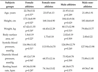
Table 1.Baseline characteristics of study participant’s athletes and non-athletes.
Subjects groups
Female athletes
Female
non-athletes Male athletes
Male non-athletes
Age, years 22.78±5.52
p=0.57* 23.07±6.13
21.97±5.41
p=0.28† 21.49±1.91
Height, cm 173.14±8.99
p=0.02* 168.14±4.90
184.41±9.04
p=0.62† 183.44±8.69
Weight, kg 67.42±11.55
p=0.30* 64.43±12.29
80.17±12.98
p=0.51† 79.00±15.77
Body surface area, m2
1.8±0.19
p=0.22* 1.73±0.18
2.02±0.19
p=0.50† 2.00±0.22
Systolic blood pressure,
mmHg
116.98±11.82
p=0.51* 113.93±14.70
124.09±12.79
p=0.24† 127.94±13.98
Diastolic blood
pressure, mmHg
69.12±7.59
p=0.94* 68.57±12.16
73.92±8.78
p=0.59† 73.89±11.95
Resting heart rate, bpm
69.34±10.98
p=0.28* 74.14±13.02
68.18±9.75
p=0.57† 69.56±7.44
Notes: the symbol “*” indicates p – value of comparison between female athletes and non-athlete’s groups
The symbol “†” - indicates p – value of comparison between male athletes and non-athlete’s groups
Table 2.Baseline characteristics of female endurance, strength and basketball players.
groups athletes athletes players
Age, years 23.08±5.69 p=0.68*
21.72±4.34
p=0.42† 23.66±4.56
Height, cm 175.41±8.13 p=0.01*
165.22±7.55
p=0.002† 179.25±7.96
Weight, kg 49.25±11.62 p=0.03*
60.89±9.08
p=0.01† 74.67±10.75 Body surface
area, m2
1.83±0.19
p=0.03*
1.67±0.15
p=0.003† 1.93±0.18 Years of professional training, years 11.63±6.38 p=0.68* 8.78±6.08
p=0.42† 12.75±5.58
Systolic blood pressure, mmHg 116.94±9.89 p=0.94* 117.11±17.86
p=0.60† 120.00±11.28
Diastolic blood pressure, mmHg 69.38±7.59 p=0.72* 68.22±7.97
p=0.55† 70.83±9.00
Resting heart
rate, bpm
69.16±10.43
p=0.82*
70.00±13.42
p=0.51† 67.92±11.67 Hours of training, hours/week 8.05±3.74 p=0.30* 7.83±3,34 p=0.22† 7.33±2.78
Notes: the symbol “*” indicates p – value of comparison between endurance and strengthathlete’s groups
The symbol “†” - indicates p – value of comparison between basketball players and strengthathlete’s
groups
Table 3.Baseline characteristics ofmale endurance, strength and basketball players.
Subjects groups Endurance athletes Strength athletes Basketball players
Age, years 20.36±4.18 p<0.001*
25.38±6.17
p<0.001† 18.83±2.46
Height, cm 186.28±8.93 p=0.01*
180.44±8.07
p<0.001† 192.21±6.44
Systolic blood
pressure, mmHg
123.62±12.81
p=0.78*
125.08±12.95
p=0.30† 127.47±13.63
Diastolic blood pressure,
mmHg
72.64±9.34 p=0.09*
76.64±6.87
p=0.05† 70.53±9.11
Resting heart rate, bpm
70.15±10.62 p=0.005*
64.00±5.83
p=0.005† 70.84±9.03
Hours of training,
hours/week
8.93±4.03 p=0.68*
7.80±2.99
p=0.49† 7.21±2.18
Notes: the symbol “*” indicates p – value of comparison between endurance and strength athlete’s groups
The symbol “†” - indicates p – value of comparison between basketball players and strength athlete’s groups
The inclusion criteria were as follows:
•
A total of 2D transthoracic echocardiography performed in Kaunas Sports Medicine Centre 2014-2015.•
Age range 16- 35 years, given the literature definition of “young” and “old” athletes as < 35 and > 35 years [1].•
Physical activity levels (athletes or non-athletes). Individuals who participated in sports for more than 4 years and 4.5 hours per week were included in the group of athletes. Individual who were active for less than 4 years and/or 4.5 hours per week were classified as non-athletes [14].•
Physical activity type (i.e., endurance and strength sports).•
No current or previous history cardiovascular diseases.•
No activity on the test day.•
Consent to participate in the study.Individuals who did not meet one or more of the inclusion criteria were excluded from the
study.
Protocol
All subjects underwent two-dimensional (2D) transthoracic echocardiography (TTE) procedure.
Prior to performing 2D TTE, subjects’ arterial blood pressure [15], heart rate, height, weight, and self-reported physical activity levels were measured .
The Ultrasound system CX50 (Philips Ultrasound, Philips Healthcare, Philips Medical Systems
Nederland, USA) – with transducer S5-1 was used in this study. Two physicians performed 2D TTE and averages for all variables of interest were computed. The measurements of aortic root and the
left ventricle were drawn upon the guidelines of the American Society of Echocardiography and the European Association of Cardiovascular Imaging [16]. The maximal diameter of the sinuses of
Valsalva was measured at end-diastole, in a strictly perpendicular plane to that of the long axis of the aorta using the edge to leading edge (L-L) convention. The aortic annulus was measured at
midsystole from inner edge to inner edge (I-I). This was done in order to obtain the rounder shape and bigger diameter of aortic annulus. All measurements were taken in the perspective that depicts
Due to the significant difference (p< 0.05) between the subjects' height and weight in the groups of athletes and non-athletes, we evaluated the relative indices with the area of the body surfaces. Relative body surface area was calculated by means of Mosteller’s (1987) formula [17] (see Figure 1).
𝐵𝑆𝐴 (𝑚2) = (ℎ𝑒𝑖𝑔ℎ𝑡(𝑐𝑚) × 𝑤𝑒𝑖𝑔ℎ𝑡(𝑘𝑔)/3600)1 2⁄
Figure 1. Formula for relative body surface calculation.
The following dimensions of the root of the aorta were evaluated as follows: sinuses of Valsalva
(VS, mm) and its relative index with body surface area (rVS, mm/m2), aortic valve annulus (AA, mm) and its relative index with body surface area (rAA, mm/m2).
For left ventricle (LV) measurements the following were used: LV end-diastolic diameter (LVEDD, mm), interventricular septum thickness in diastole (IVSTd, mm), and LV posterior wall thickness in diastole (LVPWTd, mm). Subsequently we computed: LV mass (LVM, g), LV mass index (LVMI, g/m2), LV end-diastolic diameter index (LVEDDI, mm/m2). The formula for computing the mass of the LV was drawn upon the most recent recommendations of the American Society of Echocardiography and the European Cardiovascular Association [14] (see Figure 2).
𝐿𝑉𝑀 = 0.8 × (1.04((𝐿𝑉𝐸𝐷𝐷 + 𝐿𝑉𝑃𝑊𝑇𝑑 + 𝐼𝑉𝑆𝑇𝑑3)) + 0.6 𝑔𝑟𝑎𝑚
𝐴𝑏𝑏𝑟𝑒𝑣𝑖𝑎𝑡𝑖𝑜𝑛𝑠:
𝐿𝑉𝑀 − 𝑙𝑒𝑓𝑡 𝑣𝑒𝑛𝑡𝑟𝑖𝑐𝑢𝑙𝑎𝑟 𝑚𝑎𝑠𝑠 𝐿𝑉𝐸𝐷𝐷 − 𝐿𝑉 𝑒𝑛𝑑 − 𝑑𝑖𝑎𝑠𝑡𝑜𝑙𝑖𝑐 𝑑𝑖𝑎𝑚𝑒𝑡𝑒𝑟
IVSTd – interventricular septum thickness
LVPWTd – LV posterior wall thickness
Figure 2. Formula for the mass of the LV calculation.
To determine the potential association between aorta root size and the size of the LV and blood
pressure, we computed correlational analyses between VS and LV size: LVEDD, LVM, IVSTd, LVPWTd. As well as systolic blood pressure and its correlation with rVS and LVEDDI, LVMI. The
quantitative values are given as the arithmetic mean ± standard deviation (x±SD). Student t test was used to compare the independent samples. The correlation coefficient (r) of Spearman was used for assessing the direction and strength between variables. A |r|<0.3 was considered weak, while 0.3≤|r|≤0.7 was considered as moderate and |r|>0.7 was considered strong. We deemed a p value less than 0.05 to be significant.
3. Results
3.1 Aortic root diameter
There were no significant differences between the female endurance athletes (28.99±2.32mm) and strength athletes 28.56±2.64mm (p=0.48) with regards to VS. Moreover, while comparing relative measurements with body surface area, rVS tended to increase in female strength athletes
(17.19±1.78mm/m2) in comparison to female endurance athletes (15.90±1.44mm/m2, p=0.06).
There were no significant differences of AA diameter between female endurance athletes and
strength athletes. However, rAA was significantly higher in female strength athletes than female endurance athletes (12.19±1.48mm/m2 versus 11.12±0.99mm/m2, p=0.04).
There were significant differences between the relative values of the aortic root neither at the AA nor VS between basketball players and the strength athletes. However, statistically significant
differences were observed (p=0.03) in rVS between strength athletes (17.19±1.78mm/m2) and female basketball players (15.49±1.08mm/m2). rAA in female strength athletes (12.19±1.48mm/m2) was higher than in the female basketball players (10.75±1.06mm/m2, p=0.02).
There were no significant differences with regards to the aortic root between male athletes and
non-athletes. However, a tendency for VS to increase in male athletes (VS was 32.38±2.96mm, rVS was 16.11±1.67mm/m2 and VS – 31.39±2.72mm, p=0.14, rVS – 15.80±1.58mm/m2, p=0.35, respectively) was observed. Similarly, no significant differences were obtained with regards to aortic root in male
endurance athletes and strength athletes but there was a tendency of VS to increase in the strength athletes as opposed to endurance athletes (VS – 32.08±3.01mm, rVS – 16.00±1.63mm/m2 and VS – 33.02±2.80mm, p=0.20, rVS – 16.34±1.75mm/m2, p=0.32, respectively). Finally, with regards to aortic root in male athletes, VS relative ratios tended to increase in strength athletes as opposed to basketball
players (15.47±1.38mm/m2 and 16.34±1.75mm/m2, p=0.11, respectively).
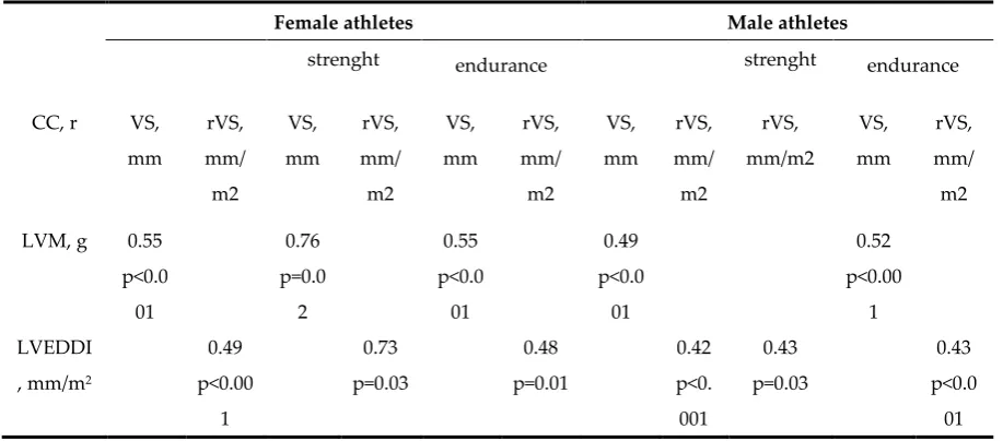
3.2 Aortic root diameter correlates with left ventricle size
Table 4. Subjects’ aortic root correlation with LV size
Female athletes Male athletes
strenght endurance strenght endurance
CC, r VS,
mm rVS, mm/ m2 VS, mm rVS, mm/ m2 VS, mm rVS, mm/ m2 VS, mm rVS, mm/ m2 rVS, mm/m2 VS, mm rVS, mm/ m2
LVM, g 0.55
p<0.0 01 0.76 p=0.0 2 0.55 p<0.0 01 0.49 p<0.0 01 0.52 p<0.00 1 LVEDDI
, mm/m2
0.49 p<0.00 1 0.73 p=0.03 0.48 p=0.01 0.42 p<0. 001 0.43 p=0.03 0.43 p<0.0 01
Notes: LV – left ventricle
LVM - left ventricular mass
LVEDDI - LV end-diastolic diameter index
VS - diameter of aorta at the sinuses of Valsalva
rVS – VS relative index with body surface area
CC - correlation coefficient
Significant and moderate correlations (r=0.32, p<0.004), (r=0.37, p=0.001) and (r=0.37, p=0,001) were found between VS and LVEDD, VS and IVSTd, VS and LVPWTd, respectively in male athletes. In male endurance athletes moderate correlations between VS and LVEDD (r=0.38, p=0.004) as well as between VS and IVSTd (r=0.39, p=0.003) were shown. Furthermore, the statistically significant moderate correlation in male endurance athletes were found between VS and LVPWTd (r=0.37,
p=0.01).
VS and LVM, IVSTd, LVPWTd or LVEDD, rVS and LVEDDI showed no significant correlations
in both male and female non-athletes. Thus, no significant correlation between VS and the systolic blood pressure was found in athletes and non-athletes.
4. Discussion
Continuous and long lasting workloads exert an overload on cardiac muscles and result in an exogenous hypertrophic pattern with normal ventricular walls and increased ventricle (especially
LV) volume [18,19]. The impact of training on cardiac structure and function depends on the type, intensity and duration of the activity, as well as previous physical activity engagement, genetics and
gender type [20–22]. Aortic dissection and rupture are occasional causes of sudden death in athletes, and incidence of such events proliferate with increasing aortic diameter in non-athletes too [3,4,7].
Classical normograms help assess aortic root diameter in the general population [23,24], yet there are no normograms for evaluating professional athletes. Therefore, the purpose of this study was to
Results from this study indicated similar aortic annulus and sinus sizes to Boraita et al. (2016). In the present data, we observed, sinus size of 28.9±2.36mm and 32.38±2.96mm for female and male athletes, respectively, while sinus values were 27.19±2.87mm, 31.39±2.72mm or female and male athletes. In our study, significant differences were found among the female athletes and female non-athletes. Specifically, aortic root diameter at sinuses in female athletes were higher as compared to
female non-athletes. The relative diameter of the aortic annulus was higher in female strength athletes compared to the endurance athletes. Similar results were observed previously in Italian athletes [26].
Furthermore, it was shown that strength sports athletes had wider aortic root diameter compared to endurance sports athletes. We also compared basketball players with strength sports athletes. We
expected that a large body surface area of the basketball players may have an effect to the relative values of the aortic root diameter. We found that the non-relative aortic diameter values of the
basketball players tended to increase in comparison to the strength athletes, but the relative size of the aortic root diameter with the body surface area (and the aortic sinus and aortic annulus) was
statistically significantly higher only for the female strength athletes compared to the female basketball players. Importantly, our study demonstrated the need to evaluate the aorta diameter changes according to relative aorta values.
In summary, our study revealed a significant moderate direct correlation between the aortic root diameter at sinuses of Valsalva and the size of the LV in Caucasian athletes. There was also a strong
correlation between the former variables in female strength athletes. Based on the present data, we can conclude that as the athletes’ left ventricle mass and cavity increase, the diameter of the aortic root at sinuses increases. This said, these correlations were not found in non-athletes (without cardiovascular system impairment). It is important to note however that the aortic diameter
enlargement at sinuses of Valsalva without LV remodelling is not associated with long-term physical load. Therefore, for female athletes, higher values of the aortic diameter at sinuses of Valsalva without the LV remodelling, were found. As such, one could recommend female athletes to take part in sports
other than the strength ones or at least to reduce the amount of the maximal strength training. There were several limitations to this study. First, we had a small size. Especially, low number
of subjects were recruited in strength and endurance sport groups. Despite the small sample size through the groups were homogenous according to physical exercise type. Secondly, there were
potential confounds such as unequal time spent training and different workload intensity among athletes. Athletes had different trainers who used diverse training methodologies, possibly affecting
adaptation to physical load and this was not taken into account within the present study. Finally, athletes have recorded the amount of training at the moment when echocardiography hence the
possibly of fluctuating training times in an athlete’s career and this was not considered in the present study either.
To conclude however, in the course of this investigation, we formulated a proposal for a
potential course of action when enlarged aorta is observed. When enlarged aorta is observed, we propose to link the increase in the diameter of the aorta with the increase in LV size for athletes. The
fact that sport remodels LV is well-known, however the present study is pioneer in that its results suggest both redesigned aorta and remodelled LV in athletic populations. In cases when the LV is
5. Conclusions
It has been determined that the diameter of the aorta at sinuses is significantly higher in female athletes compared to female non-athletes. Aortic annulus’ relative diameter is greater in female
strength athletes as compared to female endurance athletes. Female strength athletes also showed higher relative values of the aortic root (both at annulus and at sinuses) compared to female
basketball players.
The diameter of the aortic root at sinuses had a positive correlation with the size of the left
ventricle in athletes. Specifically, as the athlete's LV mass and cavity increases, the diameter of the
aortic root at sinuses also increases
.
Author Contributions: Conceptualization, R.Z.S; Methodology, R.Z.S; Validation, R.S. and K.B.; Formal Analysis, K.B.; Investigation, R.Z.S., M.B. and J.B.; Resources, R.Z.S., R.S., K.B. and J.B.; Data Curation, K.B. and M.B.; Writing – Original Draft Preparation, M.B., R.Z.S. and A.S.; Writing – Review & Editing, A.S. and R.Z.S.; Visualization, M.B., R.Z.S. and A.S.; Supervision, R.Z.S. and A.S.; Project Administration, R.Z.S.
Funding: This research received no external funding.
Acknowledgments: The authors would like to thank the participants for their valuable contributions and support. We also thank for the special editing assistance provided by Dr. Selen Razon.
Conflicts of Interest: The authors declare no conflict of interest.
References
1. Leischik, R. Endurance sport and cardiac injury. Kardiologia Polska2014, 72(7), 587–597, DOI: 10.5603/KP.a2014.0089. Available online:
https://ojs.kardiologiapolska.pl/kp/article/download/KP.a2014.0089/7804 (accessed on 14 07 2018).
2. Marijon, E.; Tafflet, M.; Celermajer, D.S.; et al. Sports-related sudden death in the general population.
Circulation 2011, 124(6), 672–681, DOI:10.1161/CIRCULATIONAHA.110.008979. Availabe online:
https://www.ahajournals.org/doi/full/10.1161/CIRCULATIONAHA.110.008979 (acessed on 12 07 2018).
3. Corrado, D.; Basso, C.; Rizzoli, G.; Schiavon, M.; Thiene, G. Does Sports Activity Enhance the Risk of
Sudden Death in Adolescents and Young Adults? J. Am. Coll. Cardiol 2003, 42(11), 1959–1963,
DOI:10.1016/j.jacc.2003.03.002. Available online:
https://pdfs.semanticscholar.org/d24f/b2f3205f48f15293e9eed932f7ee692c2685.pdf (accessed on 10 07
2018).
4. Maron, B.J.; Thompson, P.D.; Ackerman, M.J.; et al. Recommendations and considerations related to
preparticipation screening for cardiovascular abnormalities in competitive athletes: 2007 Update: A
scientific statement from the American Heart Association council on nutrition, physical activity, and
metabol. Circulation 2007, 115(12), 1643–1655, DOI: 10.1161/CIRCULATIONAHA.107.181423. Available
online:
https://www.ahajournals.org/doi/abs/10.1161/CIRCULATIONAHA.107.181423?url_ver=Z39.88-2003&rfr_id=ori%3Arid%3Acrossref.org&rfr_dat=cr_pub%3Dpubmed (accessed on 10 07 2018).
5. Solberg, E.E.; Gjertsen, F.; Haugstad, E.; Kolsrud, L. Sudden death in sports among young adults in
Norway. Eur J Cardiovasc Prev Rehabil 2010, 17(3), 337–341, DOI: 10.1097/HJR.0b013e328332f8f7.
Available online: http://journals.sagepub.com/doi/abs/10.1097/HJR.0b013e328332f8f7 (accesed on 10 06
2018).
6. Aparci, M.; Erdal, M.; Isilak, Z.; et al. Enlargement of the aorta: An occupational disease? Exp Clin
(accessed on 10 06 2018).
7. Iskandar, A.; Thompson, P.D. A meta-analysis of aortic root size in elite athletes. Circulation 2013,
127(7), 791–798, DOI: 10.1161/CIRCULATIONAHA.112.000974. Available online:
https://www.ahajournals.org/doi/abs/10.1161/CIRCULATIONAHA.112.000974?url_ver=Z39.88-2003&rfr_id=ori:rid:crossref.org&rfr_dat=cr_pub%3dpubmed (accessed on 10 06 2018).
8. Yim, E.S. Aortic root disease in athletes: Aortic root dilation, anomalous coronary artery, bicuspid
aortic valve, and Marfan’s syndrome. Sports Med 2013, 43(8), 721–732, DOI: 10.1007/s40279-013-0057-6.
Available online: https://link.springer.com/article/10.1007%2Fs40279-013-0057-6 (accessed on 09 05
2018).
9. Green, D.J.; Spence, A.; Rowley, N.; Thijssen, D.H.J.; Naylor, L.H. Vascular adaptation in athletes: is
there an “athlete’s artery”? Exp Physiol2012, 97(3), 295–304, DOI: 10.1113/expphysiol.2011.058826. Available online: https://physoc.onlinelibrary.wiley.com/doi/abs/10.1113/expphysiol.2011.058826
(accessed on 09 05 2018).
10. Beckers, P.J.; Denollet, J.; Possemiers, N.M.; Wuyts, F.L.; Vrints, C.J.; Conraads, V.M. Combined
endurance-resistance training vs. endurance training in patients with chronic heart failure: A
prospective randomized study. Eur Heart J 2008, 29(15), 1858–1866, DOI: 10.1093/eurheartj/ehn222.
Available online: https://academic.oup.com/eurheartj/article-lookup/doi/10.1093/eurheartj/ehn222
(accessed on 05 05 2018).
11. Grazioli, G.; Sanz, M.; Montserrat, S.; Vidal, B.; Sitges, M. Echocardiography in the evaluation of
athletes. F1000Res 2015, 15(4), 151, DOI:10.12688/f1000research.6595.1. Available online:
https://www.ncbi.nlm.nih.gov/pmc/articles/PMC4516021/ (accessed on 05 05 2018).
12. Zeppilli, P.; Vannicelli, R.; Santini, C.; et al. Echocardiographic size of conductance vessels in athletes
and sedentary people. Int J Sports Med1995, 16,38–44, DOI: 10.1055/s-2007-972961.
13. Pelliccia, A.; Di Paolo, F.M.; Quattrini, F.M. Aortic root dilatation in athletic population. Prog
Cardiovasc Dis2012, 54(5), 432–437, DOI: 10.1016/j.pcad.2012.01.004. Available online:
https://www.clinicalkey.com/#!/content/playContent/1-s2.0-S0033062012000059?returnurl=https:%2F%2Flinkinghub.elsevier.com%2Fretrieve%2Fpii%2FS00330620
12000059%3Fshowall%3Dtrue&referrer=https:%2F%2Fwww.ncbi.nlm.nih.gov%2F (accessed on 04 05
2018).
14. Patterson, E. IPAQ scoring protocol - International physical activity questionnaire. Ipaq 2010. Available online: https://sites.google.com/site/theipaq/scoring-protocol (accessed on 05 05 2018).
15. Hasdemir, H.; Yildiz, M.; Metin, G.; et al. Aortic properties and atrial electrophysiology in the young
and old football players. Rev Assoc Med Bras (1992)2011, 57(3), 280–285, DOI:
http://dx.doi.org/10.1016/S2255-4823(11)70059-5.
16. Lang, R.M.; Badano, L.P.; Mor-Avi, V.; et al. Recommendations for cardiac chamber quantification by
echocardiography in adults: An update from the American society of echocardiography and the
European association of cardiovascular imaging. Eur Heart J Cardiovasc Imaging 2015, 16(3), 233–271,
DOI: 10.1093/ehjci/jev014. Available online:
https://academic.oup.com/ehjcimaging/article-lookup/doi/10.1093/ehjci/jev014 (accessed on 04 05 2018).
17. Mosteller. R.D. Simplified Calculation of Body Suface Area. N Engl J Med 1987, 317(17), 1098, DOI:
10.1056/NEJM198710223171717.
18. Goodman, J.M.; Liu, P.P.; Green, H.J. Left ventricular adaptations following short-term endurance
https://www.physiology.org/doi/abs/10.1152/japplphysiol.00258.2004?url_ver=Z39.88-2003&rfr_id=ori%3Arid%3Acrossref.org&rfr_dat=cr_pub%3Dpubmed& (accessed on 22 06 2018).
19. Venckunas, T.; Raugaliene, R.; Jankauskiene, E. Structure and function of distance runners’ heart.
Medicina (Kaunas, Lithuania) 2005, 41(8), 685–692.
20. DuManoir, G.R.; Haykowsky, M.J.; Syrotuik, D.G.; Taylor, D.A.; Bell, G.J. The effect of high-intensity
rowing and combined strength and endurance training on left ventricular systolic function and
morphology. Int J Sports Med 2007, 28(6), 488–494, DOI: 10.1055/s-2006-955897. Available online:
https://www.thieme-connect.com/DOI/DOI?10.1055/s-2006-955897 (accessed on 02 05 2018).
21. Gates, P.E.; Tanaka, H.; Graves, J.; Seals, D.R. Left ventricular structure and diastolic function with
human ageing. Relation to habitual exercise and arterial stiffness. Eur Heart J 2003, 24(24), 2213–2220,
DOI: 10.1016/j.ehj.2003.09.026.
22. Mahdiabadi, J.; Gaeini, A.A.; Kazemi, T.; Mahdiabadi, M.A. The effect of aerobic continuous and
interval training on left ventricular structure and function in male non-athletes. Biol Sport 2013, 30(3),
207–211, DOI: 10.5604/20831862.1059302. Available online:
https://www.ncbi.nlm.nih.gov/pmc/articles/PMC3944570/pdf/JBS-30-1059302.pdf (accessed on 01 07
2018).
23. Roman, M.J.; Devereux, R.B.; Kramer-Fox, R.; O’Loughlin, J. Two-dimensional echocardiographic
aortic root dimensions in normal children and adults. Am J Cardiol 1989, 64(8), 507–512, DOI:
0002-9149(89)90430-X[pii]. Available online:
https://www.sciencedirect.com/science/article/pii/000291498990430X (accessed on 08 05 2018).
24. Zaidi, A.; Ghani, S.; Sharma, R.; et al. Physiological right ventricular adaptation in elite athletes of
African and afro-caribbean origin. Circulation 2013, 127(17), 1783–1792,
doi:10.1161/CIRCULATIONAHA.112.000270. Available online:
https://www.ahajournals.org/doi/abs/10.1161/CIRCULATIONAHA.112.000270?url_ver=Z39.88-2003&rfr_id=ori:rid:crossref.org&rfr_dat=cr_pub%3dpubmed (accessed on 08 09 2018).
25. Boraita, A.; Heras, M.E.; Morales, F.; et al. Reference Values of Aortic Root in Male and Female White
Elite Athletes According to Sport. Circ Cardiovasc Imaging2016, 9(10), DOI:10.1161/CIRCIMAGING.116.005292. Available online
https://www.ahajournals.org/doi/abs/10.1161/CIRCIMAGING.116.005292?url_ver=Z39.88-2003&rfr_id=ori%3Arid%3Acrossref.org&rfr_dat=cr_pub%3Dpubmed (accessed on 02 07 2018).
26. D’Andrea, A.; Cocchia, R.; Riegler, L.; et al. Aortic Root Dimensions in Elite Athletes. Am J Cardiol2010, 105(11), 1629–1634, DOI: 10.1016/j.amjcard.2010.01.028. Available online: