Article
Valproic Acid Induces Endocytosis-Mediated
Doxorubicin Internalization and Shows Synergistic
Anti-Proliferation Effects in Hepatocellular
Carcinoma Cells
Subbroto Kumar Saha, Yingfu Yin, Kyeongseok Kim, Gwangmo Yang, Ahmed Abdal Dayem, Hye Yeon Choi and Ssang-Goo Cho *
Department of Stem Cell and Regenerative Biotechnology and Incurable Disease Animal Model & Stem Cell Institute (IDASI), Konkuk University, Seoul 05029, Korea; subbroto@konkuk.ac.kr (S.K.S.);
yfy_21@hotmail.com (Y.F.Y); proproggs@naver.com (K.S.K) slayersgod@nate.com (G.-M.Y.); ahmed_morsy86@yahoo.com (A.A.D.); hyeon.choi24@gmail.com (H.Y.C.)
* Correspondence: ssangoo@konkuk.ac.kr; Tel.: +82-2-450-4207
Abstract: We evaluated the mono- and combination-therapy effects of valproic acid (VPA) and doxorubicin (DOX) in hepatocellular carcinoma (HCC) and identified a specific and efficient, synergistic anti-proliferative effect of the VPA and DOX combination in HCC cells, especially HepG2 cells; this effect was not apparent in MIHA cells, a normal hepatocyte cell line. The calculation of the coefficient of drug interaction confirmed the significant synergistic effect of the combination treatment. Concurrently, the synergistic apoptotic cell death caused by the VPA and DOX combination treatment was confirmed by Hoechst nuclear staining and western blot analysis of caspase-3 and poly (ADP-ribose) polymerase (PARP) activation. Co-treatment with VPA and DOX enhanced reactive oxygen species (ROS) generation and autophagy, which were clearly attenuated by ROS and autophagy inhibitors, respectively. Furthermore, as an indication of the mechanism underlying the synergistic effect, we observed that DOX internalization, which was induced in the VPA and DOX combination-treated group, occurred via by the caveolae-mediated endocytosis pathway. Taken together, our study uncovered the potential effect of the VPA and DOX combination treatment with regard to cell death, including induction of cellular ROS, autophagy, and the caveolae-mediated endocytosis pathway. Therefore, these results present novel implications in drug delivery research for the treatment of HCC.
Keywords: valproic acid; doxorubicin; reactive oxygen species; autophagy; cell death; caveolae endocytosis pathway
1. Introduction
According to the World Health Organization (WHO), hepatocellular carcinoma (HCC) is the second most common cause of cancer-related deaths worldwide. Conventional therapeutics for HCC, including chemotherapy and radiotherapy, have very limited efficacy [1]. HCC has hypervascular characteristics and thus, a standard treatment such as transarterial chemoembolization (TACE) is feasible [2]. However, locally recurrent nodules or advanced infiltrative HCCs may occur infrequently after TACE treatment; in these cases, TACE or a systemic chemotherapy is ineffective for the treatment of HCC. Thus, there is an urgent need to develop an alternative HCC therapy.
Valproic acid (VPA), a potent and specific histone deacetylase (HDAC) inhibitor, is another established and widely used antiepileptic drug that has been employed for the treatment of epileptic seizures [8]. Moreover, it has been used as an anticonvulsant and a mood-stabilizing drug, similar to lithium, and also has neuroprotective effects in neurodegenerative conditions [9-12]. VPA was recognized as a hepatotoxic drug [13, 14]; additionally, several studies have demonstrated that VPA treatment led to growth inhibition or apoptosis or both in a range of cancer cells [15-18], including HCC cells [19-21].
In this study, we assessed the combination treatment of VPA and DOX in HCC and attempted to determine the underlying mechanisms of the synergistic effect of VPA and DOX and the VPA-mediated internalization of DOX in HepG2 cells.
2. Results
2.1. Combination treatment of VPA and DOX synergistically inhibits viability of human HCC cells
Normal hepatocytes (MIHA) and HCC cells (HepG2 and SNU475) were separately treated with VPA or DOX for 48 h. The results showed that HCC cell viability was inhibited significantly in a dose-dependent manner while little or no effect was observed in MIHA cells (Figures 1B and C). Furthermore, to determine the synergistic dose of VPA and DOX, we calculated the coefficient of drug interaction (CDI) for a single dose of VPA (5 mM) and different doses of DOX in HepG2 cells (Table 1) [22], which revealed that the combination of 5 mM VPA and 250 nM DOX generated the lowest CDI value and therefore showed the greatest synergistic effect on the viability of HepG2 cells. Hence, MIHA, HepG2, and SNU475 cells were treated with VPA (5 mM) and DOX (250 nM) for 48 h. The combination treatment showed a significantly synergistic cytotoxic effect (approximately 90%) in HCC cells, especially HepG2 cells (p < 0.001), whereas no synergy, or a lower synergistic effect, was observed in MIHA cells (Figure 1D). As VPA is an HDAC inhibitor (HDI), we assessed the effect of a different HDI, 2 mM sodium butyrate [23], on the viability of HepG2 cells. Sodium butyrate did not demonstrate any synergistic effect with DOX in HepG2 cells (Figure 1E). Therefore, VPA is suggested to exhibit an HDAC-independent synergistic effect with DOX on the viability of HepG2 HCC cells.
Table 1. The coefficient of drug interaction (CDI) was calculated at the indicated concentration of valproic acid (VPA) and doxorubicin (DOX) by using the equation CDI = AB / (A×B). Here, AB is the ratio of the absorbance of the combination treatment group to that of the control group; A or B is the ratio of the absorbance of the single drug group to that of the control group. Hence, a CDI value <1 indicates synergism; = 1 additive; or >1 antagonism. A CDI value <0.7 indicates significant synergism [22].
Order Doxorubicin (nM) Valproic
Acid (mM) CDI
1 10 5 0.86
2 50 5 0.84
3 100 5 0.77
4 250 5 0.16
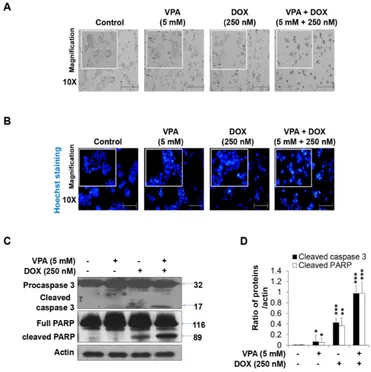
2.2. Combination treatment of VPA and DOX synergistically induces apoptotic cell death in HepG2 cells
The VPA and DOX combination treatment led to more severe changes in cell morphology (Figure 2A) than that observed for treatment with the individual drugs. Next, we conducted Hoechst nuclear staining and revealed that apoptotic nuclear condensation and fragmentation significantly increased upon the VPA and DOX combination treatment in HepG2 cells in comparison with that reported for the monotherapies (Figure 2B). In addition, cleaved caspase-3 and PARP cleavage increased significantly in the combination-treated group while VPA or DOX alone had no effect or only a slight effect (Figures 2C and D), which confirmed the synergistic cytotoxicity of the VPA and DOX combination treatment in HCC.
2.3. Combination treatment of VPA and DOX synergistically induces ROS and autophagy generation in HepG2 cells
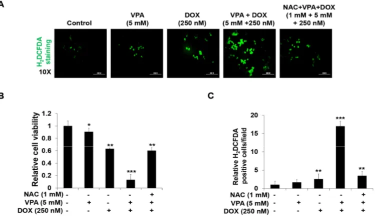
The VPA and DOX combination treatment led to an increased ROS generation (Figure 3A) compared with that reported for treatment with the individual drugs. We also found that the addition of NAC (1 mM), a ROS scavenger, suppressed the synergistic induction of apoptosis (Figure 3B) and ROS generation (Figure 3C) in HepG2 cells, which indicated that the VPA and DOX combination treatment might induce synergistic cytotoxicity through the modulation of ROS generation.
To determine the effect of the VPA and DOX combination treatment on autophagy, we used the AO staining method and found that the number of acidic organelles significantly increased following the VPA and DOX combination treatment, while treatment with either VPA or DOX alone led to very slight AO staining (Figures 4A-C). Additionally, we found that pre-incubation with 3-MA, an autophagy inhibitor, led to an apparent decrease in the synergistic induction of apoptosis (Figure 4B) and autophagy generation (Figure 4C) by the VPA and DOX combination treatment in HepG2 cells, which suggested that the combination treatment might exert a potential synergistic cytotoxic effect by regulating the autophagy pathway.
Figure 3. Combination treatment of valproic acid (VPA) and doxorubicin (DOX) synergistically enhanced reactive oxygen species (ROS) generation in HepG2 cells. (A) The 2',7'-dichlorofluorescein diacetate (H2DCFDA) fluorescence probe was used to determine ROS generation in HepG2 cells at
Figure 4. Combination treatment of valproic acid (VPA) and doxorubicin (DOX) synergistically augmented the autophagy of HepG2 cells. (A) Acridine orange (AO) staining was used to detect acidic vesicles in HepG2 cells at the indicated concentration of VPA and DOX monotherapies and combination treatment after incubation for 48 h. Images were taken using fluorescence inverted microscopy; (B) The viability of HepG2 cells was analyzed after 48-h incubation in the indicated experimental condition by using EZ-Cytox assay; (C) Percentage (%) of AO-positive cells were counted in different fields (containing at least 40 cells per field). Three independent experiments were performed and results reported as the mean ± standard deviation (SD). **p < 0.01, ***p < 0.001 compared with the control group.
2.4. VPA induces internalization of DOX in HepG2 cells through caveolae-mediated endocytosis
To understand the mechanism underlying the synergistic effect of the VPA and DOX combination treatment, we measured the cellular internalization of DOX by evaluating the fluorescence of the internalized DOX. We found that DOX internalization increased significantly upon the VPA and DOX combination treatment compared to that observed with DOX alone (Figures 5A and B). Then, we quantified the intracellular DOX concentration using a fluorescence plate-reader and confirmed that the intracellular DOX concentration markedly increased following the VPA and DOX combination treatment (Figure 5C).
Subsequently, we tried to clarify which endocytosis pathway was responsible for this phenomenon and used three different receptor-mediated endocytosis pathway inhibitors: CPZ, MβCD, and LY. We observed that DOX internalization was suppressed by MβCD pre-treatment compared to that observed with the other inhibitors (Figure 5F). The intracellular DOX concentration was confirmed to be dramatically decreased upon MβCD pre-treatment (Figure 5G), which strongly indicated that VPA might enhance DOX internalization into the cell predominantly through the caveolae-mediated endocytosis pathway.
Figure 5. Valproic acid (VPA) induces cellular doxorubicin (DOX) internalization and mediates the caveolae endocytosis pathway in HepG2 cells. (A) Cellular DOX internalization images were captured of the indicated treated cells using fluorescence inverted microscopy; (B) DOX uptake was measured in the indicated treated cells at excitation and emission wavelengths of 470 and 570 nm, respectively, by using a spectrofluorometer; (C) Intracellular DOX concentration was measured in the indicated treated cells at excitation and emission wavelengths of 470 and 570 nm, respectively, by using a spectrofluorometer; (D and E) DOX uptake and intracellular DOX concentration were measured in the indicated treated cells at the indicated temperature by using a spectrofluorometer; (F and G) DOX uptake and intracellular DOX concentration were measured in indicated treated cells with pre-incubated different endocytosis pathway inhibitors (CPZ, 10 μM; MβCD, 3 mM; and LY, 20
3. Discussion
HCC treatments using traditional radio- and chemotherapies are sometimes inefficient, partly because of their severe hepatotoxicity. In our study, we described the specific and efficient, synergistic anti-proliferative and apoptotic effect of the VPA and DOX combination in HCC cells, especially HepG2 cells (Figures 1 and 2). Recent studies have stated that the combination treatment of FDA-approved anti-HCC drugs such as DOX, sorafenib, cisplatin, interferon alpha-2b, and fluorouracil could be safely used for HCC patients [3, 4, 29], but the combination treatment showed a limited response rate (approximately 15–20%). Although DOX, as well as sorafenib, were shown to cause cell death, partially by enhancing apoptosis in HCC cells [3, 4, 29], the exact mechanism underlying the pharmacological synergy has not yet been determined. Moreover, VPA was reported to sensitize anaplastic thyroid carcinoma (ATC) cells to DOX, which caused apoptosis via the induction of histone hyperacetylation or apoptosis-related gene expression [30-32]. Concurrently, several studies demonstrated that VPA showed synergistic effects with well-known anticancer drugs, such as aspirin, flavopiridol, mitomycin C, cisplatin, adriamycin, and DOX, and could induce cell death in various cancer cells [19, 30, 33, 34]. The synergistic anticancer effect of VPA with other drugs was primarily considered to occur through histone acetylation and alteration of apoptosis related gene expression, but the underlying mechanisms of the synergistic effect and drug internalization into the cell remain unknown. In our study, calculations of the CDI confirmed the dramatically significant synergistic effect of the VPA and DOX combination, specifically in HepG2 cells (Table 1). Moreover, we revealed that VPA might exert an HDAC-independent synergistic effect with DOX on the viability of HepG2 cells. As the mechanism underlying the synergistic effect, we observed that DOX internalization, which was induced by the VPA and DOX combination treatment, occurred via the caveolae-mediated endocytosis pathway. We believe this presents novel implications for drug delivery research into the treatment of HCC.
Hoechst nuclear staining and western blot analysis of caspase-3 and PARP activation confirmed the synergistic apoptotic cell death induced by the VPA and DOX combination treatment (Figure 2). Moreover, the combination treatment resulted in an increased ROS generation and autophagy, which were clearly attenuated by ROS and autophagy inhibitors, respectively (Figures 3 and 4). Oxidative stress and autophagy have been shown to cause cell death in various types of cancers [35-38]. A previous study demonstrated that the apoptosis of solid tumor and leukemia cells was induced by the generation of ROS following treatment with an HDAC inhibitor [35]. VPA, a well-known HDAC inhibitor, induced ROS generation in several cancer cells, which was attenuated by NAC treatment [36, 39]. Concurrently, it enhanced oxidative stress in cells by increasing glutathione (GSH) levels [36]; this supported the involvement of HDAC inhibitor-mediated oxidative stress in anticancer treatment. However, our study revealed that VPA might have an HDAC-independent synergistic effect with DOX on the viability of HepG2 cells (Figure 1).
Several studies demonstrated that autophagy was induced by VPA treatment through the downregulation of the AKT/mTOR pathway in prostate cancer [40]. Moreover, it increased autophagy-mediated lymphoma cell chemo-sensitivity through IP3-mediated PRKAA activation, which was HDAC-independent [37]. Thus, we aimed to investigate whether VPA and DOX monotherapies and the combination treatment induced autophagy in HepG2 cells. AO staining is an established method for the detection of acidic compartments/vesicles in the cell cytoplasm [41]. Our observation in the cells treated with the monotherapy was consistent with previously described studies [37, 38]. In addition, the synergistic effect observed with the combination treatment led to marked changes in cell morphology and the formation of acidic vesicles; this effect was diminished by pre-treatment of the cells with 3-MA (Figure 4), which suggested that the synergistic effect on cell death by VPA and DOX monotherapies and the combination treatment might result, at least partially, from the induction of autophagy.
treatment of VPA and DOX exhibited a dramatic synergistic effect over VPA or DOX monotherapy at a clinically relevant dose. As VPA is an epilepsy drug, it may conjugate with various potent drugs, similar to other epilepsy drugs [44]. Freely available DOX can be imported to the cell by diffusion methods [24], while DOX conjugated with other chemicals or particles could pass into the cells via a cellular surface receptor [24, 25]. To explain the above studies, we hypothesized that VPA interacted with DOX and formed a particle or complex chemical-like structure, which might be the cause of increased DOX internalization. Thus, we evaluated different cellular surface receptor-mediated pathways: the clathrin-, caveolae-, and macropinocytosis-mediated DOX internalization pathways [25, 45, 46]. As expected, we observed that DOX internalization increased drastically in the VPA-DOX combination group compared with that in the free DOX group (see Figures 5 A-C). Specifically, in the current study, we found that pre-treatment with different inhibitors of cellular surface receptor-mediated endocytosis had a different effect on DOX internalization (see Figures 5 D-F). Specifically, MβCD (a caveolae-mediated endocytosis inhibitor) exhibited a dramatic inhibition of DOX internalization among the other inhibitors (Figure 5). Therefore, the synergistic effect of the VPA and DOX combination treatment might be regulated mainly through the caveolae-mediated endocytosis pathway, which consequently induced ROS and autophagy-mediated cell death. Further studies are needed to reveal the synergistic effect of the VPA and DOX combination treatment with regard to the endocytosis-mediated DOX internalization pathway.
4. Materials and Methods
4.1. Cell culture and reagents
The human normal hepatocyte cell line MIHA and the HCC cell line SNU475 were kindly gifted to us by Prof. Suk Woo Nam (The Catholic University, Seoul, Republic of Korea) and the HCC cell line HepG2 was purchased from ATCC (Manassas, VA, USA). All cell lines used in this study were grown in Roswell Park Memorial Institute (RPMI)-1640 or Dulbecco’s modified Eagle’s medium (DMEM)-high glucose media (Sigma-Aldrich, Saint Louis, MO, USA) supplemented with 10% fetal bovine serum (FBS) (Hyclone, UT, USA) and 1% penicillin-streptomycin (Invitrogen, CA, USA). The cells were incubated in humidified conditions with 5% CO2 at 37 ºC. Mycoplasma contamination of all cell lines was tested using BioMycoX®Mycoplasma PCR Detection Kit (Cellsafe, Suwon, Republic
of Korea) and short tandem repeat (STR) profiling was performed for authentication.
Valproic acid sodium salt (VPA) [Figure 1A (i)], doxorubicin hydrochloride (DOX) [Figure 1A (ii)], sodium butyrate, N-acetylcysteine (NAC), 3-methyladenine (3-MA), chlorpromazine (CPZ), methyl-β-cyclodextrin (MβCD), LY 294002 (LY), Hoechst 33258, and acridine orange hydrochloride hydrate (AO) were acquired from Sigma-Aldrich (St. Louis, MO, USA). 2',7'-Dichlorodihydrofluorescein diacetate (H2DCFDA) was purchased from Molecular Probes™ (OR, USA).
4.2. Cell viability assay
MIHA, HepG2, and SNU475 cells (1 × 10⁴ cells/well) were seeded in 96-well plates and grown overnight to confluence. The cells were then treated with the indicated dose of VPA, DOX, and the VPA and DOX combination for 48 h at 37 °C in an atmosphere of 5% CO2. After incubation for 48 h, the medium was exchanged with a fresh medium containing EZ-Cytox (Daeil Lab Service, Seoul, South Korea) and incubated for an additional 3–4 h at 37 °C in an atmosphere of 5% CO2. The absorbance was measured at 450 nm by using a microplate reader Bio-Rad x-MarkTM spectrophotometer (Bio-Rad, PA, USA) and the cell viability was calculated by comparing the viability of treated cells with that of non-treated cells, as previously described [47].
4.3. Hoechst staining for apoptotic cell detection
treated with VPA, DOX, and the VPA and DOX combination for 48 h. The cells were washed with PBS and incubated with 1 μg/mL Hoechst 33342 staining solution for 10 min. After the incubation, the cells were washed with PBS again and imaged using Nikon Eclipse TE2000-U fluorescence inverted microscopy (Nikon, Tokyo, Japan). Under the fluorescence microscope, the apoptotic cells appeared condensed and displayed fragmented nuclei.
4.4. Western blot analysis
The incubated cells were lysed using a lysis buffer [1% Triton X-100 (Sigma-Aldrich), 100 mM Tris-HCl (pH 7.5), 10% glycerol (Amresco, Solon OH, USA), 50 mM sodium fluoride (Sigma-Aldrich), 10 mM NaCl, 1 mM phenylmethylsulfonyl fluoride (PMSF; Sigma-Aldrich), 1 mM p-nitrophenyl phosphate (Sigma-Aldrich), and 1 mM sodium orthovanadate (Sigma-Aldrich)] and centrifuged at 13,000 rpm at 4 °C for 15 min. The protein supernatant was quantified using the Bradford protein assay reagent (Bio-Rad), and the proteins were resolved by either 10% or 12% sodium dodecyl sulfate polyacrylamide gel electrophoresis (SDS-PAGE). The proteins were then transferred onto nitrocellulose membranes (Bio-Rad), which were blocked with Tris-buffered 5% skimmed milk for 1 h and then incubated overnight with appropriate primary antibodies against caspase 3 (anti-rabbit, SC-7148 (1:1000)), PARP (anti-rabbit, SC-7150 (1:1000)), and actin (anti-mouse, SC-8432 (1:10000)) (Santa Cruz Biotechnology, Dallas, TX, USA) at 4 °C. After incubation with the primary antibody, the membranes were washed three times with Tris-buffered saline supplemented with Tween 20 (TBST) at room temperature followed by a 2-h incubation with anti-mouse (SC-2005, 1:1000) or anti-rabbit (SC-2004, 1:1000) secondary antibody conjugated with horse radish peroxidase (HRP) (Santa Cruz Biotechnology). The membranes were then washed three times with TBST and the protein signals were developed using an enhanced chemiluminescence (ECL) kit (Amersham Bioscience, Piscataway NJ, USA), as described previously [49]. The intensity of proteins expression was measured and normalized by actin expression using ImageJ software (National Institute of Health, Bethesda, MA, USA).
4.5. Reactive oxygen species (ROS) generation analysis
Cells (1 × 10⁵ cells/well) were seeded in 12-well plates and grown overnight to confluence. The indicated cells were then pre-incubated with the ROS scavenger, NAC (1 mM). After a 1-h pre-incubation period, the cells were treated with VPA, DOX, and the VPA and DOX combination for 48 h. Intracellular ROS levels were then analyzed using the fluorescent probe H2DCFDA [47]. Briefly, 10 μM H2DCFDA was added to the cells, which were incubated for 30 min at 37 °C in the dark. The cells were then washed twice and incubated with PBS. The fluorescent images were captured using a Nikon Eclipse TE2000-U fluorescence inverted microscope (Nikon). The ROS-generating cells were counted in different fields (containing at least 40 cells per field) and calculated relative to the control group for each experimental condition.
4.6. AO staining for autophagy detection
Cells (3 × 10⁵ cells/well) were grown overnight in 6-well plates to confluence. The cells were then incubated with VPA, DOX, and the combination of VPA and DOX for 48 h with or without pre-incubation with 3-MA. After incubation, cells were treated with 5 μg/mL AO (Sigma) in serum-free medium for 10 min. Then, the cells were washed twice with PBS and fluorescent images were captured by a Nikon Eclipse TE2000-U fluorescence inverted microscope (Nikon). Subsequently, AO-stained cells were counted in different fields (containing at least 40 cells per field) and presented as the percentage (%) of AO positive cells for each experimental condition [50].
4.7. DOX internalization analysis
membrane-bound DOX and DOX uptake was observed in cells via fluorescent microscopy and a fluorescence microplate reader (GeminiEM, Sunnyvale, CA, USA) with excitation and emission wavelengths of approximately 470 nm and 570 nm, respectively. For the quantitative analysis of the internalized DOX, cells were lysed with a protein lysis buffer and fluorescence was measured using the fluorescence microplate reader (GeminiEM) with excitation and emission wavelengths of approximately 470 nm and 570 nm, respectively. To normalize the intracellular DOX concentrations, the DOX concentration was divided by the protein concentration, as previously described [51].
4.8. Determination of the endocytosis pathways
Cells (1 × 10⁵ cells/well) were seeded in 6-well plates and grown to 60–70% confluence. To investigate the endocytosis pathway, the cells were cultured at different temperatures (37 °C, 25 °C, and 4°C) in the presence of DOX (1 μM) or the VPA and DOX combination (DOX concentration, 1 μM) for 3 h. It has been established that the incubation of cells at 4 °C could block endocytosis [28]. Concurrently, cells were pre-treated for 1 h with various kinds of specific endocytosis inhibitors: CPZ (10 μM), an inhibitor of clathrin-mediated endocytosis [51]; MβCD (3 mM), an inhibitor of caveolae-mediated endocytosis [51]; and LY (20 μM), an inhibitor of macropinocytosis [46]. After incubation for 3 h with DOX or the VPA and DOX combination, the cells were washed twice with PBS and the fluorescent intensity of DOX in the cells was evaluated using the fluorescence microplate reader (GeminiEM) with excitation and emission wavelengths of approximately 470 nm and 570 nm, respectively. For the quantitative analysis of the internalized DOX, the cells were lysed with a protein lysis buffer and the fluorescence was measured by the fluorescence microplate reader (GeminiEM) with excitation and emission wavelengths of approximately 470 nm and 570 nm, respectively. To normalize the intracellular DOX concentrations, the DOX concentration was divided by the protein concentration.
4.9. Statistical analysis
All experiments were conducted independently at least three times and the results were shown as the mean ± standard deviation (SD). Data were analyzed using GraphPad InStat version 3 program (Graphpad, San Diego, CA, USA). For statistical analyses, analysis of variance (ANOVA) was performed with a Bonferroni adjustment to compare the treated group with the non-treated group. A value was considered statistically significant when p < 0.05.
5. Conclusions
Overall, as an indication of the synergistic mechanism, our study demonstrated that the combination treatment of VPA and DOX was effective in the induction of cell death of HCC through the regulation of ROS and autophagy. Moreover, DOX internalization was mediated by the caveolae endocytosis pathway. Therefore, our study uncovered the potential effect of the VPA and DOX combination treatment with regard to cell death, including induction of cellular ROS generation, autophagy, and the caveolae-mediated endocytosis pathway. Our results might also indicate the potential role of the combination treatment of VPA and DOX by helping us understand their synergistic effect on HCC cell death through DOX internalization. These results may offer important insights into drug delivery research.
Acknowledgments: This work was supported by a grant from the National Research Foundation (NRF) funded by the Korean government (2013M3A9D3045880 and 2015R1A5A1009701).
Author Contributions: S.K.S. and S.G.C. designed experiments; S.K.S. performed most of the cellular and molecular experiments and analyzed the results; Y.F.Y., K.S.K., G.M.Y., A.D., and H.Y.C. partially contributed to the cellular and molecular experiments and analyzed the results; S.K.S. and S.G.C. wrote the manuscript. All authors reviewed and approved the manuscript.
Abbreviations
VPA valproic acid
DOX doxorubicin
HCC hepatocellular carcinoma
CDI coefficient of drug interaction PARP poly(ADP-ribose) polymerase ROS reactive oxygen species
TACE transarterial chemoembolization HDAC histone deacetylase
ATCC american type culture collection RPMI roswell park memorial institute DMEM dulbecco's modified eagle medium
FBS fetal bovine serum
STR short tandem repeat NAC N-acetylcysteine 3-MA 3-methyladenine CPZ chlorpromazine MβCD methyl-β-cyclodextrin LY LY294002
AO acridine orange
H2DCFDA 2', 7'-dichlorodihydrofluorescein diacetate
PBS phosphate-buffered saline PMSF phenylmethylsulfonyl fluoride
SDS-PAGE sodium dodecyl sulfate polyacrylamide gel electrophoresis TBST tris-buffered saline tween 20
HRP horse radish peroxidase ECL enhanced chemiluminescence SD standard deviation
ANOVA analysis of variance
HDEI histone deacetylase inhibitor
ATC anaplastic thyroid carcinoma
GSH glutathione
References
1. WHO, GLOBOCAN 2012: Estimated cancer incidence, mortality and prevalence worldwide in 2012. Lyon, France: International Agency for Research on Cancer 2014.
2. Attwa, M. H.; El-Etreby, S. A. Guide for diagnosis and treatment of hepatocellular carcinoma. World J. Hepatol. 2015, 7, 1632-1651.
3. Cao, H.; Phan, H.; Yang, L. X. Improved chemotherapy for hepatocellular carcinoma. Anticancer Res. 2012, 32, 1379-1386.
4. Yeo, W.; Mok, T. S.; Zee, B.; Leung, T. W.; Lai, P. B.; Lau, W. Y.; Koh, J.; Mo, F. K.; Yu, S. C.; Chan, A. T.; Hui, P.; Ma, B.; Lam, K. C.; Ho, W. M.; Wong, H. T.; Tang, A.; Johnson, P. J. A randomized phase III study of doxorubicin versus cisplatin/interferon alpha-2b/doxorubicin/fluorouracil (PIAF) combination chemotherapy for unresectable hepatocellular carcinoma. J. Natl. Cancer Inst. 2005, 97, 1532-1538.
5. Fan, L. L.; Song, B.; Sun, G. P.; Ma, T.; Zhong, F.; Wei, W. Endoplasmic Reticulum Stress-Induced Resistance to Doxorubicin Is Reversed by Paeonol Treatment in Human Hepatocellular Carcinoma Cells. PloS one 2013, 8, e62627.
6. Xiang, Q. F.; Zhang, D. M.; Wang, J. N.; Zhang, H. W.; Zheng, Z. Y.; Yu, D. C.; Li, Y. J.; Xu, J.; Chen, Y. J.; Shang, C. Z. Cabozantinib reverses multidrug resistance of human hepatoma HepG2/adr cells by modulating the function of P-glycoprotein. Liver Int. 2015, 35, 1010-1023.
8. Perucca, E. Pharmacological and therapeutic properties of valproate: a summary after 35 years of clinical experience. CNS drugs 2002, 16, 695-714.
9. Carriere, C. H.; Kang, N. H.; Niles, L. P. Neuroprotection by valproic acid in an intrastriatal rotenone model of Parkinson's disease. Neuroscience 2014, 267, 114-121.
10. Hu, J. P.; Xie, J. W.; Wang, C. Y.; Wang, T.; Wang, X.; Wang, S. L.; Teng, W. P.; Wang, Z. Y. Valproate reduces tau phosphorylation via cyclin-dependent kinase 5 and glycogen synthase kinase 3 signaling pathways. Brain Res. Bull. 2011, 85, 194-200.
11. Penas, C.; Verdu, E.; Asensio-Pinilla, E.; Guzman-Lenis, M. S.; Herrando-Grabulosa, M.; Navarro, X.; Casas, C. Valproate reduces CHOP levels and preserves oligodendrocytes and axons after spinal cord injury. Neuroscience 2011, 178, 33-44.
12. Yi, J.; Zhang, L.; Tang, B.; Han, W.; Zhou, Y.; Chen, Z.; Jia, D.; Jiang, H. Sodium valproate alleviates neurodegeneration in SCA3/MJD via suppressing apoptosis and rescuing the hypoacetylation levels of histone H3 and H4. PloS one 2013, 8, e54792.
13. Schmid, M. M.; Freudenmann, R. W.; Keller, F.; Connemann, B. J.; Hiemke, C.; Gahr, M.; Kratzer, W.; Fuchs, M.; Schonfeldt-Lecuona, C. Non-fatal and fatal liver failure associated with valproic acid. Pharmacopsychiatry 2013, 46, 63-68.
14. Silva, M. F.; Aires, C. C.; Luis, P. B.; Ruiter, J. P.; L, I. J.; Duran, M.; Wanders, R. J.; Tavares de Almeida, I. Valproic acid metabolism and its effects on mitochondrial fatty acid oxidation: a review. J. Inherit. Metab. Dis. 2008, 31, 205-216.
15. Nie, D.; Huang, K.; Yin, S.; Li, Y.; Xie, S.; Ma, L.; Wang, X.; Wu, Y.; Xiao, J. Synergistic/additive interaction of valproic acid with bortezomib on proliferation and apoptosis of acute myeloid leukemia cells. Leuk. Lymphoma 2012, 53, 2487-2495.
16. Sidana, A.; Wang, M.; Shabbeer, S.; Chowdhury, W. H.; Netto, G.; Lupold, S. E.; Carducci, M.; Rodriguez, R. Mechanism of growth inhibition of prostate cancer xenografts by valproic acid. J. Biomed. Biotechnol. 2012, 2012, 180363.
17. Xie, C.; Edwards, H.; Lograsso, S. B.; Buck, S. A.; Matherly, L. H.; Taub, J. W.; Ge, Y. Valproic acid synergistically enhances the cytotoxicity of clofarabine in pediatric acute myeloid leukemia cells. Pediatr. Blood Cancer 2012, 59, 1245-1251.
18. Yamauchi, Y.; Izumi, Y.; Asakura, K.; Fukutomi, T.; Serizawa, A.; Kawai, K.; Wakui, M.; Suematsu, M.; Nomori, H. Lovastatin and valproic acid additively attenuate cell invasion in ACC-MESO-1 cells. Biochem. Biophys. Res. Commun. 2011, 410, 328-332.
19. Li, X.; Zhu, Y.; He, H.; Lou, L.; Ye, W.; Chen, Y.; Wang, J. Synergistically killing activity of aspirin and histone deacetylase inhibitor valproic acid (VPA) on hepatocellular cancer cells. Biochem. Biophys. Res. Commun. 2013, 436, 259-264.
20. Machado, M. C.; Bellodi-Privato, M.; Kubrusly, M. S.; Molan, N. A.; Tharcisio, T., Jr.; de Oliveira, E. R.; D'Albuquerque, L. A. Valproic acid inhibits human hepatocellular cancer cells growth in vitro and in vivo. J. Exp. Ther. Oncol. 2011, 9, 85-92.
21. Tatebe, H.; Shimizu, M.; Shirakami, Y.; Sakai, H.; Yasuda, Y.; Tsurumi, H.; Moriwaki, H. Acyclic retinoid synergises with valproic acid to inhibit growth in human hepatocellular carcinoma cells. Cancer Lett. 2009, 285, 210-217.
22. Hao, J. Q.; Li, Q.; Xu, S. P.; Shen, Y. X.; Sun, G. Y. Effect of lumiracoxib on proliferation and apoptosis of human nonsmall cell lung cancer cells in vitro. Chin. Med. J. 2008, 121, 602-607.
23. Zhang, J.; Yi, M.; Zha, L.; Chen, S.; Li, Z.; Li, C.; Gong, M.; Deng, H.; Chu, X.; Chen, J.; Zhang, Z.; Mao, L.; Sun, S. Sodium Butyrate Induces Endoplasmic Reticulum Stress and Autophagy in Colorectal Cells: Implications for Apoptosis. PloS one 2016, 11, e0147218.
24. Cai, S.; Alhowyan, A. A.; Yang, Q.; Forrest, W. C.; Shnayder, Y.; Forrest, M. L. Cellular uptake and internalization of hyaluronan-based doxorubicin and cisplatin conjugates. J. Drug Target 2014, 22, 648-657. 25. Majumdar, S.; Tejo, B. A.; Badawi, A. H.; Moore, D.; Krise, J. P.; Siahaan, T. J. Effect of modification of the physicochemical properties of ICAM-1-derived peptides on internalization and intracellular distribution in the human leukemic cell line HL-60. Mol. Pharm. 2009, 6, 396-406.
26. Marsh, M.; McMahon, H. T. The structural era of endocytosis. Science 1999, 285, 215-220. 27. Mukherjee, S.; Ghosh, R. N.; Maxfield, F. R. Endocytosis. Physiol. Rev. 1997, 77, 759-803.
29. Bruix, J.; Raoul, J. L.; Sherman, M.; Mazzaferro, V.; Bolondi, L.; Craxi, A.; Galle, P. R.; Santoro, A.; Beaugrand, M.; Sangiovanni, A.; Porta, C.; Gerken, G.; Marrero, J. A.; Nadel, A.; Shan, M.; Moscovici, M.; Voliotis, D.; Llovet, J. M. Efficacy and safety of sorafenib in patients with advanced hepatocellular carcinoma: subanalyses of a phase III trial. J. Hepatol. 2012, 57, 821-829.
30. Catalano, M. G.; Fortunati, N.; Pugliese, M.; Poli, R.; Bosco, O.; Mastrocola, R.; Aragno, M.; Boccuzzi, G. Valproic acid, a histone deacetylase inhibitor, enhances sensitivity to doxorubicin in anaplastic thyroid cancer cells. J. Endocrinol. 2006, 191, 465-472.
31. Kim, T. H.; Yoo, Y. H.; Kang, D. Y.; Suh, H.; Park, M. K.; Park, K. J.; Kim, S. H. Efficacy on anaplastic thyroid carcinoma of valproic acid alone or in combination with doxorubicin, a synthetic chenodeoxycholic acid derivative, or lactacystin. Int. J. Oncol. 2009, 34, 1353-1362.
32. Rho, J. H.; Kang, D. Y.; Park, K. J.; Choi, H. J.; Lee, H. S.; Yee, S. B.; Yoo, Y. H. Doxorubicin induces apoptosis with profile of large-scale DNA fragmentation and without DNA ladder in anaplastic thyroid carcinoma cells via histone hyperacetylation. Int. J. Oncol. 2005, 27, 465-471.
33. Kwak, M. S.; Yu, S. J.; Yoon, J. H.; Lee, S. H.; Lee, S. M.; Lee, J. H.; Kim, Y. J.; Lee, H. S.; Kim, C. Y. Synergistic anti-tumor efficacy of doxorubicin and flavopiridol in an in vivo hepatocellular carcinoma model. J. Cancer Res. Clin. Oncol. 2015, 141, 2037-2045.
34. Wang, D.; Jing, Y.; Ouyang, S.; Liu, B.; Zhu, T.; Niu, H.; Tian, Y. Inhibitory effect of valproic acid on bladder cancer in combination with chemotherapeutic agents in vitro and in vivo. Oncol. Lett. 2013, 6, 1492-1498.
35. Eot-Houllier, G.; Fulcrand, G.; Magnaghi-Jaulin, L.; Jaulin, C. Histone deacetylase inhibitors and genomic instability. Cancer Lett. 2009, 274, 169-176.
36. Han, B. R.; You, B. R.; Park, W. H. Valproic acid inhibits the growth of HeLa cervical cancer cells via caspase-dependent apoptosis. Oncol. Rep. 2013, 30, 2999-3005.
37. Ji, M. M.; Wang, L.; Zhan, Q.; Xue, W.; Zhao, Y.; Zhao, X.; Xu, P. P.; Shen, Y.; Liu, H.; Janin, A.; Cheng, S.; Zhao, W. L. Induction of autophagy by valproic acid enhanced lymphoma cell chemosensitivity through HDAC-independent and IP3-mediated PRKAA activation. Autophagy 2015, 11, 2160-2171.
38. Park, J. H.; Choi, S. H.; Kim, H.; Ji, S. T.; Jang, W. B.; Kim, J. H.; Baek, S. H.; Kwon, S. M. Doxorubicin Regulates Autophagy Signals via Accumulation of Cytosolic Ca2+ in Human Cardiac Progenitor Cells. Int. J. Mol. Sci. 2016, 17, 1680.
39. Ungerstedt, J. S.; Sowa, Y.; Xu, W. S.; Shao, Y.; Dokmanovic, M.; Perez, G.; Ngo, L.; Holmgren, A.; Jiang, X.; Marks, P. A. Role of thioredoxin in the response of normal and transformed cells to histone deacetylase inhibitors. Proc. Natl. Acad. Sci. U. S. A. 2005, 102, 673-678.
40. Xia, Q.; Zheng, Y.; Jiang, W.; Huang, Z.; Wang, M.; Rodriguez, R.; Jin, X. Valproic acid induces autophagy by suppressing the Akt/mTOR pathway in human prostate cancer cells. Oncol. Lett. 2016, 12, 1826-1832. 41. Paglin, S.; Hollister, T.; Delohery, T.; Hackett, N.; McMahill, M.; Sphicas, E.; Domingo, D.; Yahalom, J. A
novel response of cancer cells to radiation involves autophagy and formation of acidic vesicles. Cancer Res. 2001, 61, 439-444.
42. Graziani, G.; Tentori, L.; Portarena, I.; Vergati, M.; Navarra, P. Valproic acid increases the stimulatory effect of estrogens on proliferation of human endometrial adenocarcinoma cells. Endocrinology 2003, 144, 2822-2828.
43. Takai, N.; Desmond, J. C.; Kumagai, T.; Gui, D.; Said, J. W.; Whittaker, S.; Miyakawa, I.; Koeffler, H. P. Histone deacetylase inhibitors have a profound antigrowth activity in endometrial cancer cells. Clin. Cancer Res. 2004, 10, 1141-1149.
44. Herzog, A. G.; Farina, E. L.; Blum, A. S. Serum valproate levels with oral contraceptive use. Epilepsia 2005, 46, 970-971.
45. Rejman, J.; Oberle, V.; Zuhorn, I. S.; Hoekstra, D. Size-dependent internalization of particles via the pathways of clathrin-and caveolae-mediated endocytosis. Biochem. J. 2004, 377, 159-169.
46. Yao, W.; Li, K.; Liao, K. Macropinocytosis contributes to the macrophage foam cell formation in RAW264.7 cells. Acta. Biochim. Biophys. Sin. 2009, 41, 773-780.
48. Pitchaimani, A.; Renganathan, A.; Cinthaikinian, S.; Premkumar, K. Photochemotherapeutic effects of UV-C on acridine orange in human breast cancer cells: potential application in anticancer therapy. RSC Advances 2014, 4, 22123-22128.
49. Saha, S. K.; Choi, H. Y.; Kim, B. W.; Dayem, A. A.; Yang, G. M.; Kim, K. S.; Yin, Y. F.; Cho, S. G. KRT19 directly interacts with beta-catenin/RAC1 complex to regulate NUMB-dependent NOTCH signaling pathway and breast cancer properties. Oncogene 2017, 36, 332-349.
50. Arthur, C. R.; Gupton, J. T.; Kellogg, G. E.; Yeudall, W. A.; Cabot, M. C.; Newsham, I. F.; Gewirtz, D. A. Autophagic cell death, polyploidy and senescence induced in breast tumor cells by the substituted pyrrole JG-03-14, a novel microtubule poison. Biochem. Pharmacol. 2007, 74, 981-991.
51. Hu, Q.; Sun, W.; Qian, C.; Wang, C.; Bomba, H. N.; Gu, Z. Anticancer Platelet-Mimicking Nanovehicles. Adv. Mater. 2015, 27, 7043-7050.