Nowadays smartphone utilization for disease diagnosis and remote health care applications has become promising due to their ubiquity. Here, a novel convolutional neural network method for detecting keratoconus that is wholly implemented on a smartphone is proposed. The proposed method provides accurate detection of over 72.9% for all stages of keratoconus. Preliminary results indicate 90%, 83%, 64% and 52% detection rate for severe, advanced, moderate and mild stages of disease, respectively.
Keywords: Keratoconus, Smartphone, Cornea, Convolutional Neural Network.
INTRODUCTION
Keratoconus (KC) represents a progressive corneal thinning. Keratoconus is a severe corneal dystrophy in the United States and 1 in every 2,000 Americans has the disease nowadays [1-3]. Keratoconus causes the middle cornea to become thinner and slowly bulge outwards, turning cornea into a rounded-cone shape that affects the focal point that impairs vision [4]. Such anomalous curvature weakens the refraction of the cornea. As the cornea turns out to be increasingly unpredictable fit as a fiddle, it causes dynamic myopia and sporadic astigmatism making extra issues, for example, mutilated and obscured vision. Early conclusion of keratoconus is critical in members looking for medical procedure since it can forestall movement of the pathology after medical procedure and make it symptomatic, which makes the requirement for analytic tests that give early and high affectability
When corneas shape changes, it produces vision impairment and eventually blindness. Fortunately, the keratoconus progression can be controlled if it diagnosed in early stages using a corneal surgery technique called corneal crosslinking. Currently, keratoconus can be detected by several clinical exams and equipment’s: optical coherence tomography (OCT) [4], video keratography [5], Scheimpflug camera [6] and laser interferometry [7].
The most generally utilized current clinical strategies to recognize, analyze, and screen the development of keratoconus morphology are modernized videokeratoscopy (corneal geology), corneal tomography, and OCT [9]. The corneal geography is a non-intrusive restorative imaging system which gauges the surface bend of the cornea and is of basic significance in deciding the nature of vision. In any case, geography gadgets are huge, costly and not versatile. Besides, it must be worked by a prepared expert. Right now, propose cell phone-based recognition keratoconus strategy which recognizes keratoconus in a moderate and simple manner with keeping up its identification execution contrasted with the customary geography-based location technique [1, 5].
The most widely used current clinical exams for diagnosing keratoconus and monitor its progression is videokeratoscopy ( also known as corneal topography) [8]. Corneal topography is an imaging method which measures the elevation and curvature of cornea building 3D elevation and sagittal curvature map. However, topography devices heavy, bulky devices and they require an eye care specialist to operate. Keratoconus detection using smartphones have been proposed by researchers using support vector machine (SVM) classifier [9-11]. In [9] the researchers they used a specially made gadget with SVM classifier using smartphone to detect keratoconus. Detecting keratoconus using corneal curvature features has been used in several researches [12, 13]. In [9] the authors have used a novel method for detecting keratoconus from side images acquired by smartphone and they have used a threshold base classification method to distinguish different stages of the disease by measuring the anterior curvature slope.
Here, a smartphone-based detection method for diagnosing keratoconus in an affordable and easy way with maintaining its detection performance compared to the conventional topography-based detection method is proposed. Our proposed smartphone-based method is expected to be distributed to deprived areas being short of medical services since smartphone can be used anywhere and anytime.
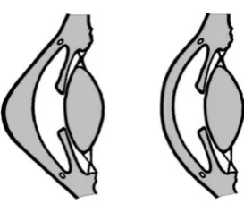
Figure 1.Normal cornea (right) and cornea with keratoconus (left)
Methodology: For data acquisition a OnePlus 3 phone was used to capture images from the eyes in this study as shown in the Figure 1. OnePlus has a 16-megapixel (3264×2448) primary camera on the rear side. For this study we use the maximum resolution to acquire the data.
The experiment involved 100 eyes from 50 volunteer participants, where 50 eyes were diagnosed with keratoconus and 50 eyes as normal. We also had the corneal topography exams from all the 100 eyes. All participants data were acquired at Dena eye clinic and an ophthalmologist diagnosis was used as a gold standard validation.
The data acquisition procedure started with recording a panoramic image from the participant's eye as shown in Figure 1 and importing the images from corneal topography device into the smartphone. The participants were asked not to move during the measurement procedure. We didn’t use smartphones flashlight for data recording we just used ambient light with auto focusing
(a) (b) (c) (d)
Figure 2. Example of keratoconus eye and healthy eye and the data acquisition procedure: (a) Normal eye, (b) eye with keratoconus, (c) corneal topography sagittal curvature map of diseased eye, and (d) image acquisition using smartphone camera.
First stage is image cropping to crop the cornea from the face image and extract the face skin, eyelash, eyelid and sclera. Our first approach system for detecting keratoconus was using shape matching techniques but because of the different distances between the eye and (also the rotate and tilt in the cameras relatively to the eye position the smartphone camera the accuracy was significantly low, so we used another approach by measuring the slope of the corneas curvature. Behaving as if it is a signal and measuring the slope at different parts of the cornea. In the first stage the corneal anterior curvature features were acquired and combined with the results from corneal topography sagittal map they were fed the neural network for training the system to detect diseased eyes.
Image cropping is used to remove the distracting content because we must only focus on the cornea. 80% of data is used for training and 20% is left out for testing.
(c) (d)
Figure 2: Demonstration of images acquired from smartphone and corneal topography device for a diseased and healthy eye. (a) Advanced KC cornea, (b) normal cornea, (c)_corneal topography of a diseased
eye, and (d) corneal topography of healthy eye.
After the preprocessing stage, we extract the corneas curvature by using the curvature gradient slope (Figure 3).
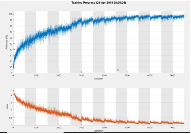
Figure 4.training processing time and error function
We finally compared the participants' cornea with the normal cornea. The advantage of our method is that it doesn't require any image normalization. The topography exam results for the same eyes are shown in Figure 4.
However, here a CNN with 6 layers including 4 convolutional layers two fully connected layers and a soft max layer is proposed. to measure the corneal curvature [10]. We implement our method and find the axial of the cornea curve as shown in Figure 3. We then find the slope of the corneal curvature in different points by splitting it into 18 points with taking samples every 10 degrees. We finally compared it with the normal cornea’s slopes.
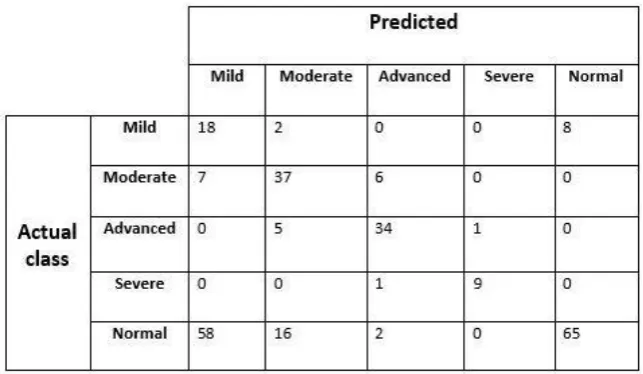
Results: We had the medical history (topography) and the ophthalmologist's diagnosis of all of the 50 participants (100 eyes) we compared their results with our method as shown in Table Ⅰ. The proposed CNN architecture was able to diagnose severe, advanced, moderate, and mild stages of keratoconus disease with accuracies of 90%, 83%, 64%, and 52% respectively. Even the highly advanced recent methods have issues detecting mild stage keratoconus due to its difficulty in separating from astigmatism. The latest computerized topography accuracy is around 90% [13]. The detection highly depends on the smartphone’s camera quality and distance, focus, light conditions and camera settings.
Conclusion:
Table Ⅰ. Keratoconus detection accuracies and 100 times for normal eyes (right)
REFERENCES
[1] R. H. Kennedy, W. M. Bourne, and J. A. Dyer, "A 48-year clinical and epidemiologic study of keratoconus," American journal of ophthalmology, vol. 101, no. 3, pp. 267-273, 1986.
[2] N. NIH, "Eye Disease Statistics," 2019. [Online]. Available: https://www.nei.nih.gov/sites/default/files/2019-04/NEI_Eye_Disease_Statistics_Factsheet_2014_V10.pdf. [3] Y. S. Rabinowitz, "Keratoconus," Survey of ophthalmology, vol. 42, no. 4, pp. 297-319, 1998.
[4] Y. Li, W. Chamberlain, O. Tan, R. Brass, J. L. Weiss, and D. Huang, "Subclinical keratoconus detection by pattern analysis of corneal and epithelial thickness maps with optical coherence tomography," Journal of Cataract & Refractive Surgery, vol. 42, no. 2, pp. 284-295, 2016.
[5] F. Cavas-Martínez, E. De la Cruz Sánchez, J. N. Martínez, F. F. Cañavate, and D. Fernández-Pacheco, "Corneal topography in keratoconus: state of the art," Eye and vision, vol. 3, no. 1, p. 5, 2016.
[6] U. De Sanctis, A. Missolungi, B. Mutani, L. Richiardi, and F. M. Grignolo, "Reproducibility and repeatability of central corneal thickness measurement in keratoconus using the rotating Scheimpflug camera and ultrasound pachymetry," American journal of ophthalmology, vol. 144, no. 5, pp. 712-718. e1, 2007.
[7] F. Jongsma, J. De Brabander, and F. Hendrikse, "Review and classification of corneal topographers," Lasers in medical science, vol. 14, no. 1, pp. 2-19, 1999.
[8] F. Versaci and G. Vestri, "Instrumentation for Diagnosis of Keratoconus," in Keratoconus: Springer, 2017, pp. 53-63.
[9] B. Askarian, F. Tabei, A. Askarian, and J. W. Chong, "An affordable and easy-to-use diagnostic method for keratoconus detection using a smartphone," in Medical Imaging 2018: Computer-Aided Diagnosis, 2018, vol. 10575: International Society for Optics and Photonics, p. 1057512.
[10] B. Askarian, J. W. Chong, and F. Tabei, "DIAGNOSTIC TOOL FOR EYE DISEASE DETECTION USING SMARTPHONE," ed: US Patent App. 16/294,902, 2019.
[11] B. Askarian, F. Tabei, G. A. Tipton, and J. W. Chong, "Novel Keratoconus Detection Method Using Smartphone," in 2019 IEEE Healthcare Innovations and Point of Care Technologies, (HI-POCT), Bethesda, MD, USA, 2019: IEEE, pp. 60-62.
[12] R. G. do Norte, "DESIGN AND DEVELOPMENT OF AN ULTRAPORTABLE CORNEAL TOPOGRAPHER FOR SMARTPHONES AS A LOW COST NEW TOOL FOR PREVENTING BLINDNESS CAUSED BY KERATOCONUS," International Journal of Latest Research in Science and Technology, vol. 4, no. 3, pp. 72-76, 2015.
[13] M. M. Daud, W. M. D. W. Zaki, A. Hussain, and H. A. Mutalib, "Detection of keratoconus in anterior segment photographed images using corneal curvature features," Indonesian Journal of Electrical Engineering and Computer Science, vol. 13, no. 3, pp. 1191-1198, 2019.